Introduction
Bone tissue, the primarily structural material of vertebrate animals, has a complex structure in a mineral–biopolymer system and its components are primary hydroxyapatite (HAP) and collagen [Reference Martin1]. HAP with a Ca/P stoichiometric ratio of 1.66–1.67 exists in bone tissue, mainly displays a rod-shaped structure with a width of about 20 nm and length of 40–60 nm and is arranged along collagen fibers [Reference Eliaz, Sridhar, Kamachi Mudali and Raj2]. Recently, HAP is widely used in orthopedics, implant materials, and drug delivery systems because of its biocompatibility, osteoconduction, and compositional similarity to that of mineralized bone [Reference Yi, Wang, Tian, Shu, Yang, Xiao, Li and Liao3, Reference Abdulkareem, Memarzadeh, Allaker, Huang, Pratten and Spratt4, Reference Zhang, Yan, Jin, Jia, Zhou and Lv5]. Additionally, hydroxyapatite nanoparticles (nHAPs) are capable of inhibiting the propagation of the cancer cells [Reference Tang, Yuan, Liu, Wu, Lu and Qian6, Reference Ghosh, Nandi, Kundu, Datta, De, Roy and Basu7]. The preparation of HAP has attracted considerable attention owing to its excellent properties, and the particle sizes of HAP can be made up to nano scale (less than 100 nm), which is more similar to the structure in the bone tissue. Up to now, many researchers have developed various synthetic methods, such as combustion synthesis [Reference Xiao, Zhou, Chen, Jiang, Zhang, Luo and Zhang8], spray freezing method [Reference Ben-Arfa, Salvado, Ferreira and Pullar9], sol–gel method [Reference Wang, Hon, Chen, Yen, Hung, Ko and Shih10], hydrolysis method [Reference Jokic, Mitric, Radmilovic, Drmanic, Petrovic and Janackovic11], hydrothermal method [Reference Mondal, Bardhan, Mondal, Dey, Mukhopadhyay, Roy, Guha and Roy12], and synthesis from biogenic sources [Reference Xu, Zeiger and Suslick13], to obtain HAP with different morphologies. However, combustion synthesis and hydrothermal method need a high reaction temperature and complex process, while combustion synthesis and spray freezing method need high-level equipment, which will lead to high cost. Besides, long preparation time exists in hydrothermal method and sol–gel method. With the development of the society, all walks of life are increasingly pursuing the concept of green environmental protection and sustainable development, which is also applicable to scientific research industry. Therefore, researchers should take efforts to develop a synthetic method with advantages of high efficiency and environmentally friendly to adapt to the new concept of sustainable development.
In recent years, sonochemical method has been applied to prepare nanomaterial and extract active ingredients from plants due to its high efficiency and environmental-friendliness [Reference Qi, Zhu, Wu, Sun, Jiang, Zhang, Wu and Chen14, Reference Wong, Li, Li, Razmovskinaumovski and Chan15, Reference Sadat-Shojai, Khorasani, Dinpanah-Khoshdargi and Jamshidi16]. The advantages of sonochemical method for synthesis of nanomaterial include improving the phase purity and accelerating the reaction rate [Reference Rouhani, Taghavinia and Rouhani17, Reference Giardina and Fanovich18]. The preparation of nHAPs via ultrasonic method has been reported, but it was limited to the single-frequency ultrasonic method and the preparation process needs a further aging process [Reference Wong, Li, Li, Razmovskinaumovski and Chan15, Reference Liu and Hsieh19, Reference Tatake and Pandit20]. The previous investigations have demonstrated that the acoustic cavitation effect and energy efficiency of the dual-frequency ultrasonic systems can be significantly enhanced compared with the single-frequency ultrasound [Reference Deng, Yu, Liu and Bi21, Reference Zeng, Qiu and Lu22, Reference Zhang23]. The dual-frequency ultrasonic method can be carried out to understand that a second sound wave is introduced into the cavity dynamics when compared with single-frequency ultrasound [Reference Zeng, Qiu and Lu22]. The introduction of a second sound wave can reduce the formation of standing waves, improve the dispersion of reagent, and make the cavitational activity distribution more uniform in the liquid medium [Reference Zeng, Qiu and Lu22]. The dual-frequency ultrasonic method can be performed by combining an ultrasonic horn and an ultrasonic bath simultaneously. There are some investigations focus on increasing extraction of active ingredients from plants by the dual-frequency ultrasonic method [Reference Holzwarth and Gibson24, Reference Kavitha, Subramanian, Vinoth, Narayanan, Venkatesh and Esakkiraja25]. However, the preparation of nHAPs in the dual-frequency ultrasonic system has been rarely reported to date.
Here, to develop a novel approach to rapidly synthesize nHAPs, the dual-frequency ultrasonic method was applied to prepare nHAPs. To obtain the optimal preparation conditions, the effect of ultrasonic power and time of the dual-frequency ultrasonic system on morphology and phase composition of nHAPs were investigated. The possible formation mechanism of nHAPs in the dual-frequency ultrasonic systems was briefly explored. Furthermore, tetracycline hydrochloride (TH) was used as a drug model to study the loading and release behaviors of the nHAPs. To verify cytocompatibility and hemocompatibility of nHAPs, the in vitro cytotoxicity evaluation and hemolytic experiment were carried out on the synthesized nHAPs.
Results and discussion
Characterization of the obtained powder
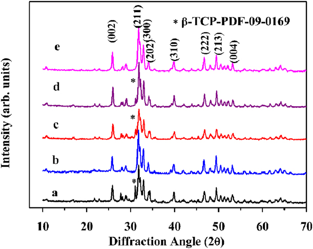
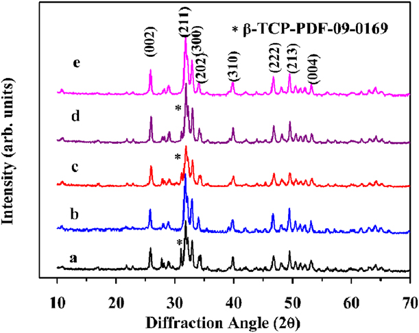
Figure 1 shows X-ray diffraction (XRD) pattern of the products synthesized with different ultrasonic parameters in the dual-frequency ultrasonic system. The major phase peaks were identified at (002), (004), (211), (300), (202), (310), (222), and (213) planes in all these samples by comparing with standard data of the HAP reference (JCPDS-09-0432). No trace of secondary crystalline phase was observed in U280T30 and U350T30, confirming the high purity of the two products. Stoichiometry of 1.66 was also obtained in samples U280T30 and U350T30, but non-stoichiometry appeared in other samples. It reveals that ultrasonic time and power affected the phase composition and stoichiometry of the as-synthesized samples. One weak phase peak of tricalcium phosphate (β-TCP, JCPDS-09-0169) was observed in U280T10 and U280T45, indicating the presence of β-TCP phase. As the power was fixed at 280 W, longer or shorter ultrasonic time was not contributed to the synthesis of single-phase HAP crystals. Besides, a characteristic peak of â-TCP was also observed in U140T30, but this phenomenon was absent in U280T30 and U350T30. It reveals that pure HAP crystals cannot be obtained at 140 W for 30 min. It may attribute to subdued cavitation effect at low power.
XRD patterns of the samples: (a) U280T10, (b) U280T30, (c) U280T45, (d) U140T30, and (e) U350T30.
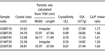
The crystallite sizes (D) and fraction of crystallinity (X c) assigned to the (002) plane of HAP crystals were calculated by Eqs. (1) and (2) and presented in Table I. The results indicate that the effect of ultrasonic time and power on crystallite sizes is not significant. There was no apparent relationship between sonication parameters and crystallite sizes. However, the difference in crystallinity appeared in different samples. The crystallinity of U280T30 reached to a maximum value of 0.99, higher than other samples. However, the crystallinity decreased as prolonging sonication time and increasing ultrasonic power. Therefore, single-phase HAP crystals with stoichiometry and high crystallinity can be obtained by ultrasonic irradiation at 280 W for 30 min.
The crystallite sizes (D), fraction of crystallinity (X c), SSA values, Ca/P molar ratio, and particle sizes of samples.

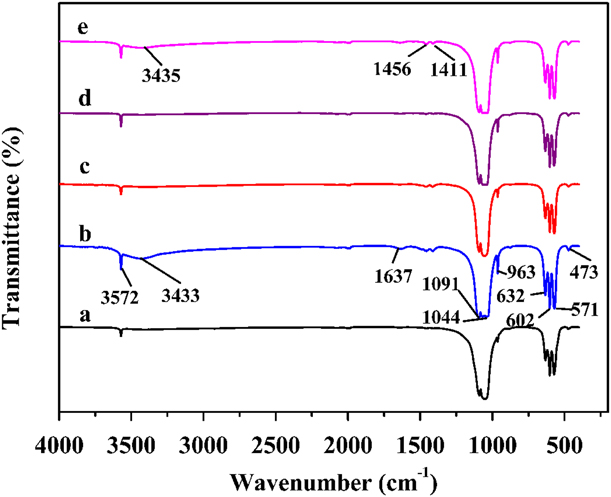
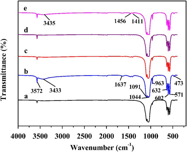
Furthermore, the Fourier transform infrared (FT-IR) spectroscopy was carried out to characterize the functional groups of samples produced in dual-frequency ultrasonic system. As shown in Fig. 2, the representative FT-IR spectrum showed characteristic functional groups of nHAPs in all these products, but the peaks were sharper in U280T30 than any other samples. The peaks at 1091 cm−1 and 1044 cm−1 could be assigned to the ν3 mode of P–O bonds, whereas the bands at around 602 cm−1 and 571 cm−1 were corresponding to the ν4 mode of PO43− group [Reference Jankoviæ, Erakoviæ, Mitriæ, Matiæ, Juraniæ, Tsui, Tang, Miškoviæ-Stankoviæ, Rhee and Park26]. The bands at 963 cm−1 and 473 cm−1 were ascribed to the ν1 and ν2 modes, respectively [Reference Castro, Kuhn, Jensen, Ferreira, Rocha, Vicente and Teixeira27, Reference Sadat-Shojai, Khorasani and Jamshidi28]. The bands of hydroxyl group of HAP were observed at 3572 and 632 cm−1, indicating the presence of HAP phase [Reference Eliaz, Sridhar, Kamachi Mudali and Raj2, Reference Barbosa, Messmer, Brazil, Marciano and Lobo29]. In addition, the peaks of adsorbed water were determined at around 1637 and 3433 cm−1 due to the bending and stretching modes [Reference Castro, Kuhn, Jensen, Ferreira, Rocha, Vicente and Teixeira27]. The presence of adsorbed water may be attributed to the products dried at a low temperature (80 °C). Besides, the presence of the CO32− group in the samples was verified, since the new stretch was observed at 1456 and 1411 cm−1 [Reference Sadat-Shojai, Khorasani and Jamshidi28]. It indicates that phosphate or hydroxyl sites of the HAP were substituted by the CO32− groups. This phenomenon was associated with the carbon dioxide reaction with alkaline precursor during the preparation process [Reference Castro, Kuhn, Jensen, Ferreira, Rocha, Vicente and Teixeira27, Reference Poinern, Brundavanam, Mondinos and Jiang30].
FT-IR spectra of the samples: (a) U280T10, (b) U280T30, (c) U280T45, (d) U140T30, and (e) U350T30.
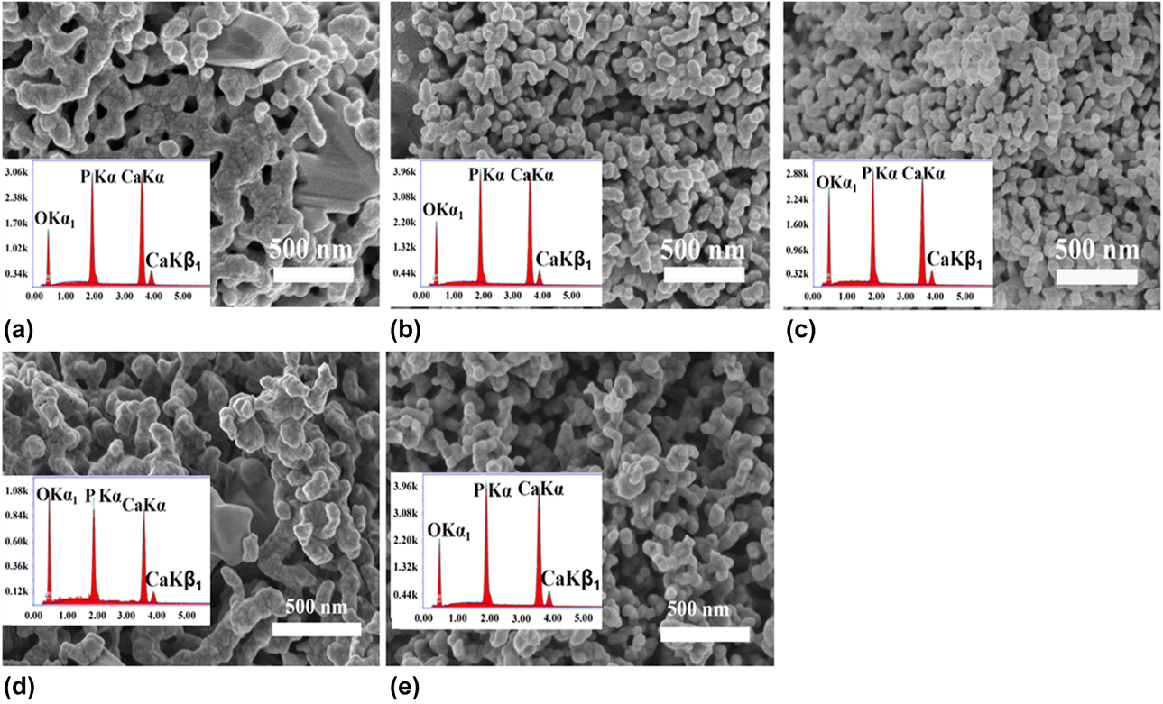
The morphology of the aggregated nanoparticles obtained by the dual-frequency ultrasonic method was observed by field-emission scanning electron microscopy (FE-SEM) micrographs. As shown in Fig. 3, a batch of nanoparticles developed to form a cluster core, forming a coral-like morphology. Irregular chunk structure and serious particles agglomeration were observed in U280T10 and U140T30, but the other samples showed a clear rod-shaped structure. As the sample sonicated at 280 W for 30 min, the obtained product displayed a uniform rod-shaped structure with less agglomeration. However, reunion was aggravating as further prolonging ultrasonic time to 45 min (namely U280T45). It demonstrates that irregular chunk structure could be reduced via prolonging sonicated time, but it should be noted that reunion may increase as the sonicated time is more than 30 min. As the sonication time was set to 30 min, irregular chunk structure appeared in U140T30 but not existed in U280T30 and U350T30. It reveals that irregular structure can be reduced via increscent power. The EDS spectra of the representative samples are shown in Figs. 3(a)–3(e) (inset) and the Ca/P molar ratio is presented in Table I. The Ca/P molar ratio of U280T30 and U350T30 were 1.66, close to the stoichiometric ratio of natural HAP. It indicates that the nHAPs were obtained at these operating conditions without second phase, which was also approved by the FT-IR and XRD analysis.
FE-SEM and EDS images (inset) of representative samples: (a) U280T10, (b) U280T30, (c) U280T45, (d) U140T30, and (e) U350T30.

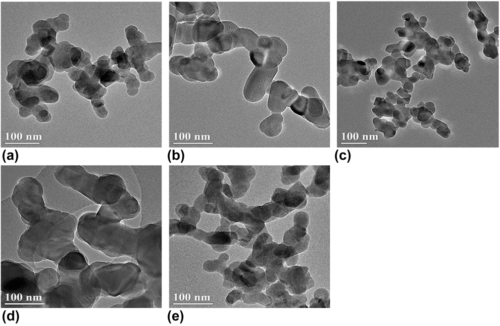
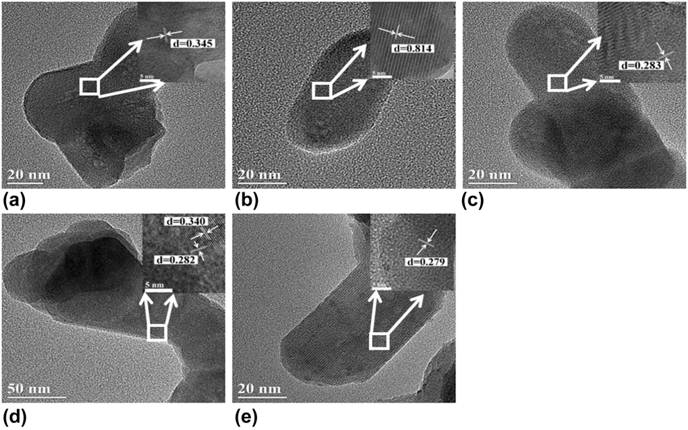
The microstructure of the product was observed by high-resolution transmission electron microscopy (HR-TEM) analysis. The TEM images of the representative samples are presented in Fig. 4. Irregular shape with severe agglomeration was observed in U280T10 and U140T30, whereas the other samples exhibited rod-shaped structure with better dispersion. As the ultrasonic power was set to 280 W, the sample sonicated for 30 min exhibited more uniform rod-like shape with better dispersion than the sample sonicated for 10 and 45 min. The results were consistent with the analysis of FE-SEM images. Therefore, ultrasonic time should be considered to prepare nHAPs with more uniform shape and fewer agglomeration. As the sample was treated by ultrasonic irradiation at 140 W for 30 min (namely U140T30), irregular aggregation was observed, indicating low ultrasonic power was not contributed to improve the dispersion of nanoparticles. The morphology showed no significant differences between U280T30 and U350T30. However, the energy consumption of U280T30 is lower than that of U350T30. Combined with the previous results, the operating parameters of ultrasonic power of 280 W and time of 30 min were considered as the optimum synthetic conditions to obtain nHAPs in the dual-frequency ultrasonic system. The particle sizes obtained from TEM images are presented in Table I. It was obvious that the particle sizes were up to nanoscale. The particle size of U280T30 was a mean width of 33.97 nm and length of 67.04 nm, smaller than that of U280T45 and U350T30. It reveals that the further extended ultrasonic time or the increased ultrasonic power was not contributed to decrease in particle size. The width and length of rod-shaped nanoparticles were (34.70 ± 1.27) and (68.67 ± 2.83) nm, respectively. The HR-TEM is shown in Figs. 5(a)–5(e). Samples U280T10 and U140T30 showed irregular structure. However, as for other samples, most of the rod-shaped particles had straight sides and regular ends. The lattice fringes of U280T10 exhibited interplanar spacing of 0.345, corresponding to the (002) plane of HAP crystal. The U280T30 showed spacing of 0.814 nm for (100) lattice plane. The lattice fringes of U140T30 showed interlayer spacing of 0.340 and 0.282 nm, matching well with the (002) and (211) planes, respectively. The U280T45 showed the same HAP crystals lattice with (211) interlayer spacing of 0.283 nm. The U350T30 sample displayed the expected spacing of 0.279 nm for the (112) lattice plane.
TEM images of representative samples: (a) U280T10, (b) U280T30, (c) U280T45, (d) U140T30, and (e) U350T30.

HR-TEM images of representative samples: (a) U280T10, (b) U280T30, (c) U280T45, (d) U140T30, and (e) U350T30.

The specific surface area (SSA) values of all samples are summarized in Table I. The SSAs of U280T10, U280T30, U280T45, U140T30, and U350T30 were 27.00, 28.86, 17.26, 14.85, and 27.44 m2/g, respectively. The sample encoded as U280T30 had the highest surface areas among all these samples, which may attributed to its smaller particle sizes and better dispersion than other samples.
The possible formation mechanism of nHAPs
Based on the above analysis, operating ultrasonic conditions of sonication time of 30 min and ultrasonic power of 280 W were considered as the optimum experimental conditions to obtain stoichiometric nHAPs with uniform rod-like shape and better dispersion. However, shorter treating time and lower ultrasonic power are not capable of providing sufficient energy to obtain nHAPs without any further aging process. The synthesis of nHAPs by ultrasonic-assisted chemical precipitation method needs further aging process [Reference Bensalah, Bekheet, Younssi, Ouammou and Gurlo31, Reference Canillas, Rivero, García-Carrodeguas, Barba and Rodríguez32]. The preparation time of HAP by traditional hydrothermal method was about 10–15 h, while the reaction temperature of combustion method was up to 500 °C [Reference Suslick and Price33, Reference Yang, Liu, Liang, Lin and Wu34]. Compared with these methods, the dual-frequency ultrasonic method exhibits a remarkable characteristics of high efficiency, time saving, and environmental-friendliness.
There is no direct interaction between the reactant and ultrasonic waves, but an indirect phenomenon provokes the rapid reaction in the liquid medium [Reference Zeng, Qiu and Lu22]. The phenomenon is referred to acoustic cavitation, which produces a specific environment in hot spots for evoking chemical reaction [Reference Bakan, Lacin and Sarac35]. The process of crystallization includes continuous formation and growth of the nucleation. Subsequently, the formed crystals are arranged and assembled to form solid particles eventually. The route of reactant addition is an important factor to guide the homogeneous nucleation. In this work, the P reactant was added to the Ca solution dropwise under magnetic stirring, in which magnetic stirring promoted nucleus formation due to increasing intermolecular collision and the slow addition rate minimized continuous nucleation. Thus, it reduced the mass reunion of particles at the nucleation stage. According to the previous investigation, cavitation activity distribution and pressure were more uniform in dual-frequency ultrasonic system than in single-frequency ultrasonic system, which lead to reducing the formation of standing waves and improving the dispersion of reagent [Reference Zeng, Qiu and Lu22]. It will contribute to increase the formation rate of homogeneous nucleus. As the introduction of ultrasonic radiation, agitation from acoustic cavitation can effectively increase the number of activated molecules and thus increase the effective collision in the liquid medium. It was contributed to accelerating reaction rate. In such conditions, the formed HAP crystals are arranged and assembled to form solid particles rapidly and effectively. Therefore, the dual-frequency ultrasonic system can be applied to successfully synthesize monodispersed nHAPs with regular shape in a rapid way. The effect of ultrasonic time and power on the composition and morphology of nHAPs may be associated with the acoustic cavitation effect.
Cytotoxicity evaluation
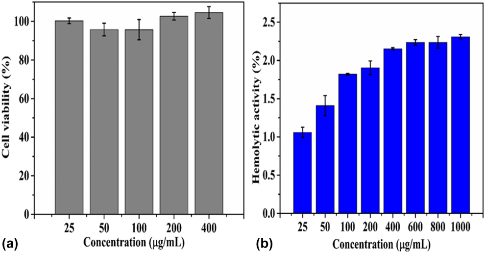
The cytocompatibility of nHAPs was screened against the normal 16 HBE cells. According to the Cytotoxicity Grading Standards of Materials from Chinese National Standards (GB/T 14233.2-2005), cytotoxicity is classified according to the level of cell viability, as shown in Table II. If cytotoxicity is divided into 0 or I, material is considered meeting the requirement of biological material. The results of cell viability of nHAPs are shown in Fig. 6(a). The cell viability (compared with cell control) after 24 h treatment with concentrations of 25, 50, 100, 200, and 400 μg/mL of nHAPs were (100.29 ± 1.50)%, (95.78 ± 3.29)%, (95.69 ± 5.24)%, (102.66 ± 2.00)%, and (104.58 ± 3.05)%, respectively. It was obvious that the cell viability of nHAPs was above 90% for all samples, for which cytotoxicity rank was considered as 0 or I . The result shows an indistinctive cytotoxic effect on 16 HBE cells within the concentration of 25–400 μg/mL, revealing a good cell compatibility of nHAPs obtained in the dual-frequency ultrasonic system and its potential application as candidates for bone application.
Cytotoxicity grading standards.

The results of (a) cytotoxicity assay and (b) hemolysis assay of nHAPs in vitro.

Hemolysis experiment
Hemolytic ratio of biomaterials is essential to know the interaction between biological materials and blood. The results of hemolytic activity of nHAPs in vitro are presented in Fig. 6(b). According to the Standard Practice for Assessment of Hemolytic Properties of Materials from Chinese National Standards (GB/T 16886.4-2003) and the ISO 10993-4 Standard, if the hemolysis ratio is below 5%, materials are considered meeting the hemolytic requirements of medical biomaterials. As shown in Fig. 6(b), the overall hemolytic activity of nHAPs at all experimental concentrations was less than 2.5%, which was acceptable according to the standard (GB/T 16886.4-2003), indicating a good hemocompatibility of nHAPs. Hemolytic ratio of HAP nanoparticles was below 2.0% at concentration of 25–200 μg/mL, while it was between 2 and 2.5% at concentration of 600–1000 μg/mL. Hemolytic ratio was found to be increasing with an increase in dosage.
Drug loading and release test
The loading of TH onto nHAPs (TH-nHAPs) was performed by physical adsorption method. The drug loading ratio and encapsulation ratio of TH-nHAPs were (26.34 ± 2.99)% and (52.68 ± 5.98)%, respectively, which were higher than the values of HAP-based drug carriers published earlier [Reference Wijesinghe, Mantilaka, Rajapakse, Pitawala, Premachandra, Herath, Rajapakse and Wijayantha36, Reference Chen, Xing and Gao37]. It was attributed to that TH molecules interacted with nHAPs through hydrogen bonding interactions due to the OH group existing in the TH molecules and the HAP. A control experiment was performed by studying the release behavior of pure TH sample without loading onto nHAPs and used to evaluate the release behavior of TH-nHAPs. The in vitro cumulative release profiles from the pure TH and TH-nHAPs samples are shown in Fig. S1. Over a period of 20 h, the drug cumulative release amount from pure TH and TH-nHAPs samples was about (99.67 ± 2.894)% and (76.61 ± 3.142)%, respectively. The drug release amount of TH-nHAPs sample was lower than that of pure TH sample due to the electrostatic adsorption between TH and HAP nanoparticles compared with pure TH sample. It was obvious that in vitro release behavior of pure TH sample showed a rapid release property compared with TH-nHAPs. There was an initial burst release for pure TH samples; cumulative release amount of the TH sample was more than 50% in the first one hour. However, the cumulative release amount of TH-nHAPs was only about 16.72% over the same period of time due to the electrostatic interaction between TH molecules and HAP. In the case of TH-nHAPs sample, the slow release was obtained after 8 h. The TH-nHAPs showed slow-release properties when compared with the pure TH sample, indicating as-synthesized nHAPs may be promising as drug carrier for drug delivery systems.
Conclusion
The present study demonstrates that the dual-frequency ultrasonic method can be utilized for one-step synthesis of monodispersed nHAPs with regular shape in a rapid way without further aging process. The ultrasonic time and power affect significantly the phase composition and morphology of nHAPs. Operating conditions of sonication time of 30 min and ultrasonic power of 280 W in the dual-frequency ultrasonic system are considered as the optimum experimental conditions to obtain stoichiometric nHAPs with uniform rod-like shape and good dispersity. However, the phase composition, morphology, and degree of crystallinity would deteriorate as further prolonging ultrasonic time and increasing ultrasonic power. The width and length of rod-shaped nHAPs are (34.70 ± 1.27) and (68.67 ± 2.83) nm, respectively. As we all know, the preparation time of the conventional hydrothermal method and the ultrasonic chemical precipitation method is generally 8–12 h, and the combustion method requires relatively complicated equipment, and the operation is not simple [Reference Bakan, Lacin and Sarac35, Reference Chen, Xing and Gao37]. Compared with the conventional hydrothermal method, ultrasonic-assisted chemical precipitation method, and combustion method previously reported, the dual-frequency ultrasonic method exhibits remarkable characteristics of high efficiency, time saving, and environmental-friendliness. The cytotoxicity evaluation and hemolysis experiment of nHAPs showed a good cytocompatibility and hemocompatibility, suggesting that the as-synthesized nHAPs with rod-shaped structure have potential application as biomaterials for bone tissue engineering. Besides, the drug loading and encapsulation efficiency of nHAPs to TH were (26.34 ± 2.99)% and (52.68 ± 5.98)%, respectively. The TH-nHAPs display slow-release properties when compared with the pure TH sample, indicating as-synthesized nHAPs may be promising as drug carrier for drug delivery system. Therefore, the dual-frequency ultrasonic method can be used as an efficient and environmentally friendly synthetic route to obtain rod-shaped nHAPs with good biocompatibility for specific biomedical applications. To the best of our knowledge, we may investigate the preparation of nHAPs by the dual-frequency ultrasonic method at the first time.
Materials
Diammonium hydrogen phosphate [(NH4)2HPO4] and calcium nitrate tetra hydrate [Ca(NO3)2·4H2O] were used as P and Ca reactants, respectively. 25% ammonium hydroxide (NH4OH) was used to adjust the pH of the reaction solutions. All chemical reagents were of analytical grade and obtained from Macklin (Shanghai, China). TH was purchased from Macklin. The dialysis bag (MD25-1M, MwCo 500 D) was purchased from MYM Biological Technology Co., Ltd. (Tianjin, China).
Methods
Experimental apparatus
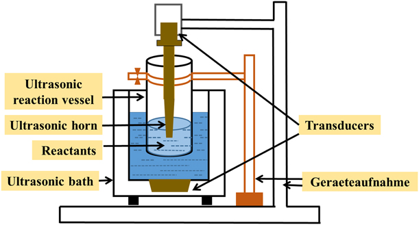
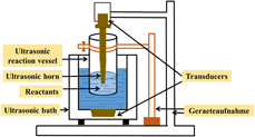
The self-assembled dual-frequency ultrasonic apparatus for synthesis of nHAPs is shown in Fig. 7. A combination of an ultrasonic horn (SCIENTZ-IID; Ningbo Science Biotechnology Co., Ltd., China) and an ultrasonic bath (KQ-400DB; Dongguan KEQIAO, China) has been considered. The ultrasonic horn generates ultrasonic waves at a frequency of 25 kHz. The ultrasonic bath was a customized cylindrical tank with frequency of 40 kHz. When the ultrasonic horn and ultrasonic bath work simultaneously, two kinds of frequencies and sound waves are produced in the systems. The advantages of using a horn as an approach for introducing a second sound wave include reducing the formation of standing, improving the dispersion of reagent and the cavitation activity distribution in the liquid medium, and enhancing the acoustic cavitation effect. The ultrasonic bath was filled with water. The cylindrical reaction vessel was used for reagent and its internal diameter and height were 6 cm and 18 cm, respectively.
Schematic illustration of experimental apparatus of the dual-frequency ultrasound.

Synthesis of nHAPs
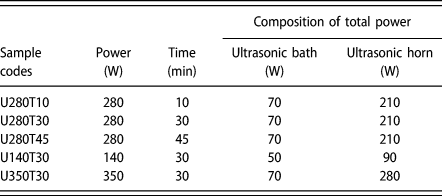
The fabrication of nHAPs was performed in the dual-frequency ultrasonic reactor. First, the P reagent was added to the Ca solution dropwise until the Ca/P molar ratio reaching about 1.67, and the reaction mixture was stirred for an hour. After this step, the milky white suspension was poured into the ultrasonic reaction vessel and treated by the dual-frequency ultrasonic irradiation. The effect of ultrasonic power and time of the dual-frequency ultrasound on morphology, phase composition, degree of crystallinity, SSA, and stoichiometry of nHAPs was investigated. The operating parameters of the dual-frequency ultrasound and sample codes are presented in Table III. The sonicated mixture was filtered and the residue was washed with distilled water and ethanol in turn. The obtained residue was dried and then the dried powder was calcined for further characterization.
The operating parameters in the dual-frequency ultrasonic system and sample codes.

Powder characterization
The phase purity, crystalline sizes, and crystallinity of calcined powder were identified by XRD analysis (D8 Advance Diffractometer; Bruker, Karlsruhe, Germany). The crystalline sizes (D) along to (002) direction of HAP crystal was calculated from the XRD line broadening using the Debye–Scherrer equation, shown as Eq. (1) [Reference Utara and Klinkaewnarong38, Reference Sun, Zheng, Hui, Fan, Song, Ma, Hua and Hui39]. The silicon powder was used to eliminate the instrumental line broadening.
 $$D = {{K\lambda } \over {\beta \cos \Theta }}\quad ,$$
where K is a constant and set to 0.89 in this study, λ is the wavelength of the Cu Kα1 radiation (λ = 0.15406 nm), D represents the crystallite size along (002) plane (nm), β stands for full width at half maximum (rad), and θ is the half Bragg diffraction angle (°).
The fraction of crystallinity (X c) of the synthesized samples was calculated by Eq. (2) [Reference Sun, Zheng, Hui, Fan, Song, Ma, Hua and Hui39, Reference Sun, Wu, Chen and Luo40].
 $${X_{\rm{c}}} = {\left( {{{0.24} \over \beta }} \right)^3}\quad .$$
The SSA of obtained powder was calculated by Brunauer–Emmett–Teller method from nitrogen adsorbed data measured from V-Sorb 2800TP device (Zhongshan Puchuan Inspection Technology Co., Ltd., Zhongshan, China). FT-IR spectra were used to characterize the structural composition using PerkinElmer Spectrometer 100 device (PERKINELMER, California). The Ca/P molar ratio was measured by an EDS spectrometer attached to a JEM-2100F. The morphology of the samples was observed by field-emission scanning electron microscopy (FE-SEM, SU8010 HITACHI, Tokyo, Japan). The microstructure was observed by HR-TEM (FEI Tecnai G2 F20 S-Twin, Hillsboro, Oregon).
Cells and cell culture
Normal human bronchial epithelial (16 HBE) cells were obtained from Cell Bank of Chinese Academy of Sciences of Shanghai (Shanghai, China) and cultured in DMEM medium (Gibco, Carlsbad, California) supplemented with 10% fetal bovine serum (Zhejiang Tianhang Biotechnology, China) at 37 °C under a humidified atmosphere containing 5% CO2.
Cytotoxicity assay
Cytocompatibility of a material is one of the most important evaluating indexes for its biological applications. The cytotoxicity assay was used to preliminarily evaluate the cytocompatibility of nHAPs and performed by Cell Counting Kit-8 method according to the previous reports [Reference Palanivelu and Ruban41, Reference Tank, Chudasama, Thaker and Joshi42]. The nHAPs obtained from the optimum experimental conditions in the dual-frequency ultrasound were utilized for further in vitro cytotoxicity testing. Briefly, the sterilized nHAPs were prepared to form a series of suspension by dispersing nanoparticles into DMEM medium. The 16 HBE cells were seeded at 96-well plates with cell density of 1 × 10⁴ cells/well and allowed to form monolayer cells in the incubator at 37 °C under a humidified atmosphere containing 5% CO2. Subsequently, the culture medium was replaced and the 16 HBE cells were treated with 25, 50, 100, 200, and 400 μg/mL suspension of nHAPs for 24 h. After 24 h treatment, the CCK-8 solution was added into each well, and the samples were further incubated for another 1.5 h at 37 °C. The optical density (OD) in each well was measured at 450 nm wavelength using microplate reader (Thermo, muLTISKANFC, Thermo Scientific, Suzhou, China). 16 HBE cells without nHAP treatment and DMEM medium were used as positive control group and blank group, respectively. The OD values of positive control group and blank group were also examined by the same procedure. The experiments were performed in triplicate for each well, and the average of OD values was considered as final OD values. Cell viability of nHAPs was calculated according to Eq. (3).
 $$\eqalign{{\rm{Cell}}\;{\rm{viability}}\;\left( \% \right){\rm{\ }} = {{\left( {{\rm{O}}{{\rm{D}}_{{\rm{test}}}} - {\rm{O}}{{\rm{D}}_{{\rm{blank}}}}} \right)} / \cr{\left( {{\rm{O}}{{\rm{D}}_{{\rm{control}}}} - {\rm{O}}{{\rm{D}}_{{\rm{blank}}}}} \right)}} \times 100\% \quad ,$$
where ODtest is the absorbance of the testing sample and ODcontrol and ODblank are the absorbance of the positive control group and blank group, respectively.
Hemolysis assay
To evaluate the blood compatibility of nHAPs obtained from the optimum experimental conditions in the dual-frequency ultrasound, the hemolysis experiment was performed according to the previously published literature with slight modifications [Reference Uskokoviæ and Desai43, Reference Qin, Zhong and Ma44]. First, fresh blood was collected from a male rabbit (purchased from Guangdong Medical Laboratory Animal Center, China; SCXK 2014-0035) and washed with NaCl solutions (0.9%, w/v) to remove impurities. The obtained erythrocytes were resuspended with the NaCl solutions to obtain a concentration of 2% (v/v) of red cell suspension (RCS). Subsequently, different concentrations of nHAPs (25, 50, 100, 200, 400, 600, 800, and 1000 μg/mL) prepared with NaCl solutions were mixed with 2% RCS, and the mixture was incubated at 37 °C for 1 h. Afterward, the mixture was centrifuged at 1000 rpm/min for 10 min and supernatant was carefully collected. The absorbance (A) of the supernatant was determined at 541 nm using ultraviolet and visible spectrophotometer (UV/Vis spectrophotometer, UV-5500PC; Shanghai Yuanxi Instrument Co., Ltd., China). Deionized water and NaCl solutions (0.9%, w/v) were used as positive and negative controls, respectively. All measurements were performed in the same procedure and performed in triplicate for each sample. The hemolysis ratio was calculated according to Eq. (4).
 $$\eqalign{\rm{Hemolysis}}\;{\rm{ratio}}\;(\% ) = {{\left( {{A_{{\rm{test}}}} - {A_{{\rm{negative}}\;{\rm{control}}}}} \right)} / \cr{\left( {{A_{{\rm{positive}}\;{\rm{control}}}} - {A_{{\rm{negative}}\;{\rm{control}}}}} \right)}} \times 100\% \quad ,$$
where A test is the absorbance of the testing sample and A negative control and A positive control are the absorbance of the negative and positive controls, respectively.
Drug loading and release test
The nHAPs obtained from the optimum experimental conditions in the dual-frequency ultrasound were used to load TH via physical adsorption [Reference Lai, Chen, Ren, Lee, Jiang and Kong45, Reference Thomas, Sharma, Rawat, Verma, Leekha, Kumar, Tyagi, Gurjar, Iqbal and Talegaonkar46]. First, the measured wavelength was determined at 354 nm via full wavelength scanning in UV/Vis spectrophotometer, and the absorbance–concentration standard curves were established. Subsequently, nHAPs (20 mg) were transferred into the TH aqueous solutions (5 mL, 2 mg/mL), and the mixture was stirred at 37 °C for 24 h. The obtained mixture was centrifuged and the TH-loaded nHAPs were rinsed by deionized water for 4 times. Supernatant were collected and measured at 354 nm using UV/Vis spectrophotometer. The drug amount of TH in supernatant was deduced via absorbance measurements. The drug loading ratio (DL) and encapsulation ratio (EL) of nHAPs were calculated according to Eq. (5) [Reference Wijesinghe, Mantilaka, Rajapakse, Pitawala, Premachandra, Herath, Rajapakse and Wijayantha36, Reference Thomas, Sharma, Rawat, Verma, Leekha, Kumar, Tyagi, Gurjar, Iqbal and Talegaonkar46]. The TH-loaded nHAPs after centrifugation were saved at −4 °C for release tests.
 $${\rm{DL}}\;(\% ) = \left[ {{{\left( {{M_0} - {M_{\rm{s}}}} \right)} / {{W_{{\rm{nHAPs}}}}}}} \right] \times 100\% \quad ,$$
 $${\rm{EL}}\;(\% ) = \left[ {{{\left( {{M_0} - {M_{\rm{s}}}} \right)} / {{M_0}}}} \right] \times 100\% \quad ,$$
where M 0 is the initial amount of TH, M s is the amount of TH in supernatant, and W nHAPs is the total amount of nHAPs in the solution.
The TH release test in vitro was performed by the dialysis method using phosphate buffered saline (PBS, pH = 7.4) solution as the dissolution medium according to the previously published literature [Reference Wijesinghe, Mantilaka, Rajapakse, Pitawala, Premachandra, Herath, Rajapakse and Wijayantha36, Reference Cho, Lee and Rhee47]. First, the TH-loaded nHAPs were placed into a dialysis bag and 2 mL of PBS solutions was added. Then, the dialysis bags were immersed in 100 mL PBS solution and then were orbitally shaken at 100 rpm at 37 °C in a thermostatic oscillator (SHZ-C; Shanghai Jingqi Instrument Co., Ltd., China). At various time intervals, release medium was taken out and fresh PBS solution was added. The absorbance of release medium was measured at 354 nm, and the cumulative release percentage was deduced according to absorbance–concentration standard curve. To verify the slow-release property of TH-loaded nHAPs, a control experiment was performed by studying the release behavior of pure TH sample without loading onto nHAPs in the same procedure. The measurements were performed in triplicate.
Supplementary material
To view supplementary material for this article, please visit https://doi.org/10.1557/jmr.2019.119.
Statements
I would like to declare on behalf of my coauthors that no any conflict of interest exits in the submission of the manuscript.
Acknowledgments
This work was supported by the Open Project Program of The Key Lab of Pollution Control and Ecosystem Restoration in Industry Clusters, Ministry of Education, South China University of Technology, Guangzhou Higher Education Mega Centre, Guangzhou 510006, PR China (No. 201701), Research and Demonstration on the Key Technology of Drinking Water and Sewage Treatment in the Northwest Desertification Area (No. 2016YFC0400707), Guangdong Provincial Department of Water Resources Project (No. 2015-20), and Guangdong Provincial Archives Project (No. YDK-141-2016).