Introduction
Although taking diverse forms, the acrosome reaction (AR) is an obligate event resulting in the unmasking of proteins required to mediate sperm–egg interactions (Primakoff & Myles, Reference Primakoff and Myles2002). In the sea urchin, the acrosome reaction requires polymerization of actin to form an acrosomal process coated with the egg-binding protein, bindin (Foltz & Lennarz, Reference Foltz and Lennarz1993). The mammalian AR is the culmination of a complex signalling web resulting in the fusion of the outer acrosomal membrane (OAM) with the overlying plasma membrane (PM) to release hydrolytic enzymes present in the acrosome (Yanagimachi, Reference Yanagimachi, Knobil and Neil1994). The hydrolytic enzymes released from the acrosome modify the mammalian sperm surface in the equatorial segment, rendering it competent to bind and fuse to the surface of the egg (Primakoff & Myles, Reference Primakoff and Myles2002).
While seemingly disparate, the AR in both examples above is postulated to be dependent on Rho GTPasemediated signalling. A RhoB orthologue is immunolocalized to the acrosome (Castellano et al., Reference Castellano, Martínez-Cadena, López-Godínez, Obregón and García-Soto1997; Cuéllar-Mata et al., Reference Cuéllar-Mata, Martínez-Cadena, López-Godínez, Obregón and García-Soto2000) as well as to the mitochondria in the midpiece of sea urchin sperm (Castellano et al., Reference Castellano, Martínez-Cadena, López-Godínez, Obregón and García-Soto1997). Castellano et al. (Reference Castellano, Martínez-Cadena, López-Godínez, Obregón and García-Soto1997) hypothesized that Rho regulates the polymerization of actin to form the acrosomal process (Castellano et al., Reference Castellano, Martínez-Cadena, López-Godínez, Obregón and García-Soto1997), a role consistent with its known activity in other cells (Van Aelst & D'Souza-Schorey, Reference Van Aelst and D'Souza-Schorey1997). A bacterial toxin, C3, that specifically inhibits Rho function (Wilde et al., Reference Wilde, Genth, Aktories and Just2000) prevents the ionophore-induced AR in the sperm of several mammalian species, suggesting that Rho is present in the sperm of those species tested (Brener et al., Reference Brener, Rubinstein, Cohen, Shternall, Rivlin and Breitbart2003).
Recently, we localized the Rho family GTPases RhoA, RhoB, Rac1 and Cdc42, to the cytoplasm and/or PM overlying the acrosome in porcine sperm. Additionally, the Rho GTPase effectors, RhoGDI and PI(4)P5K, as well as the Rho-activated kinase, ROCK-1, were present in the acrosomal region of porcine sperm, strongly suggesting the presence of a Rho-mediated signalling cascade. In support of this conclusion, the bacterial toxins, toxin B and cytotoxic necrotizing factor (CNF), and the ROCK inhibitor Y-27632, affected the zona pellucida (ZP)-induced AR in the porcine model (manuscript in preparation).
When taken together, these data strongly suggest that a Rho-mediated signalling pathway regulating the acrosome reaction may be a universally conserved mechanism among species. To address this possibility among eutherian mammals, the Rho GTPases (RhoA, RhoB, Rac1 and Cdc42), as well as their effectors RhoGDI, PI(4)P5K and ROCK-1, were immunolocalized in sperm from a variety of mammals including human, rat, ram, bull and elephant and their localization was compared with that in the boar.
Materials and methods
Antibodies
Polyclonal antibodies specific for RhoA (rabbit or goat; sc-179 and sc-179G, respectively), RhoB (sc-180), Cdc42 (sc-87), Rac1 (sc-217), RhoGDI-1 (sc-360), ROCK-1 (sc-5660) and PI(4)P5K (sc-1330) were purchased from Santa Cruz Biotechnology (Santa Cruz, CA). The secondary antibodies, Texas Red (TR)-conjugated donkey anti-goat and anti-rabbit immunoglobulins, were purchased from Jackson Immunolabs (West Grove, PA).
Semen collection and sperm preparation
Boar semen was collected using digital pressure. Semen from bulls and ram were obtained with an artificial vagina and kept at 37 °C during transport to the laboratory, where individual bull and ram samples were pooled prior to washing. Human semen was obtained by masturbation and underwent liquefaction for 20 min at 37 °C before being washed. Sperm were washed by centrifuging 1–2 ml of semen on a discontinuous Percoll step-gradient (80%, 70%, 55%, 40% Percoll in Tris-buffered medium (TBM); 20 mM Tris, 10 mM CaCl2,113 mM NaCl, 3 mM KCl, 10 mM glucose, 5 mM sodium pyruvate and 50 μg gentamicin sulfate/ml, pH 7.85) at 300 g for 20 min. The 80% layer containing the sperm pellet was diluted with TBM and washed by centrifugation for another 10 min. Sperm were then adjusted to a concentration of 4 ×10⁶ sperm/ml in TBM supplemented with 5 mg bovine serum albumin (BSA)/ml.
Epididymal sperm were collected from the rat by slicing the end off the epididymis and flushing out the sperm using a blunt needle filled with warmed Holloway's medium (94.19 mM NaCl, 4.75 mM KCl, 1.71 mM CaCl2, 1.17 mM KH2PO4, 1.17 mM MgSO4, 25.10 mM NaHCO3, 26.00 mM sodium lactate, 0.50 mM sodium pyruvate, 5.50 mM glucose, 20.00 mM HEPES, 50 μg gentamicin sulfate/ml; Holloway et al., Reference Holloway, Moore and Foster1990). Rat epididymal sperm were briefly incubated at 37 °C in 5% CO2 in air before being counted. Sperm were then diluted to a concentration of 4 × 10⁶ sperm/ml of TBM supplemented with 5 mg BSA/ml.
Semen from the African elephant Loxodonta africana was collected by rectal manual massage, extended 3:1 in Biladyl (Minitube of America, Verona, WI) and subsequently diluted 1:1 in Biladyl containing 8% dimethylsulfoxide (DMSO). The extended semen was then shipped to the laboratory at room temperature where it was aliquoted into 250 μl straws and snap-frozen for 10 min in liquid nitrogen prior to being stored in liquid nitrogen. To remove freezing medium, four or five straws of frozen elephant semen were thawed (final volume approximately 1 ml extended semen) and the extended semen was rinsed twice in 5 ml TBM by centrifugation at 300 g. Sperm were counted in a haemacytometer and the concentration was adjusted to 4 × 10⁶ sperm/ml of TBM supplemented with 5 mg BSA/ml.
Porcine, bovine, ovine and elephantine sperm were incubated for 10 min at 39 °C in 5% CO2 in air prior to fixation (Berger and Horton, Reference Berger and Horton1988). Rat and human sperm were incubated at 37 °C in 5% CO2 in air for 10 min prior to fixation.
Protein immunolocalization
Aliquots of sperm were spread on slides coated with poly-L-lysine and air-dried. Sperm were briefly fixed in ice-cold acetone and stored at −20 °C. Slides were allowed to warm to room temperature and sperm were rehydrated in water before rinsing with phosphate-buffered saline (PBS; 140 mM NaCl, 3 mM KCl, 4 mM NaH2PO4, 0.8 mM Na2HPO4, 1.5 mM MgSO4, pH 7.4). All incubations were performed in a humidified chamber. Sperm were blocked with 10% donkey serum in PBS and rinsed three times before being incubated in PBS containing either the stated primary IgG or the animal-matched serum IgG as a control. Slides were again rinsed with PBS and incubated with the appropriate TR-labelled secondary antibody. Acrosomal contents were briefly labelled with FITC-PSA and the sperm slides were rinsed once with PBS and once with water. A p-phenylenediamine-based mounting medium (5 mg p-phenylenediamine, 5 mg NaN3, 0.5 ml PBS, 4.5 ml glycerol) was applied to the slide and a coverslip was placed over the samples. Labelled sperm were viewed on either an Olympus BH-2 microscope with a Mercury Burner-100 or an Olympus BX-60 epifluorescence microscope, and photographs were taken with a CoolSnap CCD camera (Roper Scientific) utilizing C-imaging software (Compix). Antigen immunolocalization was performed a minimum of three times.
Results
RhoA
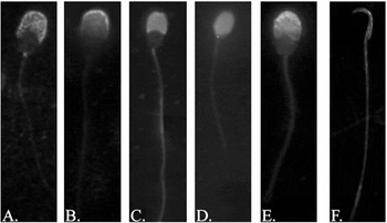
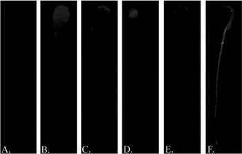
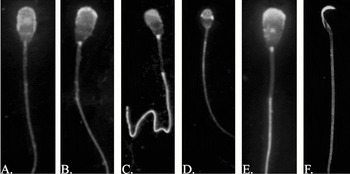
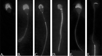
RhoA immunolocalized to the head of all sperm studied (Fig. 1). RhoA labelling was limited to the acrosomal region of porcine and bovine sperm (Fig. 1A, B); in addition to acrosomal labelling, faint post-acrosomal labelling was also seen in ovine sperm (Fig. 1C). Human and rat sperm showed full head labelling (Fig. 1D, F). Faint labelling of the tail was seen in ram and rat sperm; labelling was stronger in the rat sperm tail than the ram sperm tail, but was similar to levels observed in control sperm (Fig. 2F). The anti-RhoA polyclonal antibody strongly labelled the elephantine sperm tail as well as the acrosomal region (Fig. 1C). Human sperm also showed faint tail labelling with RhoA; however, normal serum controls also yielded occasional tail labelling. Therefore, the tail labelling seen in human sperm was determined to be non-specific. In all species, sperm with intact acrosomes showed labelling of the acrosomal region with the primary antibody whereas acrosome-reacted sperm did not (data not shown). In contrast, except for human and rat sperm tail labelling, all sperm incubated with normal serum displayed no, or significantly weaker, labelling (Fig. 2).
Immunolocalization of RhoA in mammalian sperm. The monomeric G-protein RhoA was immunolocalized in acetone-fixed boar (A), bull (B), elephant (C), human (D), ram (E) and rat (F) sperm.

Examples of normal serum control. As a negative control, sperm were treated with normal serum instead of the stated immunoglobulin. Note the minimal labelling, indicating little non-specific binding of antibody, in acetone-fixed boar (A), bull (B), elephant (C), human (D), ram (E) and rat (F) sperm.

RhoB
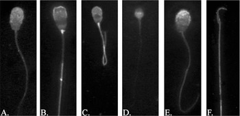
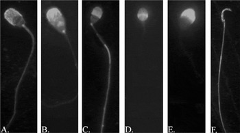
The polyclonal anti-RhoB antibody labelled the full head of porcine, elephantine, ovine and rat sperm (Fig. 3A, B, C, E, F). Bovine sperm showed RhoB labelling in the acrosomal region and in the region of the head posterior to the equatorial segment but, interestingly, not in the equatorial segment itself (Fig. 3B). Interestingly, human sperm, while having full head labelling, showed stronger labelling post-acrosomally than acrosomally (Fig. 3D). Tail labelling of RhoB was seen in all but human sperm. Bovine sperm lacked labelling in the midpiece (Fig. 3B). The labelling observed in boar and ram sperm was faint (Fig. 3A, E) but consistent and comparable labelling was not observed in control sperm treated with normal serum (Fig. 2). Acrosome-reacted sperm were not labelled over the acrosomal region of the head and sperm incubated with normal serum did not show labelling (not shown).
Immunolocalization of RhoB in mammalian sperm. The RhoB GTPase was immunolocalized in acetone-fixed boar (A), bull (B), elephant (C), human (D), ram (E) and rat (F) sperm.

Rac1
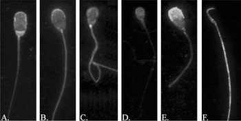
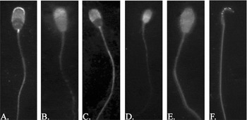
Rac1 immunolabelling was seen in the entire head and tail of sperm from all species but humans (Fig. 4), where Rac1 labelling was observed only in the head. Sperm that had undergone acrosomal exocytosis did not display acrosomal labelling (data not shown). Control sperm immunolabelled with normal serum did not display any significant labelling (Fig. 2).
Immunolocalization of Rac1 in mammalian sperm. The Rho family GTPase, Rac1, was immunolocalized in acetone-fixed boar (A), bull (B), elephant (C), human (D), ram (E) and rat (F) sperm.

Cdc42
Anti-Cdc42 labelling was observed in the acrosomal region of sperm from boar, bull, elephant, human, ram and rat (Fig. 5). Faint immunolabelling was observed post-acrosomally in the boar and ram sperm (Fig. 5A, E) but not other sperm. In addition to the head, Cdc42 was present in the principal piece of the tail of all sperm studied. However, sperm from all species showed either no or very faint labelling of the midpiece of the tail, suggesting that Cdc42 is not present in that region of the tail. No significant labelling was seen in control sperm treated with normal serum (Fig. 2). Acrosome-reacted sperm incubated with the anti-Cdc42 polyclonal antibody were not labelled in the acrosomal region of the sperm head (not shown).
Immunolocalization of Cdc42 in mammalian sperm. The G-protein Cdc42 was immunolocalized in acetone-fixed boar (A), bull (B), elephant (C), human (D), ram (E) and rat (F) sperm.

RhoGDI
The polyclonal anti-RhoGDI antibody specifically labelled the acrosomal region of bovine, elephantine, ovine and rat sperm (Fig 5B, C, E, F). RhoGDI also strongly immunolabelled the principal piece of the sperm tail except in porcine sperm (Fig. 6A). In contrast to the staining of the acrosomal region and principal piece of the tail observed in most species, RhoGDI strongly labelled the entire head and tail of human sperm (Fig. 6D) and only the acrosomal region in porcine sperm. In all species, sperm incubated with normal serum did not show significant labelling (Fig. 2) and acrosomal exocytosis prevented acrosomal labelling of sperm (data not shown).
Immunolocalization of RhoGDI-1 in mammalian sperm. The Rho family GTPase regulatory protein RhoGDI-1 was immunolocalized in acetone-fixed boar (A), bull (B), elephant (C), human (D), ram (E) and rat (F) sperm.

PI(4)P5K
Labelling of phosphatidylinositol kinase, PI(4)P5K, was strong in the acrosomal region of porcine, elephantine and ovine sperm, but was somewhat less strong post-acrosomally (Fig. 7A, C, E). Bovine, human and rat sperm all had full head labelling (Fig. 7B, D, F), although human sperm had more intense immunolabelling of the equatorial segment (Fig. 7D) than was present in either bovine or rat sperm. Additionally, boar and rat sperm had strong labelling of the full tail (Fig. 7A, F), whereas bull sperm had fainter labelling (Fig. 7B). This antibody strongly labelled the principal piece of the elephant sperm tail (Fig. 7C), but labelling was not present in the tail of human or ram sperm (Fig. 7D, E). Sperm were not immunolabelled by normal serum (Fig. 2) and sperm that had been acrosome-reacted were not labelled in the acrosomal region by the polyclonal anti-PI(4)P5K antibody (not shown).
Immunolocalization of PI(4)P5K in mammalian sperm. The Rho family effector inositol kinase, PI(4)P5K, was localized in acetone-fixed boar (A), bull (B), elephant (C), human (D), ram (E) and rat (F) sperm.

ROCK-1
The Rho effector ROCK-1 was present in the head of all sperm studied. In porcine and elephantine sperm, ROCK-1 was primarily localized to the acrosomal region of the sperm head and the full tail, although faint labelling was observed post-acrosomally (Fig. 8A, C). ROCK-1 may be differentially expressed in regions of the tail of porcine and elephantine sperm as the principal piece appeared to be more strongly labelled than the midpiece (note the lighter labelling of the midpiece than the principal piece in Fig. 8A and C). However, these differences were not uniform within a population of sperm from either species. The anti-ROCK-1 polyclonal antibody labelled the entire head of bovine and human sperm as well as faintly labelling the tail of both. Expression was stronger acrosomally than post-acrosomally in bovine sperm, whereas in human sperm, post-acrosomal labelling is stronger than acrosomal (Fig. 8B, D). ROCK-1 immunolabelled the entire body of ram and rat sperm (Fig. 8E, F). Distinctly brighter patches of labelling were frequently, but not always, observed acrosomally in rat sperm (Fig. 7F). These spots may represent natural in vivo clustering of antigen in 'raft-like' domains or may be an artefactual clustering of antigen by either the antibody used or the acetone fixation. Acrosomal labelling of the ROCK-1 antigen was seen only in acrosome-intact, and not acrosome-reacted, sperm (data not shown). Control sperm incubated in the presence of normal serum had no significant labelling (Fig. 2).
Immunolocalization of ROCK-1 in mammalian sperm. The RhoA and RhoB effector ROCK-1 was immunolocalized in acetone-fixed boar (A), bull (B), elephant (C), human (D), ram (E) and rat (F) sperm.

Discussion
Rho, Rac and Cdc42, as well as the effectors RhoGDI, ROCK and PI(4)P5K, were previously immunolocalized to the head of porcine sperm, suggesting roles for them in the AR. Manipulation of the Rho GTPases and the Rho-activated kinase ROCK by bacterial toxins and the ROCK-specific compound Y-27632, respectively, substantiated the role of the Rho GTPases and their effectors in regulating acrosomal exocytosis in porcine sperm (manuscript in preparation). A RhoB orthologue may regulate formation of the acrosomal process in sea urchin sperm (Castellano et al., Reference Castellano, Martínez-Cadena, López-Godínez, Obregón and García-Soto1997). Together, these data suggest that a Rho-mediated signalling pathway resulting in the AR was highly conserved during evolution. To address this hypothesis for mammals, the Rho GTPases RhoA, RhoB, Rac1 and Cdc42 were immunolocalized in the sperm of six species. In addition to the Rho GTPases, the effectors RhoGDI-1, PI(4)P5K and ROCK-1 were also immunolocalized in mammalian sperm.
Among the six species of sperm examined here, three of the four major clades of eutherian mammals were represented, the exception being the Xenarthra clade which includes sloths, armadillos and anteaters (Murphy et al., Reference Murphy, Eizirik, Johnson, Zhang, Ryder and O'Brien2001). The Laurasiatheria clade encompasses the Certartiodactyla, which includes pigs, sheep and cattle (Murphy et al., Reference Murphy, Eizirik, Johnson, Zhang, Ryder and O'Brien2001); not surprisingly, the sperm from these three species had the most consistent labelling pattern for the Rho family GTPases and their effectors among the six species studied. This strongly suggests that within the Certartiodactyla, the hypothesized function of the Rho GTPases in acrosomal exocytosis is highly conserved.
The superordinal Euarchontoglires clade separates into two major branches: the Glires, which includes Rodentia and Lagomorpha and the Primates (Murphy et al., Reference Murphy, Eizirik, Johnson, Zhang, Ryder and O'Brien2001). Thus phylogenetic relationships suggest antigen distribution may be similar between the sperm of rats and humans. Rat sperm generally displayed global immunolabelling of the antigens examined, with the notable exception of RhoGDI which was absent in the midpiece and principal piece of the sperm tail. RhoGDI serves as a regulatory element for Rho, Rac and Cdc42, which are all present in the entire flagellum of rat sperm, prompting questions of how these GTPases are regulated in the tail. Rhophilin and ropporin are two Rho GTPase-binding proteins found in mouse that localize only to the principal piece of the mouse sperm tail. These two proteins lack kinase activity and have no known function other than acting as adaptor proteins that localize Rho GTPases to specific domains of the fibrous sheath (Fujita et al., Reference Fujita, Nakamura, Kato, Watanabe, Ishizaki, Kimura, Mizoguchi and Narumiya2000; Nakamura et al., Reference Nakamura, Fujita, Murata, Watanabe, Mori, Fujita, Watanabe, Ishizaki, Yoshida and Narumiya1999). It is thus possible that rhophilin and ropporin act as regulatory proteins in addition to being adaptor proteins.
Although representing the second major branch of the Euarchontoglires clade (Murphy et al., Reference Murphy, Eizirik, Johnson, Zhang, Ryder and O'Brien2001), human sperm showed immunolocalization patterns of Rho GTPases and effectors distinct from those observed in rat sperm; the only antigen with any similarity was Cdc42. Immunolabelling was nearly always restricted to the head of human sperm. The interesting exceptions were Cdc42 and RhoGDI, both of which strongly labelled the tail, suggesting that in human sperm, localization and function of these two proteins are coordinated. Intriguingly, several of the antigens displayed stronger post-acrosomal than acrosomal labelling. This may reflect the directionality of the AR in human sperm (i.e. from posterior to anterior; Yudin et al., Reference Yudin, Gottlieb and Meizel1988). Altogether, these data mean that though related, the orders Archonta and Glires do not share a common superordinal sperm protein distribution. This, in turn, suggests that while family groups may share common immunolabelling patterns (i.e. porcine, bovine and ovine), these consistencies may not extend to higher, ordinal levels (i.e. from Glires to Archonta). It also implies that differences in immunolabelling may extend to differences of function. Hence, phylum-level conclusions on function should be drawn cautiously.
The most distantly related clade is the Afrotheria, which includes the sixth species studied: elephant (Murphy et al., Reference Murphy, Eizirik, Johnson, Zhang, Ryder and O'Brien2001). In elephant sperm, the antibodies showed comparable immunolabelling of the acrosome and principal piece of the tail; in addition to this labelling, RhoB and Rac1 labelled midpieces as well. The similarity of Rho family GTPase localization with that of their effectors in elephant sperm suggests a tight coupling of localization, activity and regulation.
Cumulatively, these data strongly suggest that RhoA, RhoB, Rac1 and Cdc42 are present in the sperm of all placental mammals. Furthermore, the presence among all eutherian mammals of RhoGDI-1, PI(4)P5K, ROCK-1, and perhaps other Rho family effector proteins, is strongly suggested. Although the evidence is strongest among the Cetartiodactyla for a conserved role of the Rho GTPases in the AR, the presence of the Rho GTPases in the acrosomal region of the sperm of at least three of the four major branches of placental mammals suggests that a Rho-mediated signalling pathway culminating in acrosomal exocytosis was highly conserved during mammalian evolution. If the hypothesized role of the Rho orthologue present in the sea urchin sperm (Castellano et al., Reference Castellano, Martínez-Cadena, López-Godínez, Obregón and García-Soto1997) and other Ambulacraria is confirmed, it would extend the conserved mechanism of a Rho-regulated AR to all deuterostomes (Peterson, Reference Peterson2004; Winchell et al., Reference Winchell, Sullivan, Cameron, Swalla and Mallatt2002).
Acknowledgements
The authors would like to thank Nancy Dahl for providing the cryopreserved elephant semen and Martin Dally for collecting porcine and ovine sperm.