Introduction
Although the use of fresh spermatozoa recovered from the epididymis is an alternative procedure, cryopreservation of spermatozoa allows a more efficient and economical way to use that material, because it can be utilized at anytime, not just after death of an animal. Almost all of the Felidae family are considered critically endangered and vulnerable (Novell & Jackson, Reference Novell and Jackson1996; Kendall et al., Reference Kendall, Bjørnstad, Bascompte, Keitt and Fagan2000). Genetic material either from animals of economical interest or from wildlife can be lost at anytime by unexpected death of the animal. In this case, efforts can occur to avoid the total lost of that genetic material, which can be achieved by using reproductive assisted techniques. In domestic cats, special attention has been given to semen retrieval from the cauda epididymidis and deferent duct; (Axnér et al., Reference Axnér, Ström and Linde-Forsberg1998; Zambelli & Cunto, Reference Zambelli and Cunto2006; Hermansson & Axnér, Reference Hermansson and Axnér2007). Cryopreserved semen offers the same advantages as fresh semen, as well as abrogating geographic distance, which allows the use of semen from dead or injured donors, and the ability to provide sufficient material for repetitive experimental manipulation. (Villaverde, Reference Villaverde, Martins, Castro and Lopes2006). Moreover, cryopreservation of gametes is an important tool in assisted reproduction programmes; in fact, long-term storage of oocytes or spermatozoa is necessary when in vitro fertilization (IVF) or artificial insemination needs to be performed at a future date (Luvoni, Reference Luvoni2006). The susceptibility of spermatozoa to cold shock is related to the composition of the plasma membrane and differs not only among species but also among and within individuals (White, Reference White1993). The composition of feline sperm membranes is not well known.
The use of the fluorescent antibiotic chlortetracycline (CTC) for the assessment of spermatozoal functional status has been shown to be particularly useful as, in addition to the determination of the acrosomal status of the spermatozoa, it allows for the further discrimination of cells with intact acrosome into a capacitated and uncapacitated group (Fraser et al., Reference Fraser, Abeydeera and Niwa1995). More recently, other studies have demonstrated the use of this technique in the mouse (Fraser et al., Reference Fraser, Abeydeera and Niwa1995) human (Perry et al., Reference Perry, Naeeni, Barratt, Warren and Cooke1995), bull (Perry et al., Reference Perry, Naeeni, Barratt, Warren and Cooke1995) and ram (Hewitt & England, Reference Hewitt and England1998). Recently it has been reported that a significant higher proportion of cat epididymal spermatozoa after thawing showed the pattern of acrosome reaction compared with fresh or diluted semen before freezing. The proportion of acrosome-reacted spermatozoa in frozen–thawed samples indicates that many cells were damaged during freezing and thawing and, therefore, are functionally compromised (Marinoni, Reference Marinoni2001).
A critical element of the cooling process is the passage of water across the cell membranes, the rate of which depends on permeability characteristics, cell surface-to-volume ratio, temperature and the difference between extra- and intracellular osmolality (Parks, Reference Parks, Karow and Critser1997).
Domestic cat spermatozoa and oocytes have peculiar physical characteristics that increase the difficulty of developing successful of cryopreservation methods as compared with gametes of some other species. For this reason, even though a variety of procedures have been investigated, optimal cryopreservation techniques, either for spermatozoa or oocytes, are yet to be realized (Luvoni, Reference Luvoni2006). The sensitivity of cat sperm cells, in terms of loss of motility or cell membrane damage, to changes in osmolality and to different cooling rates has been examined for the purpose of defining ‘ideal’ chilling or cryopreservation procedures. Osmolality of cat semen ranges from 290 mOsm/kg to 320 mOsm/kg and the optimal diluent should be isotonic. It has been shown that swelling and membrane rupture of cat spermatozoa caused by hypotonic solutions are more detrimental than the shrinkage caused by hypertonic solutions (Pukazhenthi et al., Reference Pukazhenthi, Spindler, Wildt, Bush and Howard2002).
Moreover, sperm motility is more sensitive to changes in osmolality than is membrane integrity and removal of cryoprotectants in multiple steps with an isotonic solution minimizes the loss of sperm motility and reduces membrane disruption (Pukazhenthi et al., Reference Pukazhenthi, Spindler, Wildt, Bush and Howard2002).
The objectives of this work were to: (1) examined sperm motility, viability, morphology and acrosome integrity of frozen–thawed domestic cat epididymal spermatozoa; and (2) evaluated the same cryopreservation method on spermatozoa of wild felines, necessary for preservation of their genetic resources.
Materials and Methods
Animals
Epididymides were obtained from 20 adult domestic cats between 12 and 48 months of age during routine neutering procedure from the Veterinary Clinics of Naples University. The males weighed 2.8–5.4 kg (mean 4 ± 0.6 kg), and consisted of a range of various breeds. As it has been stated in the literature, the epididymides were obtained in every season because in domestic cats spermatogenesis is unaffected (Spindler & Wildt, Reference Spindler and Wildt1999; Wildt et al., Reference Wildt, Brown, Swanson, Knobil and Neill1999).
Health and fertility status of tomcats were confirmed by clinical examination, breeding soundness evaluation, behavioural analysis, testis morphometry and ultrasonography evaluation.
In order to restrain all cats, atropine sulphate (0.025 mg/kg) was administered intramuscularly as a premedication; anesthesia was induced by 6 mg/kg propofol and maintained by isofluorane after endotracheal intubation. An anaesthetic density of 1.9% isofluorane provided the necessary anesthetic depth (Ishikawa et al., Reference Ishikawa, Uechi, Ishikawa, Wakao and Higuchi2007).
Once induced, all patients underwent an ultrasonographic study, in order to obtain clear visualization of testicular dimension and parenchyma. In all cases, a linear high frequency probe was applied on the scrotal sac following application of acoustic coupling gel (Sanovet Pico) 10 mHz. All the images were obtained in longitudinal and transverse view.
Orchiectomy was performed following standard procedures (Howe, Reference Howe2006) and epididymides were collected from all patients.
Testes and epididymides were collected from two wild felids from Naples Zoo [Bengala tiger (Panthera tigris)] during the autopsy performed at three different dates in the Veterinary Clinics of Naples University (October 2005 and July 2006). Retrieval of the testes and epididymides were performed within 6 h post-mortem. The subjects were 25 and 29 years of age respectively.
Sperm retrieval
Each collected epididymis was immediately washed in Dulbecco's PBS with 0.0036% (w/v) sodium pyruvate, 0.1% (w/v) glucose, 0.0066% (w/v) sodium benzylpenicillin, 0.01% (w/v) streptomycin sulphate, to which 4 mg/ml bovine serum albumin (BSA) (Fraction V, Sigma) was added in a Dewar vessel at 20–24°C and transported to the laboratory within 3 h of removal.
Finely minced cauda epididymis was obtained after separation of the epididymis from the testis. The caudal portion of each epididymis was placed in 2 ml Tris extender (3.025% Tris hydroxymethyl aminomethane, 1.7% citric acid, 1.25% fructose, 0.06% sodium benzyl penicillin, 0.1% streptomycin sulphate) (Hermansson & Axnér, Reference Hermansson and Axnér2007). Visible blood vessels were removed; epididymides and proximal deferent ducts were isolated. Two sections from the caudal portions of the epididymides were placed into 1 ml of Tris extender in a Petri dish (35 × 10 mm; Falcon).
The caudal portion was dissected with a scalpel blade to release spermatozoa. After a 10-min incubation at 38°C the epididymal tissue was removed with forceps, the medium was collected in a Falcon tube and centrifuged at 700 g for 6 min. The supernatant was removed and the pellet was diluted with 200 μl fresh Tris extender. A portion of the sperm suspension (10 μl) was collected for the spermatozoa integrity, motility and viability assessments.
In order to determine sperm concentration, a 5 μl aliquot of the sample at room temperature was diluted in 20 μl of formol saline and spermatozoa were counted in 16 squares of a Bürker chamber. Afterward, the sperm pellets were diluted in a Tris egg yolk extender containing 20% (v/v) egg yolk, in an adequate volume to achieve a concentration of 40 × 106 spermatozoa/ml.
Sperm cooling
Previous study demonstrated that cat epididymal spermatozoa are very resistant to cold shock (Hermansson & Axnér, Reference Hermansson and Axnér2007). For this reason the semen was transferred to a 4°C refrigerator, and kept there for 60 min without glycerol.
Afterwards the cooled semen was then diluted with an equal part of Tris egg yolk extender with 8.0% glycerol. The final concentration of glycerol was 4.0% and the final spermatozoa concentration was 20 × 106 spermatozoa/ml.
The sperm samples were loaded into 0.25 ml mini-straws and exposed to nitrogen vapour (6 cm over the surface of liquid nitrogen) for 20 min, then immersed and stored in liquid nitrogen. After 4 weeks, samples were thawed in a warm water bath at 37°C for 30 s and were diluted with 1 ml Tris egg yolk extender and re-evaluated.
Sperm sample evaluation
The recovered and frozen–thawed sperm samples were examined using the general semen quality test (Tanaka et al., Reference Tanaka, Takagi, Nakagawa, Fujimoto, Hori and Tsutsui2000). The following sperm parameters were evaluated: volume (ml), concentration (106 spermatozoa/ml), motility (%), viability (%), abnormal spermatozoa (%), and spermatozoa with normal acrosomes (%). Pellet volume was determined using a variable volume pipette. Sperm concentration was assessed, after fixation of an aliquot of semen in buffered formol saline, with a phase-contrast microscope at ×100 magnification, using a Bürker chamber.
Spermatozoal motility was evaluated before cryopreservation and immediately post thaw (0 h) and at l-h intervals during 3 h of incubation at 38°C. The proportion of progressively motile spermatozoa from eight randomly selected fields in each sample was evaluated subjectively in a Makler chamber at 37°C using a phase-contrast microscope at ×100 magnification.
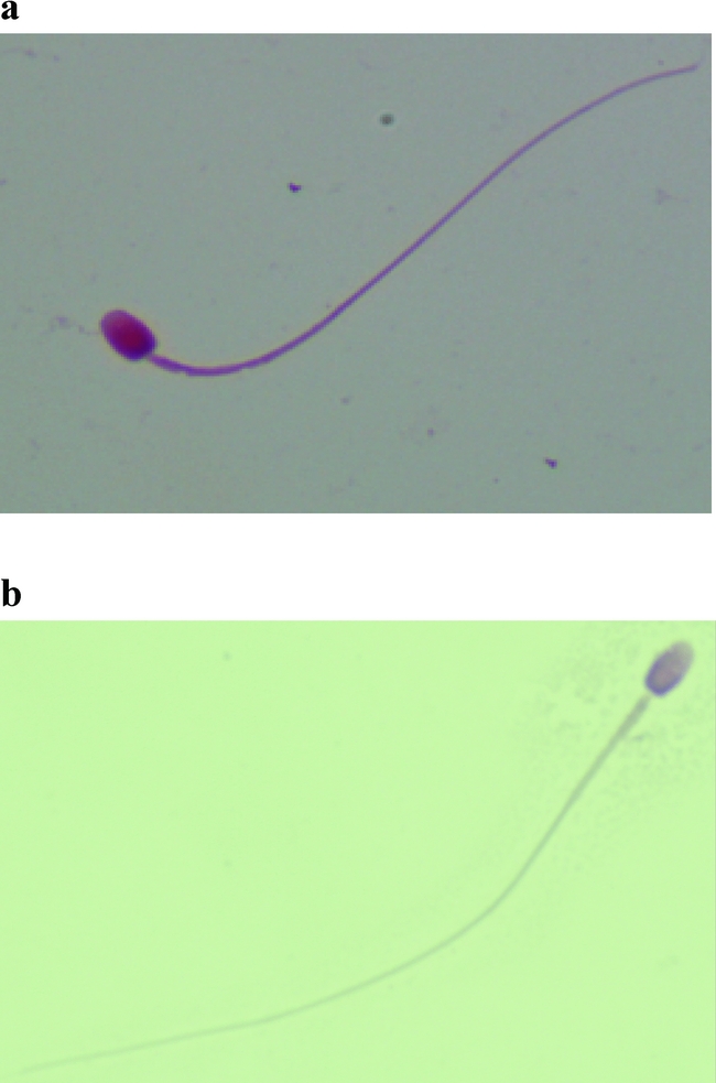
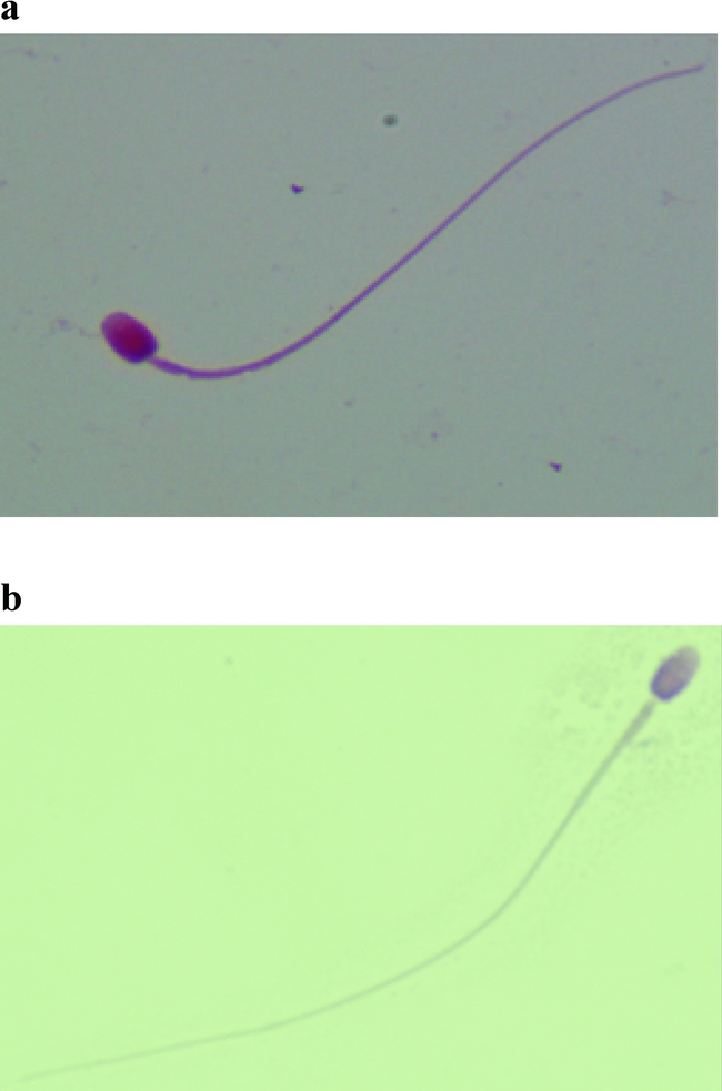
Spermatozoal viability was evaluated before cryopreservation and immediately post thaw (0 h) by eosin staining (viability stain – Europath). Stained smears were observed at an optical microscope by counting 200 sperm at ×400 magnification (Fig. 1).
(a) Tiger spermatozoa, eosin staining (×100 magnification under oil). (b) Domestic cat spermatozoa, eosin staining (×100 magnification under oil).
Morphological abnormalities of the sperm tails, cytoplasmic droplets, detached heads and abnormal mid-pieces were evaluated by counting 200 formol-saline-fixed spermatozoa under a phase-contrast microscope at ×400 magnification. For each analysis, about 5 μl of semen was used.
Spermatozoa with normal acrosome were established using fluorescent antibiotic CTC staining (Marinoni, Reference Marinoni2001).
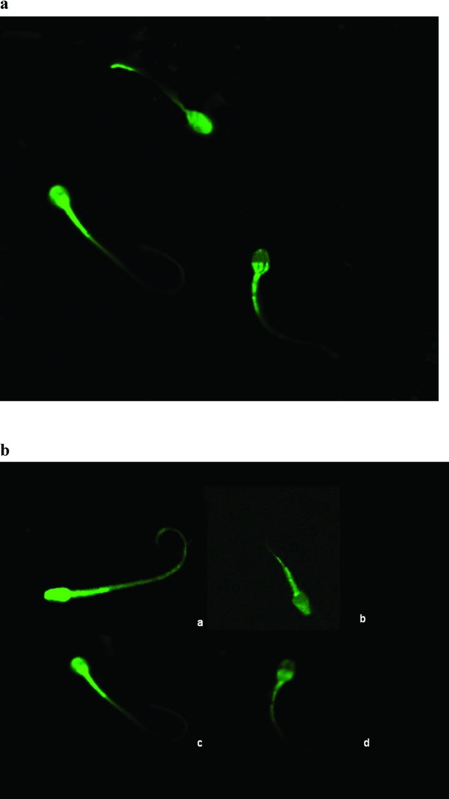
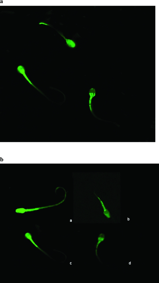
A CTC solution was made with 5 ml of CTC buffer, 2 mg of CTC (Sigma) and 4.4 mg of cysteine (Sigma). The pooled sample was overlaid with protein-free tissue culture medium 199 at a dilution of 3:1 (v/v) and was centrifuged at 300 g for 20 min. The supernatant was removed and the pellet resuspended in 45 μl of Earles medium supplemented with BSA. A 45-μl volume of CTC solution, 1 μg/ml propidium iodide (Sigma), and 8 μl of 12.5% (w/v) paraformaldehyde was added to each of the samples and mixed. A 10-μl droplet of the fixed and stained sample of spermatozoa was placed on a microscope slide and mounted with a droplet of an aqueous mounting medium (Vectashield, Vector Laboratories) to retard fading of the fluorescence. The CTC staining of the live spermatozoa was observed with a confocal laser scanning microscope LSM-510 (Zeiss). CTC was irradiated at 420 nm and detected via a 500-nm band pass filter. Propidium iodide was irradiated at 488 nm and detected via a 560-nm long pass filter. The different frames were scanned separately, with appropriate installation of the optical path for excitation and emission of each scan, according to previously published methods (Borzacchiello et al., Reference Borzacchiello, Russo, Spoleto, Roperto, Balcos, Rizzo, Venuti and Roperto2007). Characteristics of CTC-stained cells were observed, photographed and described (Fig. 2).
Domestic cat spermatozoa, CTC staining. (a) F staining pattern, uncapacitated and acrosome intact; (b) B staining pattern, capacitated and acrosome intact; (c) Acrosome reacted occurring pattern; (d) AR staining pattern, capacitated and acrosome reacted. (×100 magnification under oil).
Statistical analysis
Sperm motility, viability, and intact acrosomes were analysed using one-way analysis of variance (ANOVA) performed by GraphPad InStat, MS Windows XP version and by MedCalc version 7.3.01 after angular transformation of the data (SAS Institutes). Statistical differences were determined by the least-significant difference multiple comparison procedure (Steel & Torrie, 1980), with the level of significance set at p < 0.05. All data were expressed as means ± SD.
Results
The testicles, collected from 20 domestic cats, had a mean length of 1.8 cm (with a range of 1.3–2.5 cm) and a mean width of 1.2 cm (with a range of 0.8–2.0 cm). The mean weights observed for the left and right testes were very similar, and no correlation was observed between testis weight and body weight.
The mean testis weight found for the adult cat was approximately 1.2 g, and the mean length and width was similar with the physiological parameters of this species (Kenagy & Trombulak, Reference Kenagy and Trombulak1986).
The testicles collected at the time of the autopsy from the three wild felids had a mean length of 4.5 cm (with a range of 3.8–4.7 cm) and a mean width of 3.1 cm (with a range of 2.6–3.5 cm).
The total number of spermatozoa collected from cat cauda epididymis varied between 650.3 × 10⁶ to 23 × 106 (58.15 ± 18.3 mean) and the motility of epididymal spermatozoa at collection varied between 50 and 75% with a mean of 54.7 ± 11.3%. The motility of frozen–thawed cat epididymal spermatozoa varied between 30 and 60% with a mean of 32 ± 13.1%.
Mean ± SD for motility and viability in fresh and in frozen–thawed (1 h, 2 h and 3 h) spermatozoa are shown in Table 1 for tomcats’ spermatozoa and in Table 2 for tiger spermatozoa. In the domestic cats sample as well as in the tiger sample sperm motility and viability showed a significant reduction between fresh and frozen–thawed spermatozoa (1 h, 2 h and 3 h) (p < 0.001). Sperm motility as well as sperm viability showed a significant decline (p < 0.001) from 0 h to 1 h but did not show a decline from 1 h to 2 h and 3 h.
Characteristics of fresh and frozen–thawed domestic cat sperm obtained from the cauda epididymis immediately post thaw (0 h) and at l-h intervals during 3 h of incubation at 38°C. Mean ± SD (EP; male cats n = 20).

*p < 0.001 vs Fresh.
Characteristics of fresh and frozen–thawed tiger sperm obtained from the cauda epididymis immediately post thaw (0 h) and at l-h intervals during 3 h of incubation at 38°C. Mean ± SD (EP; Bengal tiger n = 2).

*p < 0.001 vs Fresh.
The proportion of intact acrosomes sperms (non-activated, F pattern, and activated, B pattern) showed a significant reduction (p < 0.001) between fresh sample and at the times 1 h, 2 h and 3 h). Whereas, the proportion of reacted acrosomes (AR pattern) sperms showed a significant increase (p < 0.001) from fresh sample and at the times 1 h, 2 h and 3 h after thawing. Sperm acrosal integrity showed a significant decrease in percentages between h0 and 1 h (Table 1).
Mean ± SD for motility and viability in controls and frozen–thawed spermatozoa are shown in Table 1.
Spermatozoa motility was evaluated immediately post thaw (0 h) and at l-h intervals during 3 h of incubation at 38°C. These results are presented in Table 1.
The total number of spermatozoa collected from Bengal tiger cauda epididymides varied between 57.5 × 10⁶ to 22.8 × 10⁶ (52.30 ± 17.6 mean). The motility of epididymal spermatozoa at collection from the three male felids varied between 30 and 54% with a mean of 38.3 ± 18.7%. The motility of frozen–thawed tiger epididymal spermatozoa varied between 20 and 45% with a mean of 21.5 ± 16.8%. Mean ± SD for motility and viability in controls and frozen/thawed spermatozoa are shown in Table 2. Spermatozoa motility was evaluated immediately post thaw (0 h) and at l h intervals during 3 h of incubation at 38°C. These results are presented in Table 2.
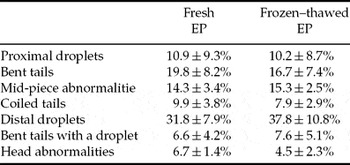
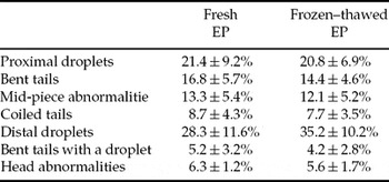
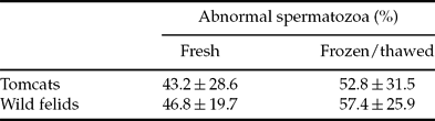
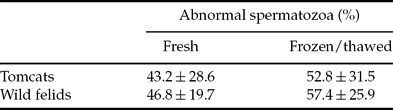
The most frequent sperm abnormalities relieved in the fresh tomcat semen were proximal droplets (10.9 ± 9.3%), bent tails (19.8 ± 8.2%), midpiece abnormalities (14.3 ± 3.4%), coiled tails (9.9 ± 3.8%), distal droplets (31.8 ± 7.9%), bent tails with a droplet (6.6 ± 4.2%) and head abnormalities (6.7 ± 1.4%). The most frequent sperm abnormalities relieved in the fresh wild felids semen were proximal droplets (21.4 ± 9.2%), bent tails (16.8 ± 5.7%), midpiece abnormalities (13.3 ± 5.4%), coiled tails (8.7 ± 4.3%), distal droplets (28.3 ± 11.6%), bent tails with a droplet (5.2 ± 3.2%) and head abnormalities (6.3 ± 1.2%) (Tables 3 and 4). Morphology showed a significant increase in percentages of abnormal cells between fresh and frozen/thawed spermatozoa (Table 5).
Sperm abnormalities relieved in the fresh and frozen–thawed tomcat semen (mean ± SD).

Sperm abnormalities relieved in the fresh and frozen–thawed wild felids semen (mean ± SD).

Rate of abnormality sperms relieved in fresh and frozen/thawed tomcats and tiger epididymal spermatozoa (mean ± SD).
The three distinct patterns observed using the CTC-staining method were first described in the mouse (Ward & Storey, Reference Ward and Storey1984; Schäfer-Somi et al., Reference Schäfer-Somi, Kluger, Knapp, Klein and Aurich2006) and were allocated an identification system as follows: F, uncapacitated and acrosome intact; B, capacitated and acrosome intact; AR, capacitated and acrosome reacted (Fig. 2). In the fresh samples, mean percentages of F, B and AR cells were 36.45, 21.25 and 6%, respectively, and in the frozen–thawed samples they were 10.65, 12.30, and 22.16%, respectively in domestic cats, and 24.55, 10.70 and 8% in the fresh samples and 7.24, 11.50, 25.40% in the tiger.
Discussion
Cryopreservation of spermatozoa in mammals is a valuable tool for preserving genetic material. Improvement of sperm freezing protocols and related technologies would increase the efficacy of gene banking from endangered felines, and the domestic cat can be used as a model for the wild felines. It has been reported that a pregnancy rate of only about 10% was obtained in the domestic cat after the use of frozen–thawed semen with vaginal insemination (Tsutsui et al., Reference Tsutsui, Wada, Anzai and Hori2003). The fertilizing ability of frozen–thawed epididymal spermatozoa has been proven (Kashiwazaki et al., Reference Kashiwazaki, Yamaguchi, Uesugi, Hishiyama, Kim, Nakatsukasa, Kojima, Okuda, Hisamatsu, Inomata and Shino2005).
Cryopreservation of epididymal spermatozoa is a potentially valuable procedure for banking genetic resource from individuals of endangered species that die accidentally, (Tsutsui et al., Reference Tsutsui, Wada, Anzai and Hori2003) a conception rate of 27.3% for eleven female cats that received unilateral intrauterine AI of 5 × 10⁷ sperm has been reported (Nakatsukasa et al., Reference Nakatsukasa, Inomata, Ikeda, Shino and Kashiwazaki2000, Hay & Goodrowe, Reference Hay and Goodrowe1993). Preceding works indicate that feline spermatozoa do not seem susceptible to cold shock in the temperature intervals usually used for liquid sperm preservation or cooling prior to cryopreservation. It could also be concluded that the addition of 20% egg yolk to the preservation extender is beneficial for preservation of sperm motility and acrosome integrity, but detrimental for plasma membrane integrity. No interaction was found between sperm type and extender, indicating that egg yolk had the same effect on both ejaculated and epididymal spermatozoa (White, Reference White1993; Pukazhenthi et al., Reference Pukazhenthi, Pelican, Wildt and Howard1999).
Furthermore, other authors have suggested that the glycerol is toxic to spermatozoa and should be added when samples are cooled in order to reduce the speed of entry. This fact is probably relevant also in felids, as in the fishing cat (Prionailurus viverrinus) post-thaw motility and acrosome integrity were higher after glycerol addition at 5°C than at 25°C (Thiangtum et al., Reference Thiangtum, Swanson, Howard, Tunwattana, Tongthainan and Wichasilpa2006).
It was concluded by Glover & Watson (Reference Glover and Watson1985) and Zambelli et al. (Reference Zambelli, Caneppele, Catagnetti and Belluzzi2002) that cat spermatozoa are clearly less susceptible to cold shock than spermatozoa from bull, a species in which freezing is routinely used worldwide with simple protocols. The tolerance of cat spermatozoa to cooling is confirmed by our findings, which are similar to the conclusions of Pukazhenthi et al. (Reference Pukazhenthi, Pelican, Wildt and Howard1999). Motility was not affected by cooling at either rate in our study. In contrary to what is expected after cold shock, the motility was in fact significantly higher in the cooled samples compared with the controls after rewarming to 38°C. It is likely that the reason for this finding was an increase in the cooled spermatozoa's longevity, induced by a lower metabolism at 4°C (Watson, Reference Watson, Morris and Clarke1981).
One of the consequences of destabilization of membranes by cooling or cryopreservation is a premature acrosome reaction that shortens the life span of the spermatozoa and reduces fertility. In fact, cryopreserved spermatozoa may be considered to be in a state resembling partially capacitated spermatozoa: their membranes have undergone similar fluidity changes as those seen during capacitation; they are permeable to calcium ions that promote both capacitation and the acrosome reaction; their viability is thus limited because capacitated spermatozoa do not have a prolonged survival: they are usually activated close to the time of meeting the oocyte. Moreover, a false acrosome reaction has also been described in thawed spermatozoa, in which acrosomal changes are associated with irreversible membrane damages (Bedford, Reference Bedford1970; Meizel, Reference Meizel and Johnson1978). The present study showed that a significant higher proportion of cat epididymal spermatozoa after thawing exhibited the pattern of acrosome reaction compared with fresh or diluted semen before freezing.
The present study showed that feline epididymal spermatozoa can be frozen in egg-yolk extender with 4.0% glycerol in 0.25 ml straws. The procedure used in the present study for epididymal cat sperm cryopreservation may be applicable for banking the genetic resources of wild felid species. However, development and improvements in reproductive technologies such as AI, in vitro fertilization (IVF), and intracytoplasmic sperm injection will be required to generate offspring from the frozen–thawed spermatozoa of wild felid species.
In conclusion, the freezing system for epididymal spermatozoa used in the present study is a workable protocol in the domestic cat that gives appropriate information for banking the genetic resources of wild felid species.