I. INTRODUCTION
The extreme physical and chemical propertiesReference Mochalin, Shenderova, Ho and Gogotsi1 as well as low toxicityReference Fu, Lee, Chen, Lim, Wu, Lin, Wei, Tsao, Chang and Fann2 of detonation nanodiamond (DND) materials have led to great interest, recently, in using these materials for both therapeutic and diagnostic applications in nanomedicineReference Kim, Zhang, Moore and Ho3 and for electrochemical supercapacitors.Reference Wang, Plylahan, Shelke, Devarapalli, Li, Subramanian, Djenizian, Boukherroub and Szunerits4 Currently, most of the nanodiamonds are manufactured through the detonation of explosives containing carbon followed by chemical purification of explosion-produced soot, which results in diamond grains with diameters in the range of single-digit-nanometers.Reference Shenderova, Panich, Moseenkov, Hens, Kuznetsov and Vieth5 Nanodiamond materials are under investigation for biomedical imaging and drug-delivery applications based on their low toxicity and demonstrated biocompatibility.Reference Fu, Lee, Chen, Lim, Wu, Lin, Wei, Tsao, Chang and Fann2 Furthermore, negatively charged nitrogen-vacancy color centers can be used for bright, photostable biolabellingReference Mohan, Tzeng, Yang, Chen, Hui, Fang and Chang6,Reference Yu, Kang, Chang, Chen and Yu7 applications based on extended red emission at ∼700 nm and near-unit fluorescence quantum yield.Reference Yu, Kang, Chang, Chen and Yu7–Reference Neugart, Zappe, Jelezko, Tietz, Boudou, Krueger and Wrachtrup9
Nanodiamond materials with high surface areas are of interest for numerous photocatalytic,Reference Zhu, Zhang, Ruther and Hamers10,Reference Kim, Dey, Seo, Kim, Lim and Lee11 photonic,Reference Vlasov, Shiryaev, Rendler, Steinert, Lee, Antonov, Voros, Jelezko, Fisenko, Semjonova, Biskupek, Kaiser, Lebedev, Sildos, Hemmer, Konov, Gali and Wrachtrup12 astrophysical,Reference Chang, Chen and Kwok13–Reference Jones15 and drug-delivery applications.Reference Kim, Zhang, Moore and Ho3 Water-based nanodiamond gels have also been demonstrated to have a significant impact on the thermodynamic behavior of water confined within nanoscale pores.Reference Korobov, Avramenko, Bogachev, Rozhkova and Ōsawa16 High-pressure, high-temperature (HPHT) synthetic approaches have been reported recently to produce high surface area diamond aerogel materials in a laser-heated diamond anvil cellReference Pauzauskie, Crowhurst, Worsley, Laurence, Kilcoyne, Wang, Willey, Visbeck, Fakra, Evans, Zaug and Satcher17 as well as mesoporous diamond using a multianvil press.Reference Mandal and Landskron18 These materials are of interest, since aerogels are lightweight, have high surface areas, and contain abundant open pores that can be readily loaded with drugs or other compounds to be used as an effective payload delivery vessel. However, HPHT processing is costly in terms of both capital equipment and processing time. Developing chemical approaches to high surface area diamond synthesis would greatly increase availability and reduce cost for a range of applied and fundamental scientific applications.Reference Brownlee, Tsou, Aleon, Alexander, Araki, Bajt, Baratta, Bastien, Bland, Bleuet, Borg, Bradley, Brearley, Brenker, Brennan, Bridges, Browning, Brucato, Bullock, Burchell, Busemann, Butterworth, Chaussidon, Cheuvront, Chi, Cintala, Clark, Clemett, Cody, Colangeli, Cooper, Cordier, Daghlian, Dai, D'Hendecourt, Djouadi, Dominguez, Duxbury, Dworkin, Ebel, Economou, Fakra, Fairey, Fallon, Ferrini, Ferroir, Fleckenstein, Floss, Flynn, Franchi, Fries, Gainsforth, Gallien, Genge, Gilles, Gillet, Gilmour, Glavin, Gounelle, Grady, Graham, Grant, Green, Grossemy, Grossman, Grossman, Guan, Hagiya, Harvey, Heck, Herzog, Hoppe, Horz, Huth, Hutcheon, Ignatyev, Ishii, Ito, Jacob, Jacobsen, Jacobsen, Jones, Joswiak, Jurewicz, Kearsley, Keller, Khodja, Kilcoyne, Kissel, Krot, Langenhorst, Lanzirotti, Le, Leshin, Leitner, Lemelle, Leroux, Liu, Luening, Lyon, MacPherson, Marcus, Marhas, Marty, Matrajt, McKeegan, Meibom, Mennella, Messenger, Messenger, Mikouchi, Mostefaoui, Nakamura, Nakano, Newville, Nittler, Ohnishi, Ohsumi, Okudaira, Papanastassiou, Palma, Palumbo, Pepin, Perkins, Perronnet, Pianetta, Rao, Rietmeijer, Robert, Rost, Rotundi, Ryan, Sandford, Schwandt, See, Schlutter, Sheffield-Parker, Simionovici, Simon, Sitnitsky, Snead, Spencer, Stadermann, Steele, Stephan, Stroud, Susini, Sutton, Suzuki, Taheri, Taylor, Teslich, Tomeoka, Tomioka, Toppani, Trigo-Rodriguez, Troadec, Tsuchiyama, Tuzzolino, Tyliszczak, Uesugi, Velbel, Vellenga, Vicenzi, Vincze, Warren, Weber, Weisberg, Westphal, Wirick, Wooden, Wopenka, Wozniakiewicz, Wright, Yabuta, Yano, Young, Zare, Zega, Ziegler, Zimmerman, Zinner and Zolensky19 Here, we report a rapid, low-cost alternative for making diamond aerogels by forming covalent bonds between individual nanocrystalline grains using a sol–gel reaction between resorcinol and formaldehyde (RF) molecular precursors.
II. EXPERIMENTAL
A. Oxidation
DND materials (ND90, Dynalene) were oxidized in ambient air within a tube furnace (Lindberg Blue) at 450 °C for 8 h to remove amorphous carbon soot and to generate chemically reactive surface oxygen functional groups (carboxylic acids, anhydrides, etc.).Reference Osswald, Yushin, Mochalin, Kucheyev and Gogotsi20,Reference Kruger, Liang, Jarre and Stegk21 It has been experimentally shown that the oxidation process serves to reduce the size of the nanodiamonds, removes graphitic material, and increases the intensity of the nitrogen-vacancy centers within the nanodiamonds.Reference Gaebel, Bradac, Chen, Say, Brown, Hemmer and Rabeau22 The final oxidized nanodiamond (OxDND) was used during synthesis and subsequent characterization of the nanodiamond aerogels (NDAGs).
B. Neutron activation analysis (NAA)
NAA was used to quantify the trace impurity content in the precursor and oxidized nanodiamonds. NAA technique is preferred for trace element quantification due to its high sensitivity and accurate, consistent, fast results with minimal sample preparation.Reference Yusuf, Krahenbuhl, Haskins and Hartman23 The samples are bombarded with neutrons to form radioactive nuclides. These radioactive nuclides decay, emitting gamma rays that can be quantitatively monitored. Decay schemes can be single events or sequential multistep progressions,Reference Soete, Gijbels and Hoste24 as is the case for Ce-133. The NAA data presented in this study were obtained using a TRIGA Mark II nuclear reactor. The samples (10 mg each) were enclosed in polyethylene capsules and were irradiated for 30 min. The study was performed at a thermal operating power of 100 kW, 4 × 10¹² neutrons/cm2 s thermal flux, and 4.8 × 10¹² neutrons/cm2 s fast and epithermal flux. Each sample was counted while positioned near the surface of a 25 cm2, trapezohedral, germanium, lithium-drifted semiconductor detector (Nuclear Diodes), which is constantly cooled by liquid nitrogen (77 K). The dead time between the end of irradiation and the start of collection was 5 min. Ratiometric analyses were performed comparing experimental activities (µCi/unit) to calculated activities per given element and reactor efficiency to determine the concentrations of metallic impurities.
C. Synthesis
NDAGs were prepared using OxDND materials based on a modified time-efficient, acid-catalyzed methodReference Mulik, Sotiriou-Leventis and Leventis25 for preparing resorcinol–formaldehyde aerogels. Briefly, 1.67 mL of high purity acetonitrile (AN) (OmniSolv) was sonicated with 17.5 mg of DNDs for 15 min in polypropylene tubes. Following dispersion of OxDND within the AN solvent, 37.6 mg of resorcinol (R, Sigma-Aldrich) and 49.4 µL of formaldehyde (F, Sigma-Aldrich, 37% w/w aqueous solution with methanol as a stabilizer) were added to the mixture and sonicated for 20 min until all the resorcinol was dissolved (1:2 molar ratio of R:F). Lastly, 4 µL of HCl (C, Sigma-Aldrich, 37% ACS reagent) was added as a catalyst and the mixture was further sonicated for 30 min (8:1 molar ratio R:C). The R:OxDND mass ratio used for the sol–gel reaction was 2:1. The mixture (sol) gelled within 30 min at room temperature during sonication. The resulting NDAG molar ratio of R:AN was 1:94. The gels were then washed with anhydrous ethanol (4× over 48 h) and supercritically dried in CO2 (E3100 critical point dryer, Quorum Technologies, Laughton, East Sussex, UK). Supercritical point drying (SCPD) was used to dry the wet gel to preserve the porosity of the solid matrix.
D. Surface area and pore volume
Surface area and pore volume measurements were performed by the Brunauer–Emmett–Teller (BET) and Barrett–Joyner–Halenda (BJH) methods, respectively, using a Nova 2200e analyzer (Quantachrome) with nitrogen gas as the adsorbate. The OxDND was degassed for 17 h at 125 °C prior to performing the BET measurement while the NDAG was loaded in the analyzer immediately following SCPD. High temperature degassing was avoided given discoloration of the aerogel that was observed at 200 °C.
E. Electron microscopy
The NDAG material was analyzed by using a FEI Sirion scanning electron microscope (SEM). Samples were prepared for SEM characterization by coating the material with a 10 nm layer of sputtered gold. The samples were also analyzed using transmission electron microscopy (TEM, FEI Tecnai G2 F20, Hillsboro, OR, 200 kV accelerating voltage) to verify the presence of nanodiamonds within the RF matrix. High-resolution bright field (HR-BF) imaging, select area electron diffraction (SAED), and high angle annular darkfield (HAADF) scanning transmission imaging were performed to characterize the NDAG microstructure.
F. Fourier transform infrared spectroscopy (FTIR)
Attenuated total reflectance FTIR (ATR-FTIR) spectroscopy was performed using a Bruker VERTEX 70 Fourier transform infrared spectrometer (Billerica, MA) with an integrated Platinum-ATR-accessory (Unit A225/Q1). The resulting data were used to identify the functional groups, impurities, and adsorbed molecules on the surface of the precursor materials and synthesized aerogels.
G. X-ray diffraction (XRD)
XRD was performed using a Bruker D8 Discover x-ray diffractometer equipped with a General Area Detector Diffraction Systems (GADDS) XRD system using a Cu Kα source. Powder samples were aligned in the system using the laser guidance system provided in the instrument.
III. RESULTS AND DISCUSSION
A. Precursor characterization
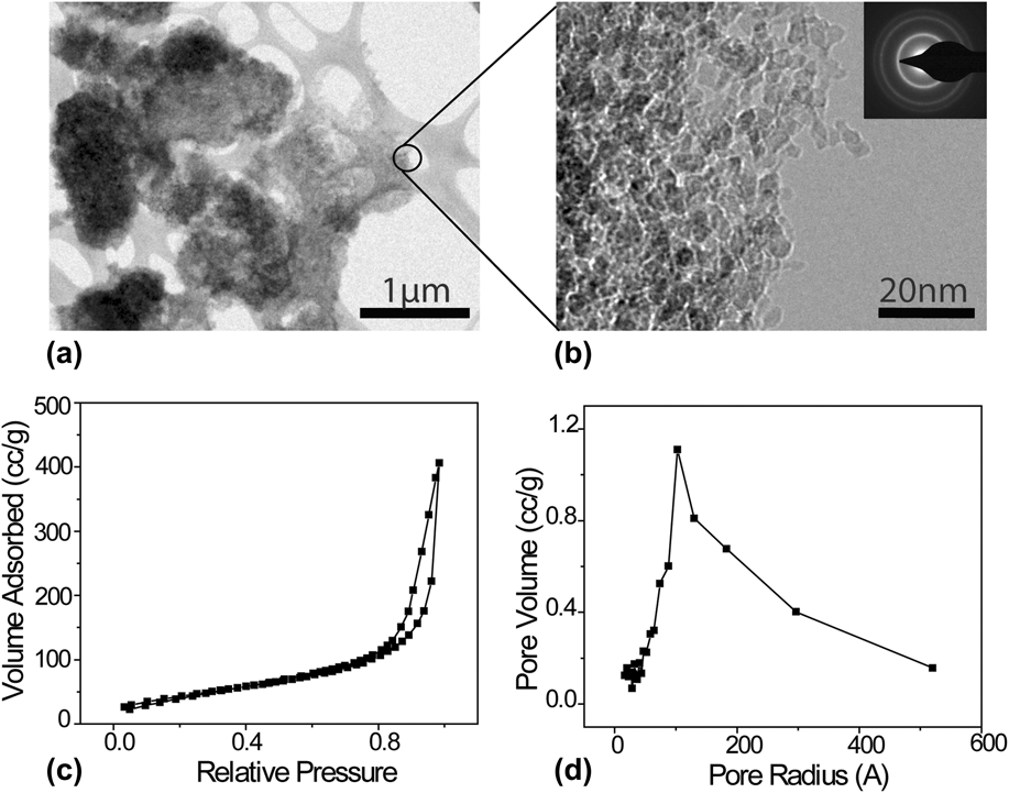
TEM images of the precursor OxDND [Figs. 1(a) and 1(b)] show that the nanodiamonds are aggregated into clusters of roughly 1 µm diameter, being made up of smaller crystalline grains of approximately 5 nm in diameter. The OxDND is polycrystalline, as verified by the SAED pattern shown in the inset of Fig. 1(b). In addition, the BET surface area of the OxDND, as calculated from the nitrogen adsorption/desorption isotherm in Fig. 1(c), is 161 m2/g, which supports the fact that the nanodiamond clusters have a high surface area. The pore volume distribution in Fig. 1(d) shows that the OxDND has an average pore volume of 0.613 cm³/g.
(a) BF-TEM of OxDND clusters. (b) BF-TEM images of OxDND at a higher magnification, showing that clusters are formed from the aggregation of smaller nanodiamond crystal grains. (c) Nitrogen adsorption isotherm. (d) Pore size distribution of OxDND.

The quality, physical/chemical properties, and potential use in specific applications of nanodiamonds depend critically on the purity of the material. The purity of the nanodiamond is determined by oxidative isolation of the diamond fraction from the synthetic detonation soot (predominately graphitic carbon) and by the remaining noncarbon impurities originating from the source of explosive materials, walls of the detonation chamber, and detonator. The variation of these noncarbon impurities is extensive and includes both metal (Fe, Ti, Al, Cr, Si, etc.) and nonmetal (S, P, B, etc.) elements by up to 8 wt%.Reference Mitev, Townsend, Paull and Nesterenko26
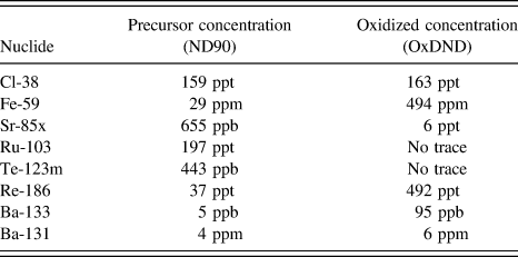
NAA was used to characterize these impurities for powder precursors used in this experiment. The NAA data in Fig. 2(a) show that several metallic impurities (rhenium, ruthenium, cerium, and iron) are present in the analyzed samples. The elements chlorine, barium, tellurium, strontium, and antimony comprise the nonmetallic impurities. In general, there is significant variation in the impurities of the precursor nanodiamonds. Iron and cerium were present in all the samples and generally increase in concentration during the oxidation process of nanodiamonds. In as-received DND material, the concentration of iron is observed to increase from 29 ppm to nearly 500 ppm following air oxidation. The mass of carbon in a given sample is reduced during the oxidation process following the formation of gas-phase CO2 and CO molecules. In contrast to carbon, iron does not form volatile oxidation by-products at this oxidation temperature (450 °C), resulting in an increase in the residual concentration of iron. Furthermore, the results indicate that the oxidation process tends to remove the trace elements ruthenium and tellurium. The list of elements identified by NAA in precursor nanodiamond (ND90) and OxDND is given in Table I.
(a) NAA of ND90 (black) and OxDND (red) precursor. (b) FTIR of ND90 (black) and OxDND (red) precursor.
List of elements identified by NAA.
FTIR spectra shown in Fig. 2(b) indicate that all of the spectra show the surface hydroxyl (O–H) vibration bands (stretch regions from 3650 to 2920 cm−1 and bend regions from 1510 to 1700 cm−1)Reference Osswald, Yushin, Mochalin, Kucheyev and Gogotsi20,Reference Ji, Jiang, Xu and Li27,Reference Jiang and Xu28 and C=O bonds of carboxylic acid (region from 1700 to 1880 cm−1).Reference Jiang and Xu28,Reference Kuznetsov, Aleksandrov, Zagoruiko, Chuvilin, Moroz, Kolomiichuk, Likholobov, Brylyakov and Sakovitch29 Also, the peak near 1100 cm−1 is related to C–O stretching vibration present in the DND and OxDND.Reference Yeap, Tan and Loh30 The small peak on the OxDND spectrum near 2324–2360 cm−1 is due to adsorbed molecules of CO2.Reference Kuznetsov, Aleksandrov, Zagoruiko, Chuvilin, Moroz, Kolomiichuk, Likholobov, Brylyakov and Sakovitch29
B. Diamond aerogel characterization
There is a dramatic lightening in color of the recovered NDAG material in comparison with pure RF aerogel, as shown in Fig. 3(a). The final color of the NDAG is pinkish-white in contrast to the deep-red color of the RF aerogel at an identical R:AN ratio. Despite the contrast in color, both RF and NDAG were similar in shape and mechanical stability and easily held under standard handling and analytic procedures. TEM SAED [Fig. 3(b), inset] confirms the presence of OxDND in the aerogel matrix, showing a polycrystalline ring pattern. HAADF TEM [Fig. 3(b)] shows that the NDAG samples exhibit a highly porous microstructure that is maintained following SCPD.
(a) Digital photograph showing side-by-side color comparison of RF and NDAGs following supercritical drying. Corresponding dark field optical microscopy images are shown below for each material at 100x magnification. (b) HAADF TEM of the nanodiamond aerogel. Insets: HR-TEM showing lattice of a nanodiamond embedded within the aerogel (top-right), and a comparison of the SAED of the nanodiamonds in the aerogel compared to the nanodiamond precursor (bottom-right).
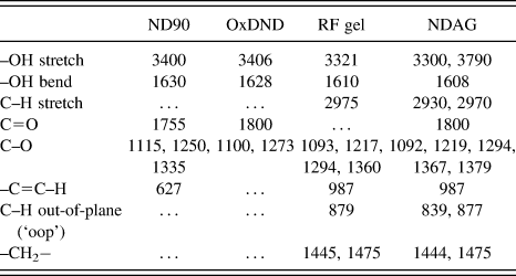
Figures 2(b) and 4(a) show FTIR spectra taken in air of the ND90 precursor, OxDND, the RF aerogel control, and NDAG samples. Due to the oxidative nature of the detonation, as well as the air oxidation of the precursor nanodiamonds, the surface of the OxDND contains various oxygen-containing groups. All samples show C–O–C stretching bands from ethers, acid anhydrides, lactones, and epoxy groups in the region 1100–1370 cm−1.Reference Lambert, Shurvell and Cooks31 Adsorbed water gives strong absorption bands in the 3500–3300 and 1620–1630 cm−1 (bending mode) regions. Carbonyl C=O peaks (1700–1865 cm−1 for carbonyls in ketones, carboxylic acids, acid anhydrides, esters, and lactones) are not observed in the pure RF gel but appear in the NDAG due to the addition of nanodiamonds, which contain these bonds. Also, neither of the precursor nanodiamonds show the presence of C–H or CH2 stretching, as opposed to the RF aerogel and NDAG, since they are not bonded with any resorcinol/formaldehyde condensation polymers. The wave numbers of the absorption peaks corresponding to each vibrational mode identified by the FTIR are shown in Table II.
(a) FTIR spectra of OxDND (red), pure RF aerogel (blue), and NDAG (black). (b) XRD spectra of OxDND (red), RF aerogel (blue), and NDAG (black).
Wave numbers of absorption peaks in FTIR spectra.
Analysis of the XRD spectra indicates the presence of amorphous hydrocarbons within both RF and NDAG samples following sol–gel cross-linking reactions. The peak at around 2θ = 43° is the characteristic (111) diffraction peak of diamond.Reference Sharda, Rahaman, Nukaya, Soga, Jimbo and Umeno32 Both aerogels exhibit an additional broader, weaker peak at 2θ = 75°, indicating that they are partially graphitic.Reference Lin and Ritter33–Reference Song, Nahm and Oh35
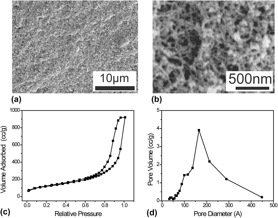
The microstructure of recovered NDAG materials was observed further using SEM. The surface of the NDAG appears to be highly porous as shown in Figs. 5(a) and 5(b). Figures 5(c) and 5(d) show the nitrogen adsorption/desorption isotherm and pore size distribution, respectively, of the NDAG with R:AN molar ratio of 1:94 and R:OxDND weight ratio of 2:1. The micropore surface area for this gel is 589 m2/g. Bulk density was approximated by dividing the measured mass of a typical sample over its macroscopic volume and was found to be ∼151 mg/cm3.
(a) SEM image of unpyrolyzed NDAG. (b) High-magnification SEM image of NDAG. (c) Nitrogen adsorption/desorption isotherm, and (d) BJH pore size distribution of NDAG.
IV. CONCLUSION
In conclusion, a fast sol–gel process is reported for preparing macroscopic quantities of NDAG materials with high surface areas based on acid-catalyzed condensation reactions in a polar, aprotic solvent (acetonitrile). Metallic impurities within detonation nanodiamond precursor materials were analyzed using NAA, and air oxidation was observed to increase the amount of iron by approximately one order of magnitude. Transmission electron microscopy was used to show that the NDAG material contains nanodiamonds dispersed throughout the resorcinol–formaldehyde matrix. Future efforts will focus on optimizing the microstructure of NDAG for loading with small-molecule pharmaceuticals, as well as tuning the density and mechanical properties for potential applications in future STARDUST missions.Reference Brownlee, Tsou, Aleon, Alexander, Araki, Bajt, Baratta, Bastien, Bland, Bleuet, Borg, Bradley, Brearley, Brenker, Brennan, Bridges, Browning, Brucato, Bullock, Burchell, Busemann, Butterworth, Chaussidon, Cheuvront, Chi, Cintala, Clark, Clemett, Cody, Colangeli, Cooper, Cordier, Daghlian, Dai, D'Hendecourt, Djouadi, Dominguez, Duxbury, Dworkin, Ebel, Economou, Fakra, Fairey, Fallon, Ferrini, Ferroir, Fleckenstein, Floss, Flynn, Franchi, Fries, Gainsforth, Gallien, Genge, Gilles, Gillet, Gilmour, Glavin, Gounelle, Grady, Graham, Grant, Green, Grossemy, Grossman, Grossman, Guan, Hagiya, Harvey, Heck, Herzog, Hoppe, Horz, Huth, Hutcheon, Ignatyev, Ishii, Ito, Jacob, Jacobsen, Jacobsen, Jones, Joswiak, Jurewicz, Kearsley, Keller, Khodja, Kilcoyne, Kissel, Krot, Langenhorst, Lanzirotti, Le, Leshin, Leitner, Lemelle, Leroux, Liu, Luening, Lyon, MacPherson, Marcus, Marhas, Marty, Matrajt, McKeegan, Meibom, Mennella, Messenger, Messenger, Mikouchi, Mostefaoui, Nakamura, Nakano, Newville, Nittler, Ohnishi, Ohsumi, Okudaira, Papanastassiou, Palma, Palumbo, Pepin, Perkins, Perronnet, Pianetta, Rao, Rietmeijer, Robert, Rost, Rotundi, Ryan, Sandford, Schwandt, See, Schlutter, Sheffield-Parker, Simionovici, Simon, Sitnitsky, Snead, Spencer, Stadermann, Steele, Stephan, Stroud, Susini, Sutton, Suzuki, Taheri, Taylor, Teslich, Tomeoka, Tomioka, Toppani, Trigo-Rodriguez, Troadec, Tsuchiyama, Tuzzolino, Tyliszczak, Uesugi, Velbel, Vellenga, Vicenzi, Vincze, Warren, Weber, Weisberg, Westphal, Wirick, Wooden, Wopenka, Wozniakiewicz, Wright, Yabuta, Yano, Young, Zare, Zega, Ziegler, Zimmerman, Zinner and Zolensky19
ACKNOWLEDGMENTS
The authors gratefully acknowledge the financial support from the University of Washington startup funding and the ACS Petroleum Research Fund. The authors also thank G. Cao for access to SCPD facilities. This work was performed under the auspices of the U.S. Department of Energy and the Fundamental & Computational Sciences Division of the Pacific Northwest National Laboratory. The PNNL is operated by Battelle under contract #DE-AC05-76RL01830. PBR acknowledges the support from the National Science Foundation Graduate Research Fellowship under Grant No. DGE-1256082.