Introduction
Anisakidosis is a zoonotic disease caused by the ingestion of larval nematodes in raw seafood such as sushi, sashimi, ceviche and pickled herring (Audicana & Kennedy, Reference Audicana and Kennedy2008; Hochberg & Hamer, Reference Hochberg and Hamer2010; Baron et al., Reference Baron, Branca, Trombetta, Punzo, Quarto, Speciale and Barresi2014). Symptoms of anisakidosis are vague, and include abdominal pain, nausea, vomiting and diarrhoea. This disease is often misdiagnosed as appendicitis, gastric ulcers or ileitis (Hochberg & Hamer, Reference Hochberg and Hamer2010; Baron et al., Reference Baron, Branca, Trombetta, Punzo, Quarto, Speciale and Barresi2014). The definitive diagnosis and management of gastric anisakidosis is generally non-operative, achieved by upper gastrointestinal endoscopy.
However, the diagnosis and management of intestinal anisakidosis can be challenging (Couture et al., Reference Couture, Measures, Gagnon and Desbiens2003; Hochberg & Hamer, Reference Hochberg and Hamer2010). Even though most patients have a self-limited illness and recover within a few days, some undergo surgery following a misdiagnosis of small bowel ischaemia or acute abdomen, due to positive peritoneal signs associated with ascites (Ishida et al., Reference Ishida, Harada, Egawa, Watabe, Ebina and Unno2007; O’ Daly et al., Reference O’ Daly, Ridgway, Keenan, Sweeney, Brophy, Hill, Evoy, O'Higgins and McDermott2009; Hochberg & Hamer, Reference Hochberg and Hamer2010; Yasunaga et al., Reference Yasunaga, Horiguchi, Kuwabara, Hashimoto and Matsuda2010). Since most intestinal anisakidosis is successfully treated non-operatively, the correct diagnosis is important for the appropriate management of these patients (Ishida et al., Reference Ishida, Harada, Egawa, Watabe, Ebina and Unno2007; Hochberg & Hamer, Reference Hochberg and Hamer2010; Shrestha et al., Reference Shrestha, Kisino, Watanebe, Itsukaichi, Hamasuna, Ohno and Tsugu2014). With the increasing popularity of eating lightly cooked or raw fish dishes, the number of patients with anisakidosis may increase (Audicana & Kennedy, Reference Audicana and Kennedy2008; Hochberg & Hamer, Reference Hochberg and Hamer2010). We report our experience with the diagnosis and management of patients with intestinal anisakidosis, and the diagnostic value of paracentesis.
Materials and methods
Patients
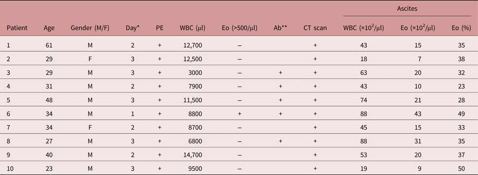
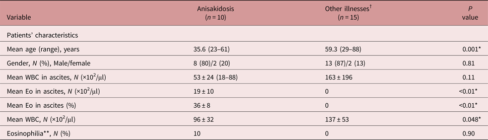
We reviewed retrospectively 16 patients diagnosed with intestinal anisakidosis at Tokyo Bay Urayasu Ichikawa Medical Center between April 2012 and December 2015. The first group included patients who were diagnosed with intestinal anisakidosis and who underwent laboratory evaluation of their ascites (table 1). The second group included patients with other abdominal conditions, associated with peritoneal signs and peritonitis, who underwent laboratory evaluation of ascites during the study period (see table 2). Epidemiological, clinical and laboratory data, as well as diagnostic and therapeutic features, and outcomes in these patients were reviewed. Patients diagnosed with liver cirrhosis were excluded from both groups. Each patient was enrolled in the study after giving written consent agreeing to treatment.
Characteristics of ten patients with intestinal anisakidosis.

*Day, onset of symptoms after eating contaminated food; Ab**, serum antibodies to Anisakis, positive (>1.5/U); PE, physical examination showing peritoneal signs; WBC, peripheral white blood cell count; Eo, peripheral blood eosinophil count (>500/μl), CT, underwent computed tomography scan of the abdomen.
Univariate analysis of clinical factors in patients with and without intestinal anisakidosis.

Eo, eosinophils; WBC, white blood cells; *, P < 0.05; Eosinophilia**, (>500/μl).
†Other illnesses: postoperative patients (n = 10), intraoperative patients (n = 3), Crohn's disease (n = 1), enteritis (n = 1).
Definitions
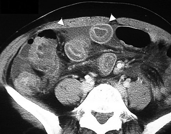
The diagnosis of intestinal anisakidosis was based on the following findings: (1) a recent history (within a week) of eating raw seafood; (2) abdominal pain with rebound tenderness; (3) computed tomography (CT) scan findings–localized submucosal oedema of the intestinal wall, dilation of the small bowel or ascites (fig. 1). All these three characteristics were included to diagnose intestinal anisakidosis. Additionally, pairs of serum IgG and IgA antibodies to Anisakis (enzyme-linked immunosorbent assay (ELISA), SRL Inc., Tokyo, Japan) were evaluated (normal range <1.50).
CT scan of intestinal anisakidosis: localized submucosal oedema of the intestinal wall, dilation of the small bowel or ascites are seen (white arrowheads).

Statistical analysis
Comparisons were performed using the non-parametric Mann–Whitney U-test for continuous variables and Student's t-test or Fisher's exact test for categorical data. All statistical analyses were performed using SPSS 23.0 (SPSS Inc., Chicago, Illinois, USA) and P < 0.05 was used to define a statistically significant difference.
Results
Patient characteristics
Sixteen patients with intestinal anisakidosis were seen during the study period. All patients underwent abdominal CT scans, which showed ascites. Ten of them (10/16, 63%) underwent laboratory evaluation of the ascites. Five of the ten patients were evaluated for paired IgG and IgA antibodies to Anisakis, and all were positive for these antibodies (table 1).
Eosinophils in the ascites
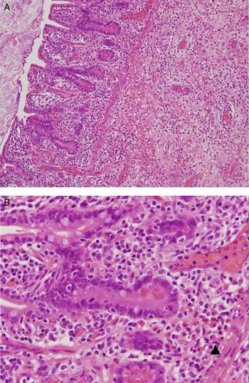
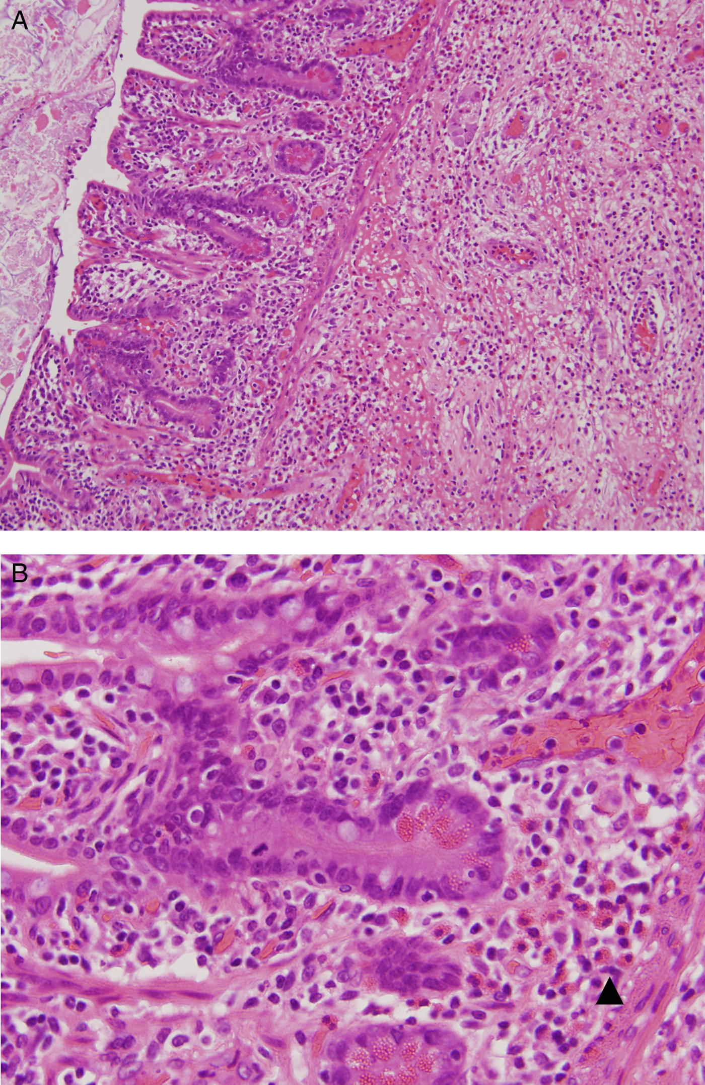
In the ten patients diagnosed with intestinal anisakidosis, all had an increased number of eosinophils in the ascites (table 1). In the 15 patients without intestinal anisakidosis, none had eosinophils in the ascites (P < 0.01) (table 2). Fifteen patients (15/16, 93.8%) diagnosed with intestinal anisakidosis recovered without complications after non-operative management. One patient underwent exploratory laparotomy for an acute abdomen and intestinal resection was performed with detection of Anisakis simplex. Eosinophil infiltration of the small bowel wall was found in the pathology specimen (fig. 2).
Microscopic findings of the resected small intestine specimen, showing: (A) proliferation and infiltration dominating around the affected area (stained with haematoxylin and eosin (H&E), ×40); (B) diffuse infiltration by eosinophils and oedema in the submucosa (black arrow) (H&E, ×600).
Discussion
The aim of this study was to investigate the association between eosinophils in ascites and the diagnosis of intestinal anisakidosis. The diagnosis of intestinal anisakidosis remains challenging in some patients (Couture et al., Reference Couture, Measures, Gagnon and Desbiens2003; Hochberg & Hamer, Reference Hochberg and Hamer2010; Shrestha et al., Reference Shrestha, Kisino, Watanebe, Itsukaichi, Hamasuna, Ohno and Tsugu2014). Although it is possible to make a diagnosis using an elevated titre of anti-Anisakis antibodies in blood, time is required to measure this, and the titre may not necessarily rise during the early phase (Ido et al., Reference Ido, Yuasa, Ide, Kimura, Toshimitsu and Suzuki1998; Sasaki et al., Reference Sasaki, Fukumori, Matsumoto, Ohmori and Yamamoto2003). Furthermore, healthy individuals who regularly eat raw fish may be false positive, and anti-Anisakis antibodies may not be a specific marker because of cross-reactions with proteins in other parasites, micro-organisms, insects and plants (Sakanari et al., Reference Sakanari, Loinaz, Deardorff, Raybourne, McKerrow and Frierson1988; Ido et al., Reference Ido, Yuasa, Ide, Kimura, Toshimitsu and Suzuki1998; Lorenzo et al., Reference Lorenzo, Iglesias, Leiro, Ubeira, Ansotegui, Garcia and Fernandez de Corres2003; Sasaki et al., Reference Sasaki, Fukumori, Matsumoto, Ohmori and Yamamoto2003). The accuracy of the diagnosis is increased by using pairs of serum antibodies. However, most of the patients get better in several days, thus the results might be useful for supporting the diagnosis of intestinal anisakidosis retrospectively, but they cannot be used for deciding the management of the patients.
Shibata and co-workers reported that intestinal anisakidosis is associated with three findings on a CT scan (Shibata et al., Reference Shibata, Ueda, Akaike and Saida2014), and we made the diagnosis of intestinal anisakidosis using these criteria on CT scans in addition to the recent history of ingestion of raw seafood and abdominal pain with rebound tenderness. The pathological appearance of A. simplex lesions shows local inflammatory lesions produced by A. simplex larvae, with a conspicuous eosinophil infiltration in the tissues surrounding the parasite (Yasunaga et al., Reference Yasunaga, Horiguchi, Kuwabara, Hashimoto and Matsuda2010; Shibata et al., Reference Shibata, Ueda, Akaike and Saida2014). Although systemic eosinophila is frequently associated with helminthic diseases, in anisakidosis this is described in fewer than 30% of cases (Audicana & Kennedy, Reference Audicana and Kennedy2008). Takei & Powell (Reference Takei and Powell2007) also reported the pathological findings of intestinal anisakidosis, including: transmural oedema in the small intestine, congestion and an inflammatory infiltrate abundant in eosinophils. Shirahama et al. (Reference Shirahama, Koga, Ishibashi, Uchida, Ohta and Shimoda1992) reported the presence of eosinophils in ascites of intestinal anisakidosis.
Based on these reports, we analysed ascites fluid collected by diagnostic paracentesis in patients with anisakidosis. The results show an increased white blood cell count, rich in eosinophils, which is characteristic of this disease (table 2). The elevated white blood cell count in the ascites is related to an inflammatory response in the abdomen. The aetiologies of peritoneal inflammatory responses associated with an elevated white blood cell count are associated with numerous causes, such as gastrointestinal perforation, pancreatitis, tuberculosis and malignancy in the peritoneum (Akriviadis & Runyon, Reference Akriviadis and Runyon1990; Soriano et al., Reference Soriano, Castellote and Alvarez2010). The fact that intestinal anisakidosis causes transmural inflammation and peritonitis has been supported since most patients have peritoneal signs on physical examination and an elevated white blood cell count in the ascites (table 2).
An elevated eosinophil count in ascites is associated with an inflammatory response. The differential diagnosis includes eosinophilic gastroenteritis, intestinal parasites, malignancy, inflammatory bowel disease, hypereosinophilic syndrome, etc. (Gomez et al., Reference Gomez, Tabel, Tuñón, Larrínaga, Alvarez, García and Olaguibel1998; Yun et al., Reference Yun, Cho, Park, Choi, Kim, Shin and Kim2007; Ramanan et al., Reference Ramanan, Blumberg, Mathison and Pritt2013). There may be an association with a systemic response, but the eosinophil count in the ascites is increased compared with peripheral blood in this study. Therefore, this finding may be very specific for intestinal anisakidosis, and helps to establish the diagnosis in patients with a history of ingestion of certain foods and characteristic findings on a CT scan.
Patients with intestinal anisakidosis often have peritoneal signs upon physical examination, which may contribute to the misdiagnosis of small bowel ischaemia, sometimes leading to exploratory laparotomy, since ascites is one of the warning signs suggesting the abdominal emergency (Ishikura et al., Reference Ishikura, Kikuchi and Ishikura1983; Sasaki et al., Reference Sasaki, Fukumori, Matsumoto, Ohmori and Yamamoto2003; Ishida et al., Reference Ishida, Harada, Egawa, Watabe, Ebina and Unno2007; O’ Daly et al., Reference O’ Daly, Ridgway, Keenan, Sweeney, Brophy, Hill, Evoy, O'Higgins and McDermott2009; Hochberg & Hamer, Reference Hochberg and Hamer2010; Yasunaga et al., Reference Yasunaga, Horiguchi, Kuwabara, Hashimoto and Matsuda2010). Paracentesis of ascites can distinguish intestinal anisakidosis from bowel ischaemia because an elevated eosinophil count is not typical in patients with bowel ischaemia. Since intestinal anisakidosis is mostly a self-limited disease, we might be able to avoid unnecessary surgery based on the paracentesis results. Eosinophilic enteritis would be another differential diagnosis in such patients; however, the clinical courses are usually different and it may be difficult to differentiate these conditions without a pathology specimen.
There are some limitations to this study. We only reviewed patients with a history of food ingestion and findings on a CT scan typical of intestinal anisakidosis. Since most patients with intestinal anisakidosis resolve spontaneously, they may not seek medical care, or they may have mild symptoms and not have a CT scan.
Secondly, an elevated eosinophil count in the ascites was very specific for intestinal anisakidosis in this study; however, other diseases, such as eosinophilic enteritis or other parasitic disease, may also cause increased eosinophil levels in ascites. In this study, we included patients who had history of digesting raw seafood and most of the patients resolved their symptoms in a short time without medical treatment, which is compatible with the clinical manifestations of intestinal anisakidosis (Shrestha et al., Reference Shrestha, Kisino, Watanebe, Itsukaichi, Hamasuna, Ohno and Tsugu2014). Since we did not check additional tests for other parasites, we cannot exclude the possibility that some parasites might cause similar symptoms; however, other parasites are unlikely to have similar symptoms and epidemiology.
Thirdly, we diagnosed intestinal anisakidosis based on history, physical examinations and CT scans, since no definitive serum markers are established to diagnose anisakidosis. Dominguez-Ortega et al. (Reference Dominguez-Ortega, Martinez-Alonso, Alonso-Liamazares, Argüelles-Grande, Chamorro, Robledo, Palacio and Martínez-Cócera2003) reported a novel way to check the serum levels of eosinophilic cationic protein to diagnose acute gastrointestinal anisakiasis, and this might be a promising and useful way to support the diagnosis. Further investigations are needed to support this new diagnostic method.
We report our experience with patients with intestinal anisakidosis. Most patients with anisakidosis have a self-limiting disease. An elevated eosinophil count in ascites is associated with intestinal anisakidosis, which might be useful to establish the diagnosis.
Financial support
This research received no specific grant from any funding agency, commercial or not-for-profit sectors.
Conflict of interest
None.
Ethical standards
This study was reviewed and approved by the Ethics Committee of the Tokyo Bay Urayasu Ichikawa Medical Center.