Introduction
Pneumatisation of the middle turbinate (concha bullosa) is the presence of an air-filled cavity within the nasal turbinate, which is believed to be associated with various sinonasal pathologies. When specifically identified, concha bullosa has been found to be present in approximately half of all patients who complain of nasal symptoms.Reference Ozcan, Selcuk, Ozkan, Akdogan and Dere1 Concha bullosa can occur unilaterally or bilaterally and are classified into three types: lamellar, bulbous and extensive concha bullosa.Reference Bolger, Butzin and Parsons2 Studies suggest that there is a developmental relationship between concha bullosa and a deviated nasal septum, which may explain the common complaint of nasal obstruction in these patients.Reference Stallman, Lobo and Som3,Reference El-Taher, AbdelHameed, Alam-Eldeen and Haridy4 Concha bullosa has also been found to be associated with chronic rhinosinusitis through obstruction of the ostiomeatal complex and ethmoid infundibulum as well as inhibition of nasal mucociliary function.Reference Smith, Edwards, Saini and Norton5–Reference Ozkiris, Karacavus, Kapusuz and Saydam8
Therefore, when concha bullosa is identified pre-operatively or intra-operatively in sinonasal surgery, it is typically addressed surgically, which has been demonstrated to aid nasal airflow and alleviate chronic rhinosinusitis.Reference Ozkiris, Karacavus, Kapusuz and Saydam8–Reference Eren, Kocak, Dogan, Ozturan, Yildirim and Tugrul10 As a result, various techniques are utilised to open the concha bullosa and reduce the bulkiness of the middle turbinate complex. These range from crushing, stripping, marsupialisation, and medial and lateral laminectomy to complete middle turbinectomy.Reference Havas and Lowinger9,Reference Eren, Kocak, Dogan, Ozturan, Yildirim and Tugrul10
Concha bullosa is typically readily identified on coronal computed topography (CT) images as a pneumatisation of the turbinate. The term concha bullosa typically refers to the middle turbinate; however, there have been cases describing involvement of the superior and inferior turbinates. Unfortunately, radiologist reporting of paranasal sinus CT images is often lacking. A study by Deutschmann et al. Reference Deutschmann, Yeung, Bosch, Lysack, Kingstone and Kilty11 found that many otolaryngologists were dissatisfied with current reporting, and the majority would like more clinically useful information. They also found that only 40 per cent of dedicated head and neck radiologists, 18 per cent of neuroradiologists and 33 per cent of other radiologists were able to correctly identify middle turbinate anatomy such as concha bullosa or a lateralised or paradoxical middle turbinate.
Artificial intelligence is a branch of computer science that studies intelligent agents that perceive their environment and take actions to maximise their chances of achieving a goal. Machine learning is the study of algorithms and models based on patterns and inferences to perform specific tasks. Deep learning is a subset of machine learning that uses layers arranged as artificial neural networks, in a similar manner to distributed communication nodes, taking resemblance to the synaptic structure of biological brains.
Convolutional neural networks are a subtype of deep learning that is well suited to image recognition. With recent advances in computational power, particularly in graphics processing units and release of user-friendly yet powerful open-source software packages, the development and implementation of artificial intelligence and convolutional neural networks have grown rapidly in many healthcare fields in recent years. Convolutional neural networks have been found to have useful diagnostic utility in various medical imaging modalities, such as identification of malignant skin lesions from dermatoscopyReference Esteva, Kuprel, Novoa, Ko, Swetter and Blau12 images and in extracting data from medical records through natural language processing.Reference Hoogendoorn, Szolovits, Moons and Numans13
Nevertheless, rhinology remains a relatively uncharted field for convolutional neural networks and deep learning, with a paucity of literature describing their use. Therefore, this proof-of-concept study aimed to determine if a convolutional neural network could be trained to diagnose the presence of concha bullosa with acceptable diagnostic accuracy. In doing so, a convolutional neural network would therefore demonstrate clinical applicability in CT sinus reporting where there is a deficiency in current standard radiologist reporting.
Materials and methods
High resolution CT scans of the paranasal sinuses taken consecutively from a single tertiary hospital radiology department were collected between 1 January 2015 and 31 December 2018 (Inteleviewer, Intelerad Medical Systems, Montreal, Canada.) A subset of approximately 80 per cent of the scans were pre-designated as training data, and the remaining 20 per cent of scans were designated as testing data. This was performed randomly using a computer random number generator.
From each scan, the images, request form and radiologist report were analysed. Information gathered from each included patient demographics of age, sex, date of scan and indication for imaging. Radiologist findings were also examined.
A single coronal slice of the bony windows that was thought to be representative of the ostiomeatal complex was used. This was determined in a similar manner to that in an image recognition paper published by Chowdhury et al., at the first coronal section through the natural maxillary ostium immediately posterior to the nasolacrimal duct.Reference Chowdhury, Smith, Chandra and Turner14
Each image was then cropped into a 250 × 250 pixel square that covered the entire middle turbinate, nasal septum, cribriform plate, lamina papyracea and medial maxillary wall. Left-sided images were flipped on their vertical axis to increase training data and maintain consistency. Each image was assessed by two blinded independent otolaryngology trainees for the presence of pneumatisation of the middle turbinate. Any discrepancies were referred for final decision by the senior author (NS), a fellowship trained rhinologist.
Ethical approval was provided by the Western Sydney Local Health District Human Research Ethics Committee prior to data collection.
Convolutional neural network training
The artificial intelligence algorithm was developed using Google's® Inception-V3 convolutional neural network base model that was trained with 1.28 million images. The classification layer was retrained in Python® programming language using a transfer learning method to interpret the coronal CT slices.
Statistical analysis
Analysis was performed using SPSS® (version 25) statistical software. Descriptive data were displayed as percentages and raw numbers for categorical data (sex, indication from report findings and presence of concha bullosa). Continuous variables were reported as means with standard deviations for data with normal distribution and reported as medians with interquartile ranges for data with non-normal distribution (age). Total diagnostic accuracy was presented as a percentage with 95 per cent confidence intervals (CIs), as well as a receiver operating characteristic curve and a calculation of the area under the curve. Inter-observer variability was assessed using the kappa statistic.
Results
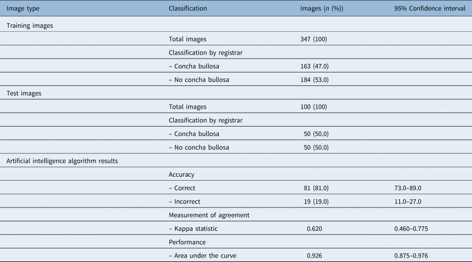
A total of 447 images of middle turbinate heads were included in the study (Table 1). Of these, 347 were used to train the developed convolutional neural network. Based on the conclusion of two blinded otolaryngology registrars, 47 per cent (n = 163) of these contained a concha bullosa at the isolated coronal CT slice, and the remaining 53 per cent (n = 184) did not contain a concha bullosa and had a normal, non-pneumatised middle turbinate.
Characteristics of training and test images for artificial intelligence convolutional neural networks to classify middle turbinate concha bullosa versus no concha bullosa from sinus computed tomography scans

The remaining 100 images were used to test the trained algorithm. Of these, 50 per cent (n = 50) were classified as having a concha bullosa based on otolaryngology registrar review, and 50 per cent (n = 50) were classified as having a normal, non-pneumatised middle turbinate. This yielded a diagnostic accuracy of 81 per cent (95 per cent CI = 73–89 per cent).
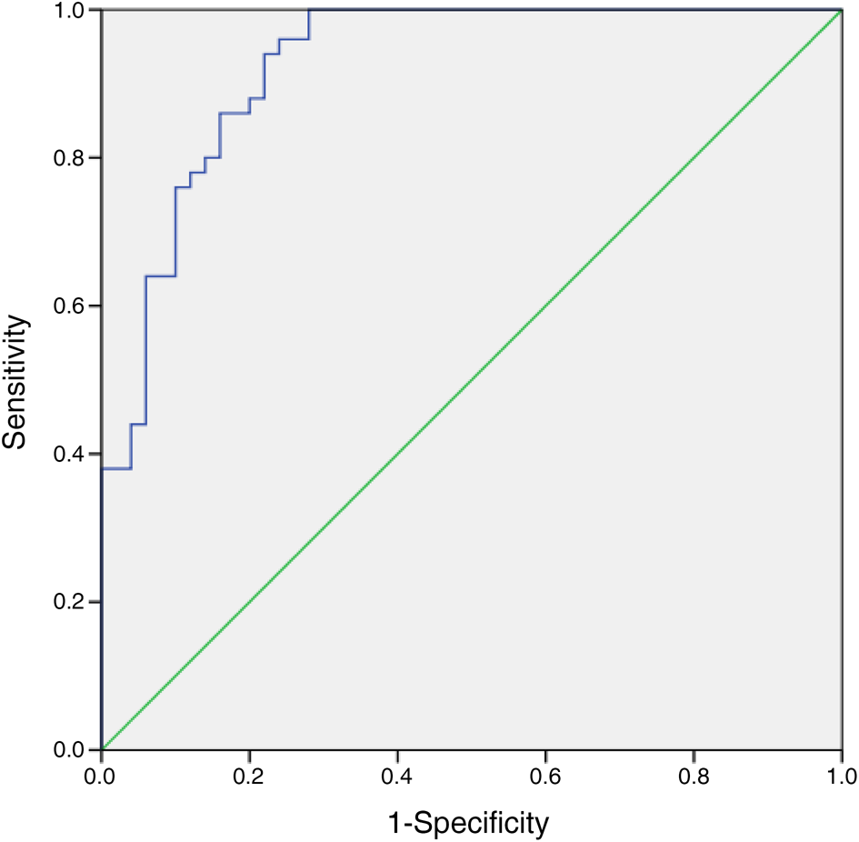
Measurement of agreement between the otolaryngology registrar and the trained convolutional neural network using Cohen's kappa statistic yielded a score of 0.62 (95 per cent CI = 0.46–0.78), reflecting a substantial agreement. Area under the curve performance was determined to be 0.93 (95 per cent CI = 0.88–0.98), reflecting excellent diagnostic accuracy (Figure 1).
Receiver operating characteristic curve of artificial intelligence convolutional neural network algorithm performance to classify middle turbinate concha bullosa versus no concha bullosa from sinus computed tomography scans.

Discussion
Artificial intelligence has been investigated in multiple health fields in recent years as a tool to assist clinicians and better fulfil the needs of patients, healthcare providers, industry and government. Although significant research has been done, it is typically in fields such as radiology, dermatology and ophthalmology, where image classification is clearly a significant aspect of clinical practice. However, artificial intelligence research in otolaryngology, and rhinology in particular, is much sparser.Reference Chowdhury, Smith, Chandra and Turner14,Reference Bur, Shew and New15
Thorough pre-operative examination of the patient's paranasal sinuses via CT imaging is routine. Concha bullosa are a specific structure of interest that should be identified pre-operatively, as there is evidence to suggest it is associated with, and predisposes patients to, sinonasal pathology. Lloyd's study of 100 patients demonstrated that concha bullosa was associated with an increased incidence of sinus infection.Reference Lloyd16 This is further supported by Calhoun et al. who reported that concha bullosa was more common in patients with sinus disease.Reference Calhoun, Waggenspack, Simpson, Hokanson and Bailey17 In contrast, some studies do not show a relationship between concha bullosa, its subtype and incidence of ostiomeatal disease.Reference Zinreich, Mattox, Kennedy, Chrisholm, Diffley and Rosenbaum18–Reference Hatipoglu, Cetin and Yüksel20
Ultimately, it is preferential to treat concha bullosa during the initial functional endoscopic sinus surgery for three main reasons: (1) surgical intervention for concha bullosa has shown to significantly improve patient symptomology (nasal obstruction, facial pain, headache, recurrent rhinitis and olfactory function);Reference Badran21,Reference Mehta, Yousuf, Wazir and Sideeq22 (2) it decreases the need to subject the patient to risks associated with general anaesthesia for a second sinus surgery to remove the concha bullosa later in life; and (3) the relatively low risk of developing synechiae (between 0–27 per cent depending on the technique) as a result of concha bullosa surgical intervention.Reference Mehta and Kaluskar23
• Deep learning convolutional neural networks are a form of artificial intelligence commonly used for image classification and diagnosis
• Although artificial intelligence has demonstrated significant promise in other medical specialties such as radiology and dermatology, limited research exists for its applicability to ENT surgery
• This study demonstrates that convolutional neural networks appear to be effective in determining whether concha bullosa is present based on computed tomography sinus scans
• This finding carries significant implications in surgical planning for sinus surgery and post-operative outcomes
However, the reporting of anatomical variants of the middle turbinate, such as concha bullosa, remains poor even for subspecialist head and neck radiologists or neuroradiologists. Furthermore, assessment of concha bullosa is not routinely included in surgeon-based checklists.Reference Eren, Kocak, Dogan, Ozturan, Yildirim and Tugrul10 This may result in concha bullosa not being identified pre-operatively and therefore not addressed intra-operatively. The role of a convolutional neural network such as the one described in this study, which can accurately identify the presence of clinically relevant concha bullosa, therefore has excellent potential clinical utility. The diagnostic accuracy yield of 82.7 per cent and area under the curve of 0.86 represents a good to excellent result according to the traditional academic point system for diagnostic tests. Nevertheless, with greater amounts of testing data, the algorithm would be reasonably expected to yield increased diagnostic accuracy. In addition, the use of segmentation augmented the diagnostic accuracy substantially, and it is therefore reasonable to assume that with increasingly advanced convolutional neural network methodology, greater accuracy may be yielded in the future.
There are several limitations to this study. Firstly, concha bullosa may be present at one slice of the middle turbinate and not another. Therefore, it is possible that a few false negative cases are present in the training data, which poses a limitation for the algorithm's use in clinical practice as some concha bullosa may be missed at a different slice. However, it is the authors’ opinion that concha bullosa at the coronal slice of the ostiomeatal complex is the most likely to result in clinical implications for obstruction causing nasal congestion or chronic rhinosinusitis. The cohort of data used to develop the algorithm is small; however, it is comparable with other image classification systems in healthcare contexts, particularly in otolaryngology.
Conclusion
A trained convolutional neural network appears to be able to reliably identify the presence of concha bullosa at a single two-dimensional coronal CT slice. This proof-of-concept study suggests that other aspects of radiological or endoscopic imaging in otolaryngology may benefit from artificial intelligence based pattern recognition. As the algorithm stands, this may provide a ‘safety net’ to prevent surgeons from forgetting to address the concha bullosa during functional endoscopic sinus surgery and ensuring patient outcomes are being maximised.
Competing interests
None declared