WHAT IS DYSLEXIA?
Dyslexia is a reading disorder in children and adults identified in part by difficulties with single-word reading and spelling (Lyon et al., Reference Lyon, Shaywitz and Shaywitz2003; Pennington, Reference Pennington2009, p. 82). Prevalence estimates range from 6 to 17% of the school age population depending largely on criteria for the severity of reading difficulties (Fletcher et al., Reference Fletcher, Lyon, Fuchs and Barnes2007, p. 105). There is male preponderance, with a ratio of about 1.5:1 but lower than historical estimates of about 3–4:1 (Pennington, Reference Pennington2009, p. 45; Rutter et al., Reference Rutter, Caspi, Fergusson, Horwood, Goodman, Maughn, Moffitt, Meltzer and Carroll2004). The origins of dyslexia are neurobiological with strong evidence for heritability, but environmental factors also shape and ameliorate risk for dyslexia; it can be prevented in many children with early intervention (Fletcher et al., Reference Fletcher, Lyon, Fuchs and Barnes2007, pp. 131–149; Pennington, Reference Pennington2009, pp. 49–57).
DEFINITIONS
Earlier definitions such as that from the World Federation of Neurology identified dyslexia as a disorder of reading in the presence of average intelligence, conventional instruction, and socioeconomic status (Critchley, Reference Critchley1970). Such definitions have been widely criticized because they mostly indicate what dyslexia is not, that is, definition by exclusion, and fail to provide inclusionary criteria (Rutter, Reference Rutter1982). Contemporary definitions have evolved through research so that dyslexia is now often defined according to a definition from the International Dyslexia Association (IDA) as “difficulties with accurate and/or fluent word recognition and by poor spelling and decoding abilities. These difficulties typically result from a deficit in the phonological component of language that is often unexpected in relation to other cognitive abilities and the provision of effective classroom instruction.” (Lyon et al., Reference Lyon, Shaywitz and Shaywitz2003). Note that not only does this definition indicate inclusionary criteria specifying that dyslexia is a word-level disorder but also that dyslexia occurs because of a specific cognitive deficit, evidence of adequate classroom instruction, and absence of other disabilities that would explain the reading problem (e.g., intellectual retardation). There is no reference to intelligence quotient (IQ) or socioeconomic status.
I argue that the example of changes in definition represents a fundamental shift in scientific understanding of learning disabilities (LDs) that has occurred over the past 25 years. A major component is the move away from general descriptions of “reading disorders” to specific types of reading problems that may involve (1) decoding single words (dyslexia), (2) the ability to read words and text automatically in the absence of a word reading problem (fluency), or (3) a comprehension problem when decoding and fluency skills are intact. A person with dyslexia typically has problems with all three domains because of the word reading bottleneck, but smaller groups of children experience difficulty primarily with fluency and/or comprehension. This distinction is important because the neuropsychological and neurobiological correlates will vary depending on the nature of the reading problem (Fletcher et al., Reference Fletcher, Lyon, Fuchs and Barnes2007).
HISTORICAL BACKGROUND: A NEUROPSYCHOLOGICAL PERSPECTIVE
To understand the evolution of the concept of dyslexia, consider Benton’s (Reference Benton and Friedlander1975) review of research on dyslexia. At this time, neuropsychologists assessed symptoms of brain dysfunction to understand the etiology of dyslexia as a prerequisite to treatment. Thus, Benton (Reference Benton and Friedlander1975) identified eight neuropsychological correlates of dyslexia, including deficits in visuoperceptual and audioperceptual functions, directional sense, right–left discrimination, finger recognition, and generalized language deficiencies. He identified putative brain mechanisms for reading disorders involving focal maldevelopment of the parietal lobes or overall organization of the cerebral hemispheres but lamented the definition issues and the apparent heterogeneity of the disorder.
Reflecting the World Federation of Neurology definition, Benton (Reference Benton and Friedlander1975) noted that criteria for dyslexia and other LDs were vague and exclusionary. Children and adults could be identified with dyslexia or LD based on a reading problem, a neuropsychological deficit, soft neurological signs, clumsiness, electrophysiological deficits, and even behavior problems, reflecting the historical origin of the concept of LD in neurologically based behavioral difficulties epitomized by the hyperactive child (Rutter, Reference Rutter1982). In addition, others observed that while directly training neurological or perceptual processes was popular, specific programs rarely showed transfer to academic skill development (Mann, Reference Mann1979). Researchers became increasingly aware of the heterogeneity of LDs (Rourke, Reference Rourke1975) and the fact that univariate comparisons showed significant differences on almost every neuropsychological variable (Doehring, Reference Doehring, Benton and Pearl1978).
These observations helped the field move toward a focus on definition and classification issues in order to understand the etiology of dyslexia. In 1985, the National Institute of Child Health and Human Development (NICHD) began a research program initially addressing definition issues from an interdisciplinary perspective in order to understand the causes of dyslexia (Lyon, Reference Lyon1999). Because of these efforts, I suggest that research has evolved to a point where there is a good understanding of how to define dyslexia, and also of the neuropsychological and behavioral correlates, and an emerging understanding of the neurobiological and environmental factors that cause this complex disorder. Sadly, this work has not penetrated to the lay public, and children continue to be identified with dyslexia for a multitude of associated signs and receive treatments that have little efficacy and deviate from scientific understanding of the disorder.
A COMPREHENSIVE MODEL OF DYSLEXIA AND OTHER LDs
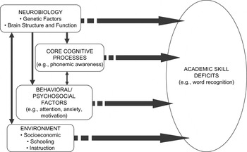
Figure 1 provides a schematic overview from Fletcher et al. (Reference Fletcher, Lyon, Fuchs and Barnes2007, pp. 2–3) of the different components I believe are necessary for a comprehensive scientific understanding of dyslexia. For all LDs, the model assumes that an achievement problem is a necessary but not a sufficient component of identification. For dyslexia, the single-word reading difficulty should be a key component of identification (Pennington, Reference Pennington2009, p. 82). If identification does not include a word-level problem, the sample becomes very heterogeneous because the cognitive and neurobiological correlates vary with different components of reading (Fletcher et al., Reference Fletcher, Lyon, Fuchs and Barnes2007, chap. 5–7). The recognition of dyslexia as a word-level problem and identification into samples on that basis may be responsible for many of the advances in research. To illustrate, I briefly discuss what is understood about identification, cognitive correlates, neurobiological factors, and environmental factors in relation to the most common LD, dyslexia.
Framework representing different sources of variability that influence academic outcomes in children with LDs. From Fletcher et al. (Reference Fletcher, Lyon, Fuchs and Barnes2007, p. 3). Reprinted with permission.

DEFINITION AND CLASSIFICATION: THE CENTRAL ROLE OF ACADEMIC DEFICITS
IQ–Achievement Discrepancy
Classification research aims to identify components of a disorder that are essential for identification. At one point, it was widely believed that academic problems needed to be referenced to IQ, epitomized by the U.S. Federal regulatory definition of LD as a discrepancy in IQ and achievement (U.S. Office of Education, 1977). This approach stemmed from the Rutter and Yule (Reference Rutter and Yule1975) Isle of Wight epidemiological studies, in which the presence of an IQ–achievement discrepancy differentiated children with “specific reading retardation” from those who were “general backwards readers.” However, Rutter and Yule (Reference Rutter and Yule1975) did not exclude children with mental retardation or brain injury. The average IQ of the group with general reading backwardness was about 2 SDs below the mean, which raises issues about the extrapolation of this concept of reading backwardness to children with IQ scores not associated with intellectual retardation. Little evidence has emerged showing that poor readers who would not be considered intellectually retarded can be meaningfully differentiated based on an IQ–achievement discrepancy classification in cognitive skills (Hoskyn & Swanson, Reference Hoskyn and Swanson2000; Stuebing et al., Reference Stuebing, Fletcher, LeDoux, Lyon, Shaywitz and Shaywitz2002), prognosis (Francis et al., Reference Francis, Shaywitz, Stuebing, Shaywitz and Fletcher1996), and intervention response (Stuebing et al., in press). Partly in response to this body of research, Congress changed Federal statutes so that schools could not be required to use IQ tests for identification of LD (U.S. Department of Education, 2004).
Dimensional Nature of Dyslexia
Unlike Rutter and Yule (Reference Rutter and Yule1975), international epidemiological studies have shown that dyslexia is dimensional and exists as the lower end of a normal continuum of reading ability (Jorm et al., Reference Jorm, Share, MacLean and Matthews1986; Rodgers, Reference Rodgers1983; Shaywitz et al., Reference Shaywitz, Escobar, Shaywitz, Fletcher and Makuch1992). Deciding reliably where on this continuum a disability resides is inherently arbitrary, which is why prevalence estimates range so broadly (Francis et al., Reference Francis, Fletcher, Stuebing, Lyon, Shaywitz and Shaywitz2005; Pennington, Reference Pennington2009, p. 47). More importantly, since dyslexia does not represent a qualitatively distinct disorder, there is no need for separate theories of success and failure in learning to read. Research can link directly to normative development, and the dimensional nature encourages a focus on reading as a cardinal attribute. Such an approach fueled research in dyslexia and other domains of LD (Shavelson & Towne, Reference Shavelson and Towne2002, pp. 38–41).
Cognitive Correlates
No single theory and body of research has had more impact on the concept of dyslexia and LD than scientific understanding of how children develop word recognition skills. Representing what Stanovich (Reference Stanovich2000) described as a “big idea” in science, this research had far-reaching implications for understanding and teaching children who are typically developing and who struggle to read. In contrast to earlier neuropsychological approaches (Benton, Reference Benton and Friedlander1975), it highlighted why a theory of dyslexia must explain the reading problem and that a focus on associated features was likely to be less productive. Efforts to explain the reading problem in people with dyslexia have been very productive.
The major breakthrough was the discovery that the link between oral language and written language resided in the phonological structure of speech (Shavelson & Towne, Reference Shavelson and Towne2002, pp. 38–41). In the late 1960s, researchers at the Haskins Laboratories were developing machines that would help people with deafness communicate. The investigators realized that speech was processed as a segmented signal, although the speaker may not recognize this segmented structure because speech sounds are merged together during production. So words like “dog,” which actually has three segments at a phonemic level, are heard as one coarticulated unit. These observations led to a major theory of speech processing and then of reading, where it was hypothesized that the segmented units of speech are also represented in print at a phonemic level through the alphabet (Liberman & Shankweiler, Reference Liberman, Shankweiler, Rieben and Perfetti1991). Thus, written language is scaffolded upon oral language, and literacy is a product of evolutionarily established human capabilities for speech (Liberman, Reference Liberman and Blachman1997). These findings anchored conceptualizations of reading disabilities in specific reading processes and language (Pennington, Reference Pennington2009, pp. 57–62; Vellutino et al., Reference Vellutino, Fletcher, Snowling and Scanlon2004).
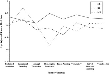
There are other cognitive and neuropsychological skills linked to dyslexia, and this relation of phonological awareness and word recognition may not fully explain why children with dyslexia have other (often comorbid) language and attention problems, math, motor, and other difficulties (Pennington, Reference Pennington2009, p. 62). As Figure 2 shows, children with dyslexia (or math disability) may differ significantly from typically developing children on almost any neuropsychological variable if the sample is large enough. If children with comorbid attention deficit hyperactivity disorder are included in the dyslexia or math disability samples, the differences are much larger (Fletcher et al., Reference Fletcher, Lyon, Fuchs and Barnes2007, pp. 56–57). There are theories involving the cerebellum, low-level vision and speech processing, and other domains (Fletcher et al., Reference Fletcher, Lyon, Fuchs and Barnes2007, pp. 96–98; Pennington, Reference Pennington2009, pp. 62–66; Vellutino et al., Reference Vellutino, Fletcher, Snowling and Scanlon2004, pp. 7–10). Harkening back to Doehring (Reference Doehring, Benton and Pearl1978), univariate theories abound and much depends on identification criteria. It is common to take a cognitive deficit and extrapolate to a brain mechanism underlying the neuropsychological deficit as well as the reading disorder, which often, as in Benton’s (Reference Benton and Friedlander1975) time, leads to the construction of reading theories to fit the neuropsychological data. None of these hypotheses has had much success in explaining or treating the word reading problem (Vellutino et al., Reference Vellutino, Fletcher, Snowling and Scanlon2004, pp. 7–10).
Neuropsychological profiles across different cognitive tests for children who are only impaired in word reading (RD) and in computational math (MD) relative to typical achievers (NL). The groups differ in shape and elevation, suggesting three distinct groups. From Fletcher et al. (Reference Fletcher, Lyon, Fuchs and Barnes2007, p. 46). Reprinted with permission.

It is possible that some of these theories could explain other aspects of a broader phenotype (e.g., fluency problems) or even why some children with dyslexia have problems in the motor system unrelated to reading (Denckla et al., Reference Denckla, Rudel, Krieger and Chapman1985). However, phonological awareness, along with rapid naming and verbal working memory, is most consistently linked to the word reading disorder regardless of comorbidities (Willcutt et al., Reference Willcutt, Pennington, Olson, Chhabildas and Huslander2005), thus helping to explain the word reading problem that is the cardinal feature of dyslexia. These skills would be at the top of Benton’s (Reference Benton and Friedlander1975) list of neuropsychological correlates in 2009. In my opinion, identifying children with word-level problems and assuming dimensionality have proven remarkably fruitful for functional neuroimaging (Fletcher et al., Reference Fletcher, Lyon, Fuchs and Barnes2007, pp. 112–123) and genetic studies (Grigorenko & Naples, in press).
NEUROBIOLOGICAL FACTORS
Brain
Studies of brain structure in dyslexia, whether through the small number of postmortem studies or structural imaging studies, have implicated a variety of regions of the brain and cerebellum. The most consistent evidence identifies perisylvian language areas as either small or symmetric relative to controls (Pennington, Reference Pennington2009, p. 53). The lack of consistency reflects the small heterogeneous samples that characterize many of these studies (Fletcher et al., Reference Fletcher, Lyon, Fuchs and Barnes2007, pp. 108–112). Future studies with larger samples and newer imaging modalities, such as diffusion tensor imaging, may lead to a more consistent set of findings (Ben-Shachar et al., Reference Ben-Shachar, Dougherty and Wandell2007).
In contrast, functional neuroimaging studies of different components of reading in children defined with dyslexia show reliable differences in activation relative to proficient readers (Price & McCrory, Reference Price, McCrory, Snowling and Hulme2005, p. 49). There is no cognitive skill more frequently imaged than word recognition, and the neural networks that support word reading are fairly well established. These systems involve bilateral basal temporal regions for feature recognition, the angular gyrus, middle and superior temporal gyri for cross-modal integration and phonological processing predominantly in the left hemisphere, and frontal regions if production is involved. The involvement of these and other brain regions will vary depending on task characteristics and level of proficiency (Price & McCrory, Reference Price, McCrory, Snowling and Hulme2005, pp. 475–483). In dyslexia, different functional imaging studies converge in identifying underactivation of the posterior regions in children with dyslexia and sometimes hyperactivity in the frontal regions. Moreover, these posterior differences predominantly normalize when intervention is successful (Meyler et al., Reference Meyler, Keller, Cherkassky, Gabrieli and Just2008; Shaywitz et al., Reference Shaywitz, Shaywitz, Blachman, Pugh, Fulbright, Skudlarski, Mencl, Constable, Holahan, Marchione, Fletcher, Lyon and Gore2004; Simos et al., Reference Simos, Fletcher, Bergman, Breier, Foorman, Castillo, Fitzgerald and Papanicolaou2002), although there is variation across studies.
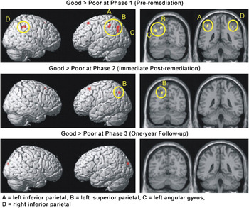
Figure 3 shows an example of common findings from a yearlong intervention of Grade 5 poor readers before and after an intense reading intervention (Meyler et al., Reference Meyler, Keller, Cherkassky, Gabrieli and Just2008). The children were identified with a reading fluency task, so there is probably more heterogeneity compared to selection with a word reading test. Using functional magnetic resonance imaging and a sentence comprehension activation task, the temporal–parietal areas were underactivated in poor readers at baseline but normalized with improved reading after intervention. Interestingly, unlike Shaywitz et al. (Reference Shaywitz, Shaywitz, Blachman, Pugh, Fulbright, Skudlarski, Mencl, Constable, Holahan, Marchione, Fletcher, Lyon and Gore2004), the occipital–temporal regions were not underactivated at baseline and did not show major shifts with intervention. Differences in the activation task and selection criteria, which involved word reading in Shaywitz et al. (Reference Shaywitz, Shaywitz, Blachman, Pugh, Fulbright, Skudlarski, Mencl, Constable, Holahan, Marchione, Fletcher, Lyon and Gore2004), likely explain the differences in the two studies. Although there are now about 15 intervention-imaging studies showing convergence around normalizing changes in the neural network supporting word recognition as well as apparent compensatory changes (Fletcher et al., Reference Fletcher, Lyon, Fuchs and Barnes2007, pp. 117–123), the idea that the neural systems underlying word recognition are malleable is most important. Learning to read literally rewrites the organization of the brain. Since we are not born to read, in contrast to speaking, instruction of some sort is necessary to engage these regions of the brain (Liberman, Reference Liberman and Blachman1997). For some people, more instruction is needed; for others, instruction will never result in reading skills in the average range.
Changes in brain activation for students with reading difficulties before a yearlong intervention, at posttest, and after a 1-year follow-up. From Meyler et al. (Reference Meyler, Keller, Cherkassky, Gabrieli and Just2008). Reprinted with permission. A, left inferior parietal; B, left superior parietal; C, left angular gyrus; D, right inferior parietal.

Genes
With a focus on specific reading processes, genetic loci associated with poor reading have been replicated in many laboratories around the world. At this point, there are nine regions of the genome and six candidate genes under active investigation (Grigorenko & Naples, in press). Dyslexia clearly has a heritable component that accounts for about 50–80% of the variance in reading outcomes. However, no major gene effects have been identified, and the contributions reflect multiple small effects. There are multiple genes involved in good and poor reading, with no dyslexia-specific genes (Pennington, Reference Pennington2009, pp. 49–52). However, there is also evidence that the genetic correlation for reading increases significantly with schooling (Samuelsson et al., Reference Samuelsson, Olson, Wadsworth, Corley, DeFries, Willcutt, Hulsander and Byrne2007), highlighting the importance of early intervention.
ENVIRONMENTAL FACTORS
Factors related to poverty and the family’s orientation to literacy represent risk factors for dyslexia. The impact of poverty on language and achievement is clearly established, but even in middle-class families where one or more parents are poor readers, literacy-related activities are often not emphasized (Pennington et al., Reference Pennington, McGrath, Rosenberg, Barnard, Smith, Willcutt, Friend, DeFries and Olson2009). However, another critically important factor is instruction. At a classroom level, the quality of reading instruction varies considerably. Meta-analytic reviews like the National Reading Panel Report (NICHD, 2000) have shown that children at risk for reading problems require instructional approaches that are more explicit, meaning that translation of the alphabetic principle into instruction through methods like phonics needs to be intentionally laid out in an organized fashion in order for at-risk children to make explicit what is inherently an implicit understanding of the relation of print and sound (Rayner et al., Reference Rayner, Foorman, Perfetti, Pesetsky and Seidenberg2002). In addition, only teaching phonics reduces transfer to other domains of reading, so more comprehensive approaches that also include reading practice to build fluency and explicit teaching of comprehension strategies and vocabulary usually result in higher levels of overall reading proficiency (Stuebing et al., Reference Stuebing, Barth, Cirino, Francis and Fletcher2008). Better outcomes are associated with earlier intervention, primarily because children fall far behind their peers when they are not able to access print (Torgesen et al., Reference Torgesen, Alexander, Wagner, Rashotte, Voeller and Conway2001).
The past decade has seen an explosion in research evaluating reading interventions using designs from which stronger causal inferences can be made (Fletcher et al., Reference Fletcher, Lyon, Fuchs and Barnes2007, pp. 129–162). These studies include evaluations of classroom programs, prevention programs, and remedial programs. While still evolving, this research had major impact on the reauthorization of the Individuals With Disabilities Education Act (IDEA in 2004), which included components permitting school districts to move away from the IQ discrepancy model of identification adopted in 1977 (U.S. Office of Education, 1977) and to implement models that focus in part on intervention response. Specifically, the regulations permit either a response to intervention process or an alternative discrepancy models depending on state guidelines (U.S. Department of Education, 2006, p. 46786):
(i) The child does not make sufficient progress to meet age or State-approved grade-level standards in one or more of the [8 domains of achievement] when using a process based on the child’s response to scientific, research-based intervention …; or
(ii) The child exhibits a pattern of strengths and weaknesses in performance, achievement, or both, relative to age, State-approved grade-level standards, or intellectual development, that is determined by the group to be relevant to the identification of a specific learning disability, using appropriate assessments …
Response to interventionmodels link prevention and remedial interventions through multitiered approaches to service delivery in schools, including (1) universal screening for reading (and math and behavior problems), (2) monitoring progress of at-risk children through frequent assessment probes using reading fluency tasks, and (3) providing increasingly intense intervention based on the child’s progress (Fletcher & Vaughn, Reference Fletcher and Vaughn2009). The data on intervention response can be used as a component of the identification process for special education, with IDEA (2004) still requiring a comprehensive evaluation that uses multiple sources of information. Recognizing that many children included in special education may be instructional casualties because of the need for better instruction, IDEA (2004) also requires evidence of adequate instruction in reading and math as one of these sources.
CONCLUSIONS: THE IMPORTANCE OF INTRACTABILITY
Returning to the issue of definition, the intervention research helps clarify what else needs to be added to the definition of LDs like dyslexia, namely evidence of adequate instructional opportunity (Fuchs & Fuchs, Reference Fuchs and Fuchs1998). Many children are at risk for dyslexia because of neurobiological and environmental factors, and even those with genetic risk may not manifest the disorder depending on their home environments and quality of instruction (Pennington et al., Reference Pennington, McGrath, Rosenberg, Barnard, Smith, Willcutt, Friend, DeFries and Olson2009). As the IDA definition of dyslexia suggests (Lyon et al., 2003), evidence of adequate instruction is another inclusionary criterion that should be included in the definition of dyslexia and other LDs.
Research in the future should focus on children whose single-word deficits are resistant to intervention, with comparisons to typical and at-risk children along the domains in Figure 1. Such studies may shed new light on the neuropsychological and neurobiological factors of historical interest to neuropsychologists. However, views in which brain dysfunction is directly assessed with neuropsychological tests (Benton, Reference Benton and Friedlander1975; Rourke, Reference Rourke1975) may need to shift because of newer conceptualizations of dyslexia as a heritable disorder that makes the brain at risk and that emerges due to interactions of neurobiological and environmental factors. The use of neuropsychological tests as a central part of identification has not been well justified, given the classification research that has been completed, which simplifies identification. In addition, there is little evidence that such assessments help plan treatment or that other forms of intervention based on neuropsychological assessment improve academic or adaptive outcomes. Despite claims to the contrary (Hale et al., Reference Hale, Fiorello, Miller, Wenrich, Teodori, Henzel, Prifitera, Saklofske and Weiss2008), there is little evidence of Aptitude × Treatment interactions for cognitive/neuropsychological skills at the level of treatment or aptitude (Reschly & Tilly, Reference Reschly, Tilly, Reschly, Tilly and Grimes1999, pp. 28–29). The strongest evidence of Aptitude × Treatment interactions is when strengths and weaknesses in academic skills are used to provide differential instruction (e.g., Connor et al., Reference Connor, Morrison, Fishman, Schatschneider and Underwood2007).
I am not encouraging acceptance of the null hypothesis but rather calling the neuropsychological research and professional communities to action. We need research that supports the assessments we do. More importantly, as neuropsychologists, we need to fully understand the interdisciplinary body of research that has changed scientific understanding of dyslexia and LD and adds to the research base outlined in Figure 1, which is most complete for dyslexia and rapidly emerging in other LDs. We need to prioritize intervention and link our scientific and professional practices to the goal of enhancing adaptive functions in children with or at risk for LDs. Neuropsychology has been at the forefront of the evolution of dyslexia as a scientific concept and should continue to make contributions by questioning what we know and working at the edges of disciplinary boundaries.
ACKNOWLEDGMENT
This study is supported in part by a grant from the NICHD, 1 P50 HD052117, Texas Center for Learning Disabilities.