Introduction
Inherited retinal degenerations (RD) represent the most frequent cause of visual dysfunction in people of working group; these conditions therefore have a significant impact on both quality of life and economics. The advent of genetic linkage, DNA sequencing and, most recently, next generation sequencing technologies, has provided an opportunity to elucidate the molecular pathogenesis of inherited ocular disorders (Estrada-Cuzcano et al., Reference Estrada-Cuzcano, Roepman, Cremers, den Hollander and Mans2012). A notable feature associated with inherited ocular conditions, as ascertained from this enormous body of research, is the significant level of genetic heterogeneity inherent in many of these disorders. The future application of exome and whole genome sequencing is set to progress the dissection of the molecular genetics of ocular disorders to a new level. Undoubtedly, this will serve to further emphasize the inherent genetic diversity of inherited ocular disorders which, historically, were categorized together. Retinitis pigmentosa (RP) is the most prevalent inherited RD, affecting approximately one in 3000 people (approximately 250,000 people in Europe alone (Shanks et al., Reference Shanks, Downes, Copley, Lise, Broxholme, Hudspith, Kwasniewska, Davies, Hankins, Packham, Clouston, Seller, Wilkie, Taylor, Ragoussis and Németh2012)) and involves a gradual loss of photoreceptor cells. While many inherited ocular disorders such as Leber congenital amaurosis (LCA), Leber hereditary optic neuropathy (LHON), choroideremia, Best disease, Stargardt disease, and retinoschisis (amongst others) are less prevalent than RP, cumulatively these disorders also represent significant patient numbers and hence have substantial economic consequences, not to mention deleterious impact on quality of life. Typically, the disorders that fall within this group of conditions result from a progressive loss of one or more retinal cell layers over years or decades (Fig. 1).
Diagrammatic representation of the mammalian retina (from http://www.nin.knaw.nl/research_groups/kamermans_group/research_line_1).

Of note, many of the disease mechanisms in operation in Mendelian forms of RD are mirrored in more common multifactorial conditions such as age-related macular degeneration (AMD), which affects approximately 10% of people over 65 years, causing devastating and sometimes rapid vision loss. Common disease features observed in AMD and RP include oxidative stress, mitochondrial dysfunction, and apoptotic cell death, amongst others (Athanasiou et al., Reference Athanasiou, Aguilà, Bevilacqua, Novoselov, Parfitt and Cheetham2013). Advances in therapeutic development for single gene forms of RD may therefore be relevant to more common, multifactorial disorders such as AMD. Knowledge regarding the molecular causes of single gene ocular disorders provides an opportunity to develop therapies targeted toward amending the primary genetic defects in these conditions. Elucidation of the biochemical mechanisms underlying the disease processes enables the identification of novel targets for therapeutic development, which, in principle, allows for modulation of common pathways perturbed in many retinopathies and hence the development of therapies with broader applicability (Athanasiou et al., Reference Athanasiou, Aguilà, Bevilacqua, Novoselov, Parfitt and Cheetham2013; Doonan et al., Reference Doonan, Groeger and Cotter2012).
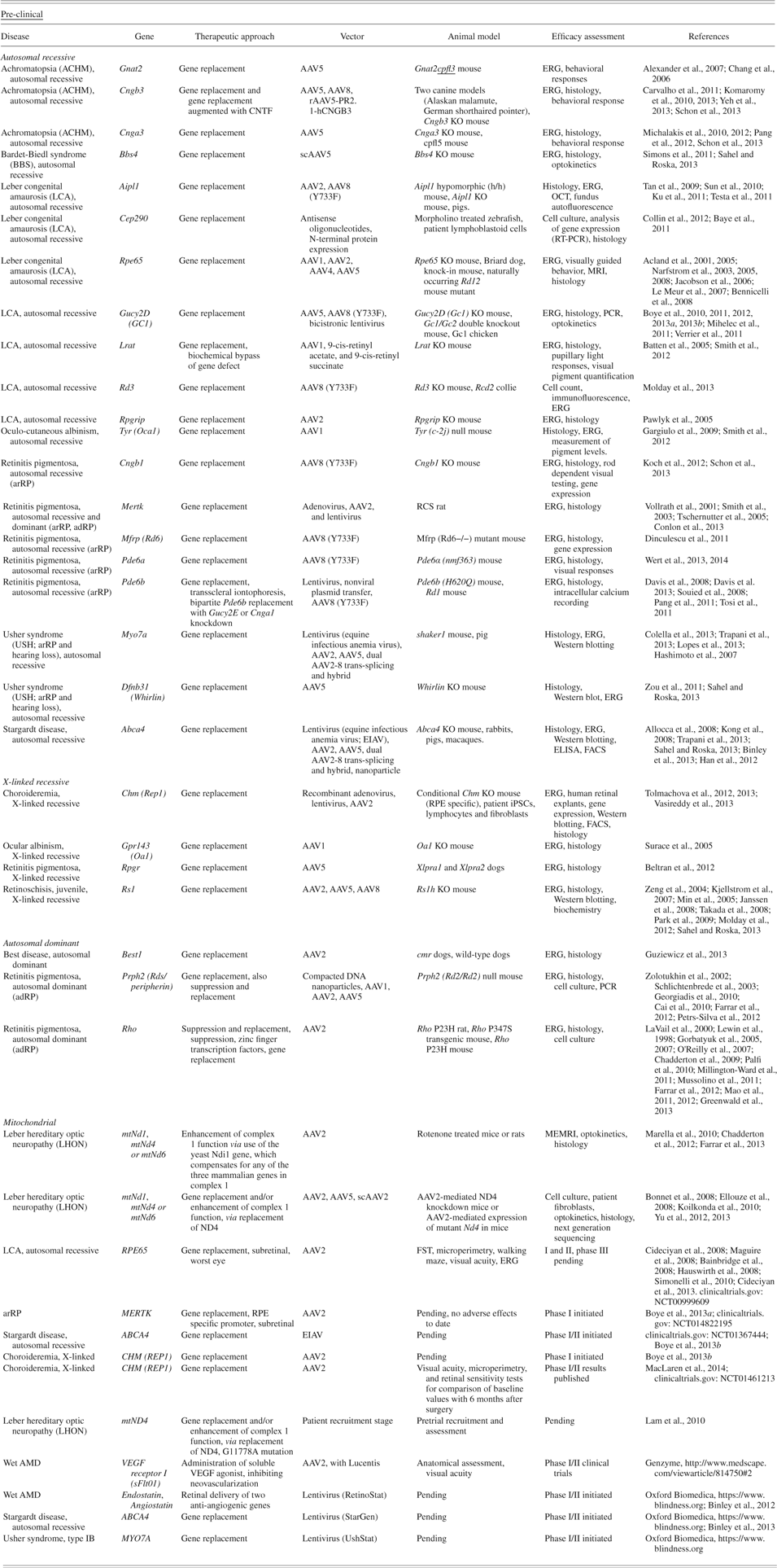
The availability of appropriate animal models that accurately simulate human disease has enabled preclinical evaluation of gene therapies for many ocular conditions, which has highlighted the essential role of animal models in clinical development (Fletcher et al., Reference Fletcher, Jobling, Vessey, Luu, Guymer and Baird2011). The utilization of human induced pluripotent stem cells (iPS cells) to model ocular diseases can also provide in vitro systems to evaluate preclinical therapies for some disorders, where animal models are either unavailable or suboptimal (Tolmachova et al., Reference Tolmachova, Tolmachov, Barnard, de Silva, Lipinski, Walker, Maclaren and Seabra2013; Vasireddy et al., Reference Vasireddy, Mills, Gaddameedi, Basner-Tschakarjan, Kohnke, Black, Alexandrov, Zhou, Maguire, Chung, Mac, Sullivan, Gadue, Bennicelli, French and Bennett2013). Of note, a number of ocular gene therapies have progressed from preclinical studies in animal models to human clinical trials (Bainbridge et al., Reference Bainbridge, Smith, Barker, Robbie, Henderson, Balaggan, Viswanathan, Holder, Stockman, Tyler, Petersen-Jones, Bhattacharya, Thrasher, Fitzke, Carter, Rubin, Moore and Ali2008; Cideciyan et al., Reference Cideciyan, Aleman, Boye, Schwartz, Kaushal, Roman, Pang, Sumaroka, Windsor, Wilson, Flotte, Fishman, Heon, Stone, Byrne, Jacobson and Hauswirth2008, Reference Cideciyan2010; Maguire et al., Reference Maguire, Simonelli, Pierce, Pugh, Mingozzi, Bennicelli, Banfi, Marshall, Testa, Surace, Rossi, Lyubarsky, Arruda, Konkle, Stone, Sun, Jacobs, Dell'Osso, Hertle, Ma, Redmond, Zhu, Hauck, Zelenaia, Shindler, Maguire, Wright, Volpe, McDonnell, Auricchio, High and Bennett2008; Simonelli et al., Reference Simonelli, Maguire, Testa, Pierce, Mingozzi, Bennicelli, Rossi, Marshall, Banfi, Surace, Sun, Redmond, Zhu, Shindler, Ying, Ziviello, Acerra, Wright, McDonnell, High, Bennett and Auricchio2010). An update on the current status of key preclinical and clinical studies for gene-based medicines targeted toward inherited retinal disorders will be provided in this review. Broadly, three major gene-based therapeutic strategies can be delineated: first, therapies targeted toward correcting the primary genetic deficit in a particular eye condition; second, therapies directed toward modulating secondary features associated with the disease pathology characteristic of a given disorder and finally, therapies focused on provision of new functions to residual cells within a degenerating retina, such as the provision of light-sensing characteristics (optogenetics) (Busskamp et al., Reference Busskamp, Picaud, Sahel and Roska2012). Gene-based approaches in these three categories for ocular disorders with recessive, X-linked recessive, dominant or mitochondrial inheritance are detailed in Table 1. While exciting advances have been made in the fields of cell-based therapies and retinal prosthesis for inherited ocular diseases (Boucherie et al., Reference Boucherie, Sowden and Ali2011; Ramsden et al., Reference Ramsden, Powner, Carr, Smart, da Cruz and Coffey2013; Shepherd et al., Reference Shepherd, Shivdasani, Nayagam, Williams and Blamey2013), these approaches fall outside the remit of the current review.
Gene therapies for inherited retinal disorders: Summary of pre-clinical research and clinical trials to date; March 2014
Gene therapies targeting primary genetic defects
The group of diseases classed as “inherited retinopathies” or RD provides the ultimate example of the application of genetic information to drive preclinical and clinical therapeutic development. Knowledge of the genetic etiologies of inherited retinopathies has grown exponentially, from the localization almost three decades ago of the first retinopathy gene to Xp11.3 (Bhattacharya et al., Reference Bhattacharya, Wright, Clayton, Price, Phillips, McKeown, Jay, Bird, Pearson and Southern1984), to the current characterization of over 150 causative RD disease genes (RetNet, https://sph.uth.edu/retnet/; OMIM, http://omim.org). In concert with disease gene identification, animal models mirroring specific forms of inherited RD have been generated, largely using transgenic methodologies such as homologous recombination (Fletcher et al., Reference Fletcher, Jobling, Vessey, Luu, Guymer and Baird2011). Equally, advances in the development of viral and nonviral vectors for gene delivery to the mammalian retina have been essential in the orchestration of preclinical studies for retinal gene therapy (Charbel & MacLaren, Reference Charbel and MacLaren2012; Vandenberghe & Auricchio, Reference Vandenberghe and Auricchio2012; Bainbridge et al., Reference Bainbridge, Tan and Ali2006). Through elegant experimentation employing directed evolution, vector design can be optimized, for example, to direct tropisms of adeno-associated virus (AAV) serotypes to particular cell types in the mammalian retina and to optimize serotypes for specific routes of administration, i.e., subretinal versus intravitreal; similarly, systemic administration has been shown to be a potential route for delivery to the retina (Dalkara et al., Reference Dalkara, Byrne, Lee, Hoffmann, Schaffer and Flannery2012, Reference Dalkara, Byrne, Klimczak, Visel, Yin, Merigan, Flannery and Schaffer2013). Rational mutagenesis has also been used to optimize AAV serotypes for particular delivery routes (Vandenberghe & Auricchio, Reference Vandenberghe and Auricchio2012; Flannery & Visel, Reference Flannery and Visel2013). Undoubtedly, optimization of AAV and other viral and nonviral vectors for gene delivery will play an important role in the future development of efficacious gene therapies for genetic eye disorders. Generally, the central nervous system (CNS) represents a key target for gene therapy given its relative immune privilege compared to many tissues (Willett & Bennett, Reference Willett and Bennett2013), hence increasing the likelihood that gene delivery vectors will be tolerated. The relative tolerance of AAV by the immune system in the CNS, in conjunction with the view that wild-type AAV is not pathogenic, suggests a significant role for AAV-mediated delivery in future gene therapies for neurodegenerative disorders. Given these features, AAV is being used extensively in gene therapy strategies, however, it should be noted that due to its packaging limitation in the region of 5 kb, this vector will not be suitable for all gene therapy applications.
It is worth highlighting that the eye, in particular, represents a very attractive target for gene therapy; it is a readily accessible bilateral system with relative immune privilege. In principle, the eye requires only single doses of small quantities of vector, thereby potentially reducing the cost associated with manufacture of the therapeutic product.
Recessively inherited retinopathies
When considering the many preclinical studies exploring gene therapies targeting a primary genetic defect, it is of value to differentiate between therapeutic strategies for disorders displaying the different modes of inheritance, that is, X-linked recessive, autosomal recessive, dominant or mitochondrial inheritance. The greatest advances have been made thus far for recessively inherited retinopathies, largely due to the relatively simple mode of action driving the primary disease process, that is, the absence of the wild-type gene and encoded protein. In this review, replacement of an absent or mutated gene will be considered together for both X-linked recessive and autosomal recessive disorders, as gene replacement represents a relevant therapeutic strategy for both categories of disease.
The seminal preclinical studies in rodents and dogs for a severe recessive retinopathy, LCA (Acland et al., Reference Acland, Aguirre, Ray, Zhang, Aleman, Cideciyan, Pearce-Kelling, Anand, Zeng, Maguire, Jacobson, Hauswirth and Bennett2001, Reference Acland, Aguirre, Bennett, Aleman, Cideciyan, Bennicelli, Dejneka, Pearce-Kelling, Maguire, Palczewski, Hauswirth and Jacobson2005; Narfstrom et al., Reference Narfström, Katz, Bragadottir, Seeliger, Boulanger, Redmond, Caro, Lai and Rakoczy2003, Reference Narfström, Vaegan, Katz, Bragadottir, Rakoczy and Seeliger2005, Reference Narfström, Seeliger, Lai, Vaegan, Katz, Rakoczy and Remé2008; Jacobson et al., Reference Jacobson, Acland, Aguirre, Aleman, Schwartz, Cideciyan, Zeiss, Komaromy, Kaushal, Roman, Windsor, Sumaroka, Pearce-Kelling, Conlon, Chiodo, Boye, Flotte, Maguire, Bennett and Hauswirth2006; Le Meur et al., Reference Le Meur, Stieger, Smith, Weber, Deschamps, Nivard, Mendes-Madeira, Provost, Péréon, Cherel, Ali, Hamel, Moullier and Rolling2007; Bennicelli et al., Reference Bennicelli, Wright, Komaromy, Jacobs, Hauck, Zelenaia, Mingozzi, Hui, Chung, Rex, Wei, Qu, Zhou, Zeiss, Arruda, Acland, Dell'Osso, High, Maguire and Bennett2008; Annear et al., Reference Annear, Bartoe, Barker, Smith, Curran, Bainbridge, Ali and Petersen-Jones2011) have paved the way for many further preclinical studies for other recessive retinopathies. In terms of academic and commercial investment in the sector, the influence of the LCA preclinical and clinical studies has been monumental. Preclinical studies for LCA more than a decade ago demonstrated clear therapeutic benefit in dogs with RPE65 deficiency after subretinal injection of an AAV2 therapeutic virus (Acland et al., Reference Acland, Aguirre, Ray, Zhang, Aleman, Cideciyan, Pearce-Kelling, Anand, Zeng, Maguire, Jacobson, Hauswirth and Bennett2001). Additional preclinical studies in rodent and dog models of RPE65-linked LCA (Jacobson et al., Reference Jacobson, Acland, Aguirre, Aleman, Schwartz, Cideciyan, Zeiss, Komaromy, Kaushal, Roman, Windsor, Sumaroka, Pearce-Kelling, Conlon, Chiodo, Boye, Flotte, Maguire, Bennett and Hauswirth2006; Bennicelli et al., Reference Bennicelli, Wright, Komaromy, Jacobs, Hauck, Zelenaia, Mingozzi, Hui, Chung, Rex, Wei, Qu, Zhou, Zeiss, Arruda, Acland, Dell'Osso, High, Maguire and Bennett2008; Annear et al., Reference Annear, Bartoe, Barker, Smith, Curran, Bainbridge, Ali and Petersen-Jones2011), employed AAV for gene delivery but utilized different promoter sequences to drive expression of the RPE65 replacement gene. These studies provided evidence of significant benefit using histology, electrophysiology, mobility in low illumination and pupillary responses as readouts and provided the impetus to drive the therapies forward to human clinical trials in Europe and the US. As with the preclinical studies, the human clinical trials employed AAV2 to deliver the RPE65 replacement gene and a single subretinal injection as the route of administration (Bainbridge et al., Reference Bainbridge, Smith, Barker, Robbie, Henderson, Balaggan, Viswanathan, Holder, Stockman, Tyler, Petersen-Jones, Bhattacharya, Thrasher, Fitzke, Carter, Rubin, Moore and Ali2008; Maguire et al., Reference Maguire, Simonelli, Pierce, Pugh, Mingozzi, Bennicelli, Banfi, Marshall, Testa, Surace, Rossi, Lyubarsky, Arruda, Konkle, Stone, Sun, Jacobs, Dell'Osso, Hertle, Ma, Redmond, Zhu, Hauck, Zelenaia, Shindler, Maguire, Wright, Volpe, McDonnell, Auricchio, High and Bennett2008; Cideciyan et al., Reference Cideciyan, Aleman, Boye, Schwartz, Kaushal, Roman, Pang, Sumaroka, Windsor, Wilson, Flotte, Fishman, Heon, Stone, Byrne, Jacobson and Hauswirth2008, 2010; Simonelli et al., Reference Simonelli, Maguire, Testa, Pierce, Mingozzi, Bennicelli, Rossi, Marshall, Banfi, Surace, Sun, Redmond, Zhu, Shindler, Ying, Ziviello, Acerra, Wright, McDonnell, High, Bennett and Auricchio2010). AAV was well tolerated in the human eye subsequent to subretinal injection and improvements in visual acuity, microperimetry, walking maze performance, and electroretinogram (ERG) were noted. Having demonstrated both safety and efficacy in early Phase I/II trials, some studies are now in Phase III trials. More recent cohorts of LCA patients treated with AAV-RPE65 have included younger children and second eye injections, which were previously shown to be safe in primates (Amado et al., Reference Amado, Mingozzi, Hui, Bennicelli, Wei, Chen, Bote, Grant, Golden, Narfstrom, Syed, Orlin, High, Maguire and Bennett2010; Annear et al., Reference Annear, Bartoe, Barker, Smith, Curran, Bainbridge, Ali and Petersen-Jones2011; Bennett et al., Reference Bennett, Ashtari, Wellman, Marshall, Cyckowski, Chung, McCague, Pierce, Chen, Bennicelli, Zhu, Ying, Sun, Wright, Auricchio, Simonelli, Shindler, Mingozzi, High and Maguire2012). In summary, the LCA studies have paved the way for the design, evaluation, and clinical testing of gene therapies for other inherited ocular conditions. Confidence in gene therapies has been further fueled by the recent market authorization in the EU of Glybera (UniQure, The Netherlands; www.uniqure.com/products/glybera/) for lipoprotein lipase deficiency, the world's first gene therapy employing AAV for delivery.
Observations with LCA have been followed by an exponential growth in preclinical gene therapy studies undertaken for X-linked and autosomal recessive RD in rodent models (Table 1). The most common form of X-linked recessive RP (XLRP) is caused by mutations in the RP GTPase regulator (RPGR) gene, which encodes a photoreceptor ciliary protein. AAV5 has been used to successfully deliver the gene to two canine models of this form of XLRP (Beltran et al., Reference Beltran, Cideciyan, Lewin, Iwabe, Khanna, Sumaroka, Chiodo, Fajardo, Román, Deng, Swider, Alemán, Boye, Genini, Swaroop, Hauswirth, Jacobson and Aguirre2012). Beneficial effects were demonstrated by retinal histology, electrophysiology, and optical coherence tomography (OCT), providing a compelling rationale for further clinical progression of this therapeutic. An autosomal gene, RPGRIP, encodes a protein which anchors RPGR in the photoreceptor cilium. Null mutations in RPGRIP cause more severe RD than those in RPGR, resulting in LCA. A gene augmentation therapy for RPGRIP1-linked LCA has been shown to be efficacious in a murine model of the disease using AAV8 for gene delivery (Pawlyk et al., Reference Pawlyk, Bulgakov, Liu, Xu, Adamian, Sun, Khani, Berson, Sandberg and Li2010; Smith et al., Reference Smith, Bainbridge and Ali2012; Lhériteau et al., Reference Lhériteau, Petit, Weber, Le Meur, Deschamps, Libeau, Mendes-Madeira, Guihal, François, Guyon, Provost, Lemoine, Papal, El-Amraoui, Colle, Moullier and Rolling2013). While RPGRIP1-LCA is significantly less prevalent than RPGR-xlRP, the results from this study likewise provide the impetus to progress such a therapeutic toward clinical trial. Other LCA genes including GUCY2D (GC1), LRAT, CEP290, RD3, and AIPL1 have now also been the subject of successful preclinical gene therapy studies (Batten et al., Reference Batten, Imanishi, Tu, Doan, Zhu, Pang, Glushakova, Moise, Baehr, Van Gelder, Hauswirth, Rieke and Palczewski2005; Tan et al., Reference Tan, Smith, Pawlyk, Xu, Liu, Bainbridge, Basche, McIntosh, Tran, Nathwani, Li and Ali2009; Sun et al., Reference Sun, Pawlyk, Xu, Liu, Bulgakov, Adamian, Sandberg, Khani, Tan, Smith, Ali and Li2010; Baye et al., Reference Baye, Patrinostro, Swaminathan, Beck, Zhang, Stone, Sheffield and Slusarski2011; Boye et al., Reference Boye, Conlon, Erger, Ryals, Neeley, Cossette, Pang, Dyka, Hauswirth and Boye2011, 2012, 2013b; Mihelec et al., Reference Mihelec, Pearson, Robbie, Buch, Azam, Bainbridge, Smith and Ali2011; Smith et al., Reference Smith, Bainbridge and Ali2012; Molday et al., Reference Molday, Djajadi, Yan, Szczygiel, Boye, Chiodo, Gregory-Evans, Sarunic, Hauswirth and Molday2013; Collin et al., Reference Collin, den Hollander, van der Velde-Visser, Bennicelli, Bennett and Cremers2012). Five types of arRP, caused by mutations in CNGB1 MERTK, MFRP PDE6A, and PDE6B, have also been investigated (Davis et al., Reference Davis, Tosi, Janisch, Kasanuki, Wang, Kong, Tsui, Cilluffo, Woodruff, Fain, Lin and Tsang2008; Souied et al., Reference Souied, Reid, Piri, Lerner, Nusinowitz and Farber2008; Koch et al., Reference Koch, Sothilingam, Garcia Garrido, Tanimoto, Becirovic, Koch, Seide, Beck, Seeliger, Biel, Mühlfriedel and Michalakis2012; Schon et al., Reference Schön, Biel and Michalakis2013; Conlon et al., Reference Conlon, Deng, Erger, Cossette, Pang, Ryals, Clément, Cleaver, McDoom, Boye, Peden, Sherwood, Abernathy, Alkuraya, Boye and Hauswirth2013; Dinculescu et al., Reference Dinculescu, Estreicher, Zenteno, Aleman, Schwartz, Huang, Roman, Sumaroka, Li, Deng, Min, Chiodo, Neeley, Liu, Shu, Matias-Florentino, Buentello-Volante, Boye, Cideciyan, Hauswirth and Jacobson2011; Wert et al., Reference Wert, Davis, Sancho-Pelluz, Nishina and Tsang2013). These genes are involved in diverse processes, including protein trafficking in the photoreceptor cilium (RPGR, RPGRIP, CEP290), catalysis of visual transduction components or components of the visual transduction cycle (GUCY2D, LRAT, CNGB1, PDE6A, PDE6B, RD3), phagocytosis of photoreceptor outer segments by the RPE (MERTK), developmental signaling (MFRP), and possible involvement in nuclear transport and/or protein folding (AIPL). Regardless of mechanism, however, gene replacement therapies have typically proven efficacious (Boye et al., 2013a).
Gene replacement has also proven effective in other diverse forms of retinal dysfunction, sometimes associated with disability in organs besides the eye. Three forms of achromatopsia (also known as rod monochromacy), which is characterized by complete loss of cone function, can be caused by recessive mutations in GNAT2, CNGB3, and CNGA3. AAV-based gene replacement has proven effective for all three genes, in diverse mouse and canine models of this disease (Alexander et al., Reference Alexander, Umino, Everhart, Chang, Min, Li, Timmers, Hawes, Pang, Barlow and Hauswirth2007; Chang et al., Reference Chang, Dacey, Hawes, Hitchcock, Milam, Atmaca-Sonmez, Nusinowitz and Heckenlively2006; Yeh et al., Reference Yeh, Goldstein, Kukekova, Holley, Knollinger, Huson, Pearce-Kelling, Acland and Komáromy2013; Schon et al., Reference Schön, Biel and Michalakis2013). Bardet-Biedl syndrome is a heterogeneous disorder resulting from mutations in one of seven genes that comprise a complex required for ciliogenesis. Symptoms include obesity, polydactyly, nephropathy, intellectual disability, and RP, resulting from dysfunction of the photoreceptor cilium. The BBS4 gene has been the subject of AAV-based ocular intervention in a BBS4 knockout mouse model: ERG, histology, and optogenetics showed improvement after gene replacement therapy (Simons et al., Reference Simons, Boye, Hauswirth and Wu2011; Sahel & Roska, Reference Sahel and Roska2013). Oculo-cutaneous albinism, which has a phenotype involving absent or reduced pigmentation of the hair, skin, and eyes, can be caused by loss of function mutations in the tyrosinase gene (TYR). Loss of function mutations results in lack of RPE pigment, abnormal presence of ganglion cells overlying the fovea, and misrouting of nerves at the optic chiasm. An AAV2-based gene replacement approach resulted in improved ocular pigmentation, ERG, and retinal histology (Gargiulo et al., Reference Gargiulo, Bonetti, Montefusco, Neglia, Di Vicino, Marrocco, Corte, Domenici, Auricchio and Surace2009; Smith et al., Reference Smith, Bainbridge and Ali2012). Choroideremia, an X-linked disorder that results in degeneration of the choriocapillaris, RPE, and photoreceptors, results from mutations in the Rab escort protein-1 (REP1). AAV-mediated therapies have been tested in preclinical studies (Tolmachova et al., Reference Tolmachova, Tolmachov, Barnard, de Silva, Lipinski, Walker, Maclaren and Seabra2013; Vasireddy et al., Reference Vasireddy, Mills, Gaddameedi, Basner-Tschakarjan, Kohnke, Black, Alexandrov, Zhou, Maguire, Chung, Mac, Sullivan, Gadue, Bennicelli, French and Bennett2013) and clinical trials initiated. Findings from one AAV2-based choroideremia trial have been reported, with improved visual acuity, microperimetry, and retinal sensitivity noted in comparison with baseline values, 6 months after surgery (MacLaren et al., Reference MacLaren, Groppe, Barnard, Cottrial, Tolmachova, Seymour, Clark, During, Cremers, Black, Lotery, Downes, Webster and Seabra2014).
One feature of AAV that limits broad application for all ocular gene therapies is its cargo capacity, with an optimal transgene size of approximately 4.7 kb of DNA. Clearly, not all genes conform to this size limit. For AAV-mediated delivery of larger genes, packaging efficiencies and heterogeneous viral populations with differing sized cargos remain obstacles. It has been suggested that successful delivery of large cargos may be, at least in part, achieved through homologous recombination of partial gene packaging to reconstitute larger transgenes (Wu et al., Reference Wu, Yang and Colosi2010). A dual AAV vector strategy to increase cargo capacity has been explored, splitting the replacement gene between two vectors, with subsequent reconstitution by trans-splicing and/or homologous recombination mechanisms (Duan et al., Reference Duan, Yue and Engelhardt2001; Reich et al., Reference Reich, Auricchio, Hildinger, Glover, Maguire, Wilson and Bennett2003; Lopes et al., Reference Lopes, Boye, Louie, Boye, Dyka, Chiodo, Fofo, Hauswirth and Williams2013; Trapani et al., Reference Trapani, Colella, Sommella, Iodice, Cesi, De Simone, Marrocco, Rossi, Giunti, Palfi, Jane Farrar, Polishchuk and Auricchio2013). Single packaging of large genes, which likely involves gene fragmentation during virion assembly, with resulting genetic heterogeneity of progeny virus, has also been tested (Lopes et al., Reference Lopes, Boye, Louie, Boye, Dyka, Chiodo, Fofo, Hauswirth and Williams2013). A question that arises from deploying strategies involving delivery of multiple vectors to reconstitute single genes, is what level of co-infection can be achieved in transduced cell populations in vivo, with two vectors, or with different gene segments from a heterogeneous viral population derived from single packaging of a “large” gene.
Both Stargardt disease (a recessive maculopathy caused by mutations in ABCA4) and Usher syndrome, Type 1B (profound deafness and RP, caused by mutations in MYO7A) have causative genes that are too large for standard AAV packaging. Single packaging of MYO7A has been compared with dual packaging (splitting the gene into two segments; Lopes et al., Reference Lopes, Boye, Louie, Boye, Dyka, Chiodo, Fofo, Hauswirth and Williams2013). This study found that AAV2 and AAV5 carrying the MYO7A gene packaged in single vectors were more effective at correcting the mutant phenotype than dual transduction with AAVs carrying the split gene (Lopes et al., Reference Lopes, Boye, Louie, Boye, Dyka, Chiodo, Fofo, Hauswirth and Williams2013). Furthermore, pathology was noted in RPE cell culture with the dual packaging approach (Lopes et al., Reference Lopes, Boye, Louie, Boye, Dyka, Chiodo, Fofo, Hauswirth and Williams2013). In contrast, in a recent study, dual packaging was successfully demonstrated for both MYO7A and ABCA4 (Trapani et al., Reference Trapani, Colella, Sommella, Iodice, Cesi, De Simone, Marrocco, Rossi, Giunti, Palfi, Jane Farrar, Polishchuk and Auricchio2013); comparisons with single packaging of the same genes were not undertaken. The issue of co-transfection percentages has been addressed for both AAV5 and AAV8 serotypes, using two vectors expressing EGFP and DsRed reporter genes, respectively; it was established that, given sufficient viral load, the majority of transduced cells are co-infected with both viruses in the retina (Fig. 2; Trapani et al., Reference Trapani, Colella, Sommella, Iodice, Cesi, De Simone, Marrocco, Rossi, Giunti, Palfi, Jane Farrar, Polishchuk and Auricchio2013; Palfi et al., Reference Palfi, Chadderton, McKee, Blanco-Fernandez, Humphries, Kenna and Farrar2012). Undoubtedly, viral co-transduction will find application not solely for delivery of larger genes, but moreover for co-delivery of multivalent therapies such as gene replacement in conjunction with neurotrophic factors or anti-apoptotic factors (Yao et al., 2012) or the suppression and replacement dual-component therapies for dominant disorders referred to below (Millington-Ward et al., Reference Millington-Ward, Chadderton, O'Reilly, Palfi, Goldmann, Kilty, Humphries, Wolfrum, Bennett, Humphries, Kenna and Farrar2011).
Co-transduction of AAV2/5 reporter vectors in the mouse retina Eyes of adult 129 wild-type mice were subretinally injected with a mixture of 1.5 × 10⁹ vg of AAV-CMVP-EGFP and 1.5 × 10⁹ vg of AAV-CMVP-DsRed. Two weeks post-delivery, eyes were fixed, cryosectioned and nuclei counterstained with DAPI. Representative microscopy images illustrating native fluorescence of reporter proteins are presented and indicate that the majority of photoreceptors express both reporter genes. A: DsRed signal, B: DsRed and EGFP signals overlaid, C: EGFP signal, D: DsRed, EGFP, and DAPI signals overlaid. GCL: ganglion cell layer, INL: inner nuclear layer, ONL: outer nuclear layer, PS: photoreceptor segment layer. Scale bar: 25 μm. (Authors unpublished data).
In contrast to AAV, lentiviral vectors offer a larger cargo capacity in the region of 10 kb. Lentiviruses are less efficient at photoreceptor transduction than AAV, but greater carrying capacity may nevertheless make them the retinal gene therapy vector of choice for genes that exceed the maximum capacity of AAV in size. In this regard, lentiviral vectors have been used to deliver replacement genes for MYO7A-linked Usher syndrome and ABCA4-linked Stargardt disease, both of which are caused by mutations in large genes (Colella et al., Reference Colella, Sommella, Marrocco, Di Vicino, Polishchuk, Garrido, Seeliger, Polishchuk and Auricchio2013; Allocca et al., Reference Allocca, Doria, Petrillo, Colella, Garcia-Hoyos, Gibbs, Kim, Maguire, Rex, Di Vicino, Cutillo, Sparrow, Williams, Bennett and Auricchio2008; Kong et al., Reference Kong, Kim, Binley, Pata, Doi, Mannik, Zernant-Rajang, Kan, Iqball, Naylor, Sparrow, Gouras and Allikmets2008; Hashimoto et al., Reference Hashimoto, Gibbs, Lillo, Azarian, Legacki, Zhang, Yang and Williams2007; Sahel and Roska, Reference Sahel and Roska2013; Binley et al., Reference Binley, Widdowson, Loader, Kelleher, Iqball, Ferrige, de Belin, Carlucci, Angell Manning, Hurst, Ellis, Miskin, Fernandes, Wong, Allikmets, Bergstrom, Aaberg, Yan, Kong, Gouras, Prefontaine, Vezina, Bussieres, Naylor and Mitrophanous2013; Han et al., 2012). Oxford Biomedica has developed lentiviral-based therapies for both of these diseases, UshGen and StarGen, and has initiated Phase I/II clinical trials. Notably, safety and biodistribution studies demonstrated retinal localization and good tolerance of the lentiviral vector with no long-term toxicity (Binley et al., Reference Binley, Widdowson, Loader, Kelleher, Iqball, Ferrige, de Belin, Carlucci, Angell Manning, Hurst, Ellis, Miskin, Fernandes, Wong, Allikmets, Bergstrom, Aaberg, Yan, Kong, Gouras, Prefontaine, Vezina, Bussieres, Naylor and Mitrophanous2013).
An additional advantage of greater lentiviral carrying capacity is the ability to provide expression of more than one gene in the same vector. Bicistronic lentiviral vectors have been developed and can be shown to successfully deliver two fluorescent proteins to transduced cells. A modified vector has also been used to deliver a replacement copy of the mutated GC1 gene to the Gucy*1 chicken retina, successfully ameliorating the symptoms of LCA in these birds (Verrier et al., Reference Verrier, Madorsky, Coggin, Geesey, Hochman, Walling, Daroszewski, Eccles, Ludlow and Semple-Rowland2011). Similarly, symptoms of RD resulting from Pde6b mutations are caused, not only by the absence of Pde6b, but also by high levels of cyclic guanosine monophosphate (cGMP) and Ca(2+), which are normally held in check by opposing regulation of Pde6b and guanylate cyclase (Gucy). Pde6b and Gucy control the opening of cyclic nucleotide-gated ion channels (CNG), and therefore calcium ion influx into photoreceptor outer segments (Tosi et al., Reference Tosi, Sancho-Pelluz, Davis, Hsu, Wolpert, Sengillo, Lin and Tsang2011). Therefore, the effects of Pde6b mutations can be controlled by either the administration of Pde6b or by the knockdown of Gucy2E and Cnga1. Lentiviral therapy combining the two approaches (Pde6b replacement with either Gucy2E or Cnga1 knockdown) has been tested (Tosi et al., Reference Tosi, Sancho-Pelluz, Davis, Hsu, Wolpert, Sengillo, Lin and Tsang2011). This could have positive implications for the treatment of dominant RD, where suppression and replacement of some dominant mutations will be required, as discussed below.
While lentiviruses have disadvantages compared with AAV in terms of the efficiency of photoreceptor transduction, these vectors are efficient at transducing the RPE, making them suitable for disease targets affecting the RPE such as LCA. Additionally, they may be used to package secreted therapeutic proteins, such as anti-angiogenic factors. Again, when administering more than one gene, the extra carrying capacity of lentiviral vectors presents a significant advantage. In this regard, Oxford Biomedica has developed a lentiviral therapy, RetinoStat, to deliver endostatin and angiostatin for the treatment of wet AMD. Safety and biodistribution studies demonstrated transient inflammation following injection, however, this was not persistent and the vector was otherwise well tolerated with persistent, long-term expression of the administered genes, which was appropriately restricted to the eye (Binley et al., Reference Binley, Widdowson, Kelleher, de Belin, Loader, Ferrige, Carlucci, Esapa, Chipchase, Angell-Manning, Ellis, Mitrophanous, Miskin, Bantseev, Nork, Miller and Naylor2012). Hence, a Phase I clinical trial has been initiated. Lentiviral vectors are therefore a useful and well-tolerated choice in instances where gene size, or the need to deliver more than one gene, is an important consideration. On the other hand, given the lower efficiency of photoreceptor transduction, it may be the case that co-transduction using multiple AAV vectors may prove more efficient in certain instances. It is helpful, in any case, that a number of tools are now available to tackle previously untreatable genetic disorders.
Dominantly inherited retinopathies
In contrast to recessive diseases caused by absence of wild-type protein, the mode of action of dominant mutations can vary between disorders and will determine the most appropriate therapeutic strategy to be adopted for therapies directed to amelioration of the primary genetic defect. Dominant disease pathology may be associated with pathological effects of the mutant protein, reduced levels of the wild-type protein (haploinsufficiency), or a combination of both mechanisms. One therapeutic scenario for dominant conditions may involve suppression of the mutant allele while maintaining expression of the wild-type allele. The group of dominant disorders requiring this approach has the added complication that often many different mutations in the same gene can give rise to the disease pathology; indeed for some dominant disorders, each individual family can have almost a unique “personal” mutation. If suppression is targeted toward the mutant allele, then possibly hundreds of different therapies will be required each targeting a specific mutation—this is technically and economically nonviable. To circumvent this, a mutation-independent strategy which corrects the primary genetic defect involving suppression and replacement has been explored (Millington-Ward et al., Reference Millington-Ward, O'Neill, Tuohy, Al-Jandal, Kiang, Kenna, Palfi, Hayden, Mansergh, Kennan, Humphries and Farrar1997, Reference Millington-Ward, Chadderton, O'Reilly, Palfi, Goldmann, Kilty, Humphries, Wolfrum, Bennett, Humphries, Kenna and Farrar2011; O’Reilly et al., Reference O'Reilly, Palfi, Chadderton, Millington-Ward, Ader, Cronin, Tuohy, Auricchio, Hildinger, Tivnan, McNally, Humphries, Kiang, Humphries, Kenna and Farrar2007; Chadderton et al., Reference Chadderton, Millington-Ward, Palfi, O'Reilly, Tuohy, Humphries, Li, Humphries, Kenna and Farrar2009; Mao et al., Reference Mao, Gorbatyuk, Rossmiller, Hauswirth and Lewin2012). The approach involves two components, suppression of both mutant and wild-type alleles together with provision of a suppression-resistant replacement gene engineered using, for example, codon-redundancy, such that transcripts from the replacement gene are refractory to suppression (Millington-Ward et al., Reference Millington-Ward, O'Neill, Tuohy, Al-Jandal, Kiang, Kenna, Palfi, Hayden, Mansergh, Kennan, Humphries and Farrar1997, Reference Millington-Ward, Chadderton, O'Reilly, Palfi, Goldmann, Kilty, Humphries, Wolfrum, Bennett, Humphries, Kenna and Farrar2011).
Rhodopsin-linked autosomal dominant RP (RHO-adRP) is the most common form of RP accounting for 25–30% of cases of autosomal dominant RP (adRP). Approximately 200 disease-causing mutations have been identified thus far in the rhodopsin (RHO) gene. The rhodopsin knockout mouse (Humphries et al., Reference Humphries, Rancourt, Farrar, Kenna, Hazel, Bush, Sieving, Sheils, McNally, Creighton, Erven, Boros, Gulya, Capecchi and Humphries1997; Lem et al., Reference Lem, Krasnoperova, Calvert, Kosaras, Cameron, Nicolò, Makino and Sidman1999) has abnormal retinal function and does not elaborate rod outer segments, thus highlighting the essential role of rhodopsin in the mammalian retina. Given these features of RHO-adRP, suppression and replacement gene therapies have been explored in transgenic mice expressing human mutant RHO transgenes, for example, in P23H and P347S mice, and significant benefit has been obtained with histological and electrophysiological readouts (Chadderton et al., Reference Chadderton, Millington-Ward, Palfi, O'Reilly, Tuohy, Humphries, Li, Humphries, Kenna and Farrar2009; Millington-Ward et al., Reference Millington-Ward, Chadderton, O'Reilly, Palfi, Goldmann, Kilty, Humphries, Wolfrum, Bennett, Humphries, Kenna and Farrar2011; O’Reilly et al., Reference O'Reilly, Palfi, Chadderton, Millington-Ward, Ader, Cronin, Tuohy, Auricchio, Hildinger, Tivnan, McNally, Humphries, Kiang, Humphries, Kenna and Farrar2007; Mao et al., Reference Mao, Gorbatyuk, Rossmiller, Hauswirth and Lewin2012). Notably, the strategy of suppression and replacement is being considered for various dominant retinopathies including RHO-adRP and peripherin-adRP (Palfi et al., Reference Palfi, Ader, Kiang, Millington-Ward, Clark, O’Reilly, McMahon, Kenna, Humphries and Farrar2006; Georgiadis et al., Reference Georgiadis, Tschernutter, Bainbridge, Robbie, McIntosh, Nathwani, Smith and Ali2010). Moreover, this concept holds broad applicability for many other dominant disorders involving mutational heterogeneity which require, or could benefit from, continued expression (or overexpression) of the wild-type protein. For example, neurodegenerative disorders such as spinocerebellar ataxia (SCA) or Huntington disease (HD) may be suited to such a therapeutic strategy (Keiser et al., Reference Keiser, Geoghegan, Boudreau, Lennox and Davidson2013). Suppression and replacement represents a promising mutation-independent gene therapy for dominant disorders targeting the primary genetic defect. However, one should note that any strategy involving dual components will require considerable optimization to ensure potent suppression of the target gene in conjunction with sufficient expression of its replacement.
Where haploinsufficiency accounts for at least some of the disease pathology in a dominant disorder, delivery of the wild-type gene alone may potentially provide benefit. Mao et al. (Reference Mao, James, Schwein, Shabashvili, Hauswirth, Gorbatyuk and Lewin2011) delivered the wild-type rhodopsin gene and obtained histological and ERG benefit in mice expressing a human mutant transgene with the dominant Pro23His mutation. More recently, AAV has been employed to deliver the BEST1 gene encoding bestrophin to a recessive canine model of Best disease, a macular degeneration that can be inherited either recessively or dominantly. It remains to be elucidated if gene augmentation alone will be relevant to both recessive and dominant forms of this condition (Guziewicz et al., Reference Guziewicz, Zangerl, Komáromy, Iwabe, Chiodo, Boye, Hauswirth, Beltran and Aguirre2013). In contrast to the above, certain wild-type proteins may not be required for normal functioning of retinal neurons. “Suppression only” involving suppression of both wild-type and mutant alleles may provide a viable therapeutic strategy for dominant diseases involving such genes. IMPDH1-linked adRP is a relatively severe form of RP and yet the Impdh1 knockout mouse has an extremely mild phenotype suggesting that absence or reduced levels of IMPDH1 may be tolerated in the mammalian retina. Therefore for IMPDH1-adRP, a suppression only strategy may provide benefit as demonstrated in an AAV-induced mouse model of IMPDH1-adRP (Tam et al., Reference Tam, Kiang, Kennan, Kenna, Chadderton, Ader, Palfi, Aherne, Ayuso, Campbell, Reynolds, McKee, Humphries, Farrar and Humphries2008). However, one should note that this may vary across species; careful investigation of IMPDH knockdown in larger animals such as pigs and/or primates will be required to provide a rationale for clinical trials in this instance.
In principle, gene suppression, whether alone, or in conjunction with gene replacement, should provide therapeutic solutions for dominantly inherited conditions, however, methods to achieve potent suppression are required. In this regard, a myriad of molecular tools is now available to orchestrate sequence-specific gene silencing, an essential component for the majority of therapeutic strategies for dominant disorders. Included in these technologies are antisense oligonucleotides, ribozymes, RNA interference (RNAi), and zinc finger nuclease (ZNF)-based methods for suppression of gene expression amongst others. RNAi, a phenomenon identified in petunia plants (Napoli et al., Reference Napoli, Lemieux and Jorgensen1990) and Caenorhabditis elegans (Fire et al., Reference Fire, Xu, Montgomery, Kostas, Driver and Mello1998) in the 1990s and demonstrated to be relevant to mammals in 2001 (Elbashir et al., Reference Elbashir, Harborth, Lendeckel, Yalcin, Weber and Tuschl2001), represented significant progress in terms of the potency of suppression. Indeed, the associated scientific teams had discovered not only a powerful technology for gene suppression, but also an endogenous cellular machinery involved in generating noncoding RNAs and central to fundamental biological processes such as development, cellular homeostasis, and chromatin remodeling, amongst others (Gurtan and Sharp, Reference Gurtan and Sharp2013; Li, Reference Li2013). RNAi typically employs short double-stranded RNA (dsRNA) of approximately 19-26 nucleotides. Using synthesized dsRNA or dsRNA expressed from plasmid or viral vectors, the endogenous cellular machinery comprising components such as the RNA-induced silencing complex (RISC), can be recruited to orchestrate potent suppression of a target gene in a sequence-specific manner (Castel & Martienssen, Reference Castel and Martienssen2013). Recently, there has been a re-emergence of interest in the field of gene correction with the evolution of systems for genome engineering including zinc finger nucleases (ZNF), TALENs, and CRISPR/Cas. These new molecular tools enable generation of site-specific nucleases for targeted correction of DNA (Gaj et al., Reference Gaj, Gersbach and Barbas2013) and hence potentially will have broad applicability for correction of both dominant and recessive genetic disorders in the future; indeed in a recent study, repair of the Usher1C gene encoding harmonin, mutations in which cause deafness in conjunction with RP, was demonstrated in cells using ZNFs (Overlack et al., Reference Overlack, Goldmann, Wolfrum and Nagel-Wolfrum2012). However, it remains to be established whether it may be important to “shut off” expression of ZNF, TALEN, or CRISPR site-specific nucleases following genomic correction in the majority of transduced cells, since prolonged expression may potentially result in nonspecific, off-target toxic effects. For example, prolonged Cre expression in the testis has been shown to result in sterility, prompting development of germline self excision methods to limit duration of Cre expression (Bunting et al., Reference Bunting, Bernstein, Greer, Capecchi and Thomas1999). Conversely, eliminating the expression of a therapy for a recessive condition prematurely may limit the utility of the treatment; the optimization of nuclease-based therapies will require careful observation over protracted timelines.
For many dominantly inherited mutations, the precise mode of action remains to be fully elucidated, representing an additional complication in the development of therapies for such disorders. It is clear that further elucidation of the modes of action of dominant mutations and their downstream effects may provide additional novel pathways, the modulation of which may provide beneficial effects (see below).
Mitochondrially inherited retinopathies
Mitochondrial dysfunction is involved in a wide range of neurodegenerative disorders such as Alzheimer disease, HD, dominant optic atrophy (DOA), and LHON amongst others. Indeed, it is increasingly evident that mitochondrial dysfunction is involved in a range of eye disorders from primary mitochondrial disorders such as LHON to more common multifactorial diseases (Barot et al., Reference Barot, Gokulgandhi and Mitra2011; Yu-Wai-Man et al., Reference Yu-Wai-Man, Griffiths and Chinnery2011). Primary mitochondrial disorders are caused directly by mutations in mitochondrial genes or nuclear genes involved in mitochondrial function. There is increasing evidence that an accumulation of mitochondrial mutations and/or mitochondrial damage can result in impaired energy metabolism. Mitochondrial damage can influence apoptotic pathways, one mechanism by which disruption of mitochondrial function can contribute to the pathogenesis of common eye disorders such as AMD and diabetic retinopathies (Barot et al., Reference Barot, Gokulgandhi and Mitra2011; Yu-Wai-Man et al., Reference Yu-Wai-Man, Griffiths, Hudson and Chinnery2009, Reference Yu-Wai-Man, Griffiths and Chinnery2011). It is perhaps not surprising that a tissue such as the retina, with the most significant energy requirements of any mammalian tissue (Ames, Reference Ames2000), may be particularly vulnerable to diminishing mitochondrial function. However, such a dependency on energy metabolism, in principle, may provide an opportunity for development of therapeutic interventions, where a shift in bioenergetics may potentially provide substantial benefit.
LHON is a mitochondrially inherited disorder affecting predominantly young males (male:female ratio, 5:1), which involves degeneration of retinal ganglion cells (RGCs), loss of central vision, and optic nerve atrophy. The disorder, affecting approximately one in 14,000 males, typically presents with vision loss in one eye, which is usually followed by second eye involvement within approximately 3 months (Yu-Wai-Man et al., Reference Yu-Wai-Man, Griffiths, Hudson and Chinnery2009, Reference Yu-Wai-Man, Griffiths and Chinnery2011). The molecular pathogenesis of LHON involves mutations in genes encoding components of the mitochondrial respiratory NADH-ubiquinone oxidoreductase complex (complex I); mutations in three of the mitochondrially encoded complex I subunit genes, ND1, ND4, and ND6, account for the majority of LHON cases (Yu-Wai-Man et al., Reference Yu-Wai-Man, Griffiths, Hudson and Chinnery2009, Reference Yu-Wai-Man, Griffiths and Chinnery2011), see www.mitomap.org/MITOMAP/MutationsLHON for details.
As for many neurodegenerative disorders, a variety of neurotrophic factors, antioxidants, and anti-apoptotics are being considered as potential therapeutics for LHON. One potential therapy being considered for LHON involves AAV-delivered ND4, effectively replacing one of the genes most commonly mutated in LHON (Ellouze et al., Reference Ellouze, Augustin, Bouaita, Bonnet, Simonutti, Forster, Picaud, Sahel and Corral-Debrinski2008; Guy et al., Reference Guy, Qi, Koilkonda, Arguello, Chou, Ruggeri, Porciatti, Lewin and Hauswirth2009). The approach involves AAV-mediated delivery and nuclear expression of the gene encoding the ND4 mitochondrially encoded subunit of complex I; in principle, the therapy would be relevant to approximately 50% of LHON patients who have mutations in the ND4 gene. Preclinical studies in rodents with ND4 therapies, in which benefit was demonstrated, have been undertaken (Ellouze et al., Reference Ellouze, Augustin, Bouaita, Bonnet, Simonutti, Forster, Picaud, Sahel and Corral-Debrinski2008; Guy et al., Reference Guy, Qi, Koilkonda, Arguello, Chou, Ruggeri, Porciatti, Lewin and Hauswirth2009) and clinical trials are currently being planned for this form of LHON. In addition, a gene therapy for LHON which has the advantage that it targets the primary underlying pathogenic mechanism (involving complex I deficiency) and is applicable to all LHON patients irrespective of the complex I subunit involved, has also been explored in rodent models and found to provide benefit (Marella et al., Reference Marella, Seo, Thomas, Matsuno-Yagi and Yagi2010; Chadderton et al., Reference Chadderton, Palfi, Millington-Ward, Gobbo, Overlack, Carrigan, O'Reilly, Campbell, Ehrhardt, Wolfrum, Humphries, Kenna and Jane Farrar2012). The therapy employs NDI1, a single nuclear gene from yeast that encodes a complex I equivalent. NDI1 is a small gene which can be readily packaged into AAV and in principle provides a replacement gene for any mutation in a complex I subunit, therefore circumventing the genetic heterogeneity inherent in LHON. Given that the Ndi1 protein is normally encoded in the nucleus, it has inherent mechanisms for mitochondrial import. NDI1 therefore circumvents barriers associated with direct mitochondrial gene delivery or with artificially engineered transport of nuclear delivered mitochondrial gene products back into mitochondria via a leader sequence, for example. In principle, the NDI1-based therapy should be relevant to any patient with LHON irrespective of which subunit of complex I is causative of the disease.
In summary, amending the primary genetic defect for recessive, dominant, and mitochondrial disorders optimally requires an appropriate animal model of an ocular disease, and a vector capable of transducing the target cell population and expressing the therapeutic gene. Given these tools, many preclinical studies have shown that it is possible to demonstrate therapeutic benefit for many forms of RD using histological and functional assays (Table 1). An issue arising as a consequence of the many studies undertaken is how to expedite the progression of specific therapies (some targeted to extremely rare disorders with small market sizes) toward clinical trial. Furthermore, careful design of clinical studies and the primary, secondary and/or surrogate endpoints to be adopted to determine efficacy in patient cohorts will be central to the successful orchestration of such clinical studies.
Gene therapies modulating secondary effects associated with disease course
An alternative therapeutic strategy to correction of the primary genetic defect is modulation of secondary effects associated with the disease course; such approaches are actively being explored (Trifunović et al., Reference Trifunović, Sahaboglu, Kaur, Mencl, Zrenner, Ueffing, Arango-Gonzalez and Paquet-Durand2012; Athanasiou et al., Reference Athanasiou, Aguilà, Bevilacqua, Novoselov, Parfitt and Cheetham2013). The observation of commonalities in patterns and modes of action of retinal pathologies provides an opportunity to develop gene therapies with broader applicability than gene-specific therapy. This is particularly relevant for retinal dystrophies with over 150 different genes implicated thus far and additional genes still to be identified (https://sph.uth.edu/retnet/). Furthermore, some ocular disorders are so rare that it may be challenging to progress therapies toward clinical trial given associated significant costs. Targeting features common to multiple disorders might provide a more readily translatable therapeutic opportunity.
Generally, the therapeutic strategies under consideration for retinopathies mirror those being explored for other neurodegenerative disorders. The convergence of disease mechanisms between retinal and brain degenerations provides an opportunity to design novel treatments that may be relevant to both these broad categories of disease (Sivak, Reference Sivak2013). Therapies being explored include provision of neurotrophic factors to extend longevity of neurons, modulation of oxidative stress known to damage cells and compromise survival, provision of anti-apoptotic molecules to modulate programmed cell death which can be inappropriately invoked in degenerating neurons and modulation of the cellular response to presence of aggregated proteins, a damaging insult for neurons. A number of examples relating to gene therapies for inherited retinopathies are listed here to provide an overview of these emerging fields, each of which is growing rapidly. In addition to provision of therapies as genes, the encoded molecules can be provided in protein form or as novel drugs designed to target the same pathways, however, these approaches fall outside the remit of the current review.
Clearly, there is a significant interplay between the therapeutic strategies being addressed. For example, modulation of oxidative stress, will in many situations, results in modulation of apoptotic programmes and vice versa. Similarly, provision of neurotrophic factors can operate via multiple mechanisms and again can potentially modulate apoptosis, oxidative stress and/or deleterious effects of protein aggregation. Therefore, the categories defined below should be interpreted loosely and serve solely to structure the types of therapies being explored to modulate secondary effects associated with retinal pathologies.
Neurotrophic factors as therapies for retinopathies
The delivery of genes encoding neurotrophic factors has been extensively explored in preclinical rodent models of retinopathies. Initial indications of the possible benefits associated with delivery of neurotrophic factors for retinopathies were observed in the Q344ter murine model of RHO-adRP (Portera-Cailliau et al., Reference Portera-Cailliau, Sung, Nathans and Adler1994) and the Rdy cat (Curtis et al., Reference Curtis, Barnett and Leon1987; Menotti-Raymond et al., Reference Menotti-Raymond, Deckman, David, Myrkalo, O’Brien and Narfström2010), a feline model of CRX-linked adRP, as early as 1998 and 1999, respectively. Therapeutic benefit in rodent models of RP, as evaluated by histology, electrophysiology, and/or functional assays, has been obtained using neurotrophic factors including rod-derived cone viability factor (RdCVF), ciliary neurotrophic factor (CNTF), glial-derived neurotrophic factor (GDNF), and members of the family of fibroblast growth factors (FGF), amongst many others (Léveillard and Sahel, Reference Léveillard and Sahel2010; Ohnaka et al., Reference Ohnaka, Miki, Gong, Stevens, Iwase, Hackett and Campochiaro2012; Touchard et al., Reference Touchard, Heiduschka, Berdugo, Kowalczuk, Bigey, Chahory, Gandolphe, Jeanny and Behar-Cohen2012). For example, transgenic (Ohnaka et al., Reference Ohnaka, Miki, Gong, Stevens, Iwase, Hackett and Campochiaro2012), viral (Buch et al., Reference Buch, MacLaren, Durán, Balaggan, MacNeil, Schlichtenbrede, Smith and Ali2006; Dalkara et al., Reference Dalkara, Kolstad, Guerin, Hoffmann, Visel, Klimczak, Schaffer and Flannery2011), and nonviral (Touchard et al., Reference Touchard, Heiduschka, Berdugo, Kowalczuk, Bigey, Chahory, Gandolphe, Jeanny and Behar-Cohen2012) delivery of GDNF have been shown to be neuroprotective for different cell types within the degenerating mammalian retina. Many of the studies with neurotrophic factors remain at a preclinical stage, however, a therapy comprising an intravitreal device with cells engineered to overexpress CNTF has been in clinical trial (Birch et al., Reference Birch, Weleber, Duncan, Jaffe and Tao2013). Results thus far indicated that the device was well tolerated although no beneficial effects for primary outcome measures including visual acuity and visual field sensitivity were obtained. Significant increases in macular volumes were observed in treated eyes (Birch et al., Reference Birch, Weleber, Duncan, Jaffe and Tao2013). It is worth noting that for any particular neurotrophic factor, the route of administration, the ability to confine the therapy to the target cell type together with tight control on dose given the possible pleiotropic effects of the therapies, will undoubtedly determine efficacy of such therapies in human patients.
Gene-based therapies modulating cellular stress as therapies for retinopathies
Disease mechanisms in common between Mendelian forms of retinopathies and more prevalent RD such as AMD includes cellular stresses such as oxidative and ER stress, stimulation of apoptotic programmes, and at times response to inappropriate aggregation of endogenous proteins; each of these processes either individually or in concert can stimulate cascades of events leading to cellular damage and sometimes cell death. One therapeutic strategy for retinopathies is modulation of levels of oxidative stress in the degenerating retina. As rod photoreceptor cells die in a retina undergoing a progressive rod–cone dystrophy, remaining cone photoreceptor cells may be subjected to increasing levels of oxygen, promoting oxidative damage to cones. In this regard, cocktails of exogenously delivered antioxidants have been injected into rodent models of RP and beneficial effects on photoreceptor cell density and function observed in treated versus control eyes (Komeima et al., Reference Komeima, Rogers and Campochiaro2007). Alternatively, a transgenic approach has been used to achieve overexpression of superoxide dismutase 2 (SOD2) and catalase resulting in reduction in oxidative damage and preservation of cone density in the rd10 mouse model of recessive RP (Usui et al., Reference Usui, Komeima, Lee, Jo, Ueno, Rogers, Wu, Shen, Lu, Oveson, Rabinovitch and Campochiaro2009); it has been suggested that co-expression of a peroxide detoxifying enzyme in the same cellular compartment as SOD may be required to optimize benefit from, for example, SOD1 overexpression (Usui et al., Reference Usui, Oveson, Iwase, Lu, Lee, Jo, Wu, Choi, Samulski and Campochiaro2011). Clearly, linkages between neurotrophic and antioxidant therapeutic strategies exist. For example, RdCVF is a thioredoxin-like protein encoded by the Nxn1 gene that mediates resistance to photo-oxidative damage and can protect cone photoreceptors in the rd1 mouse (Levéillard et al., Reference Léveillard, Mohand-Saïd, Lorentz, Hicks, Fintz, Simonutti, Forster, Cavusoglu, Chalmel, Dollé, Poch, Lambrou and Sahel2004) and P23H rat (Yang et al., Reference Yang, Mohand-Said, Danan, Simonutti, Fontaine, Clérin, Picaud, Léveillard and Sahel2009); the paralogous Nxn2 gene product RdCVF2 similarly has been found to be protective to cones (Jaillard et al., Reference Jaillard, Mouret, Niepon, Clérin, Yang, Lee-Rivera, Aït-Ali, Millet-Puel, Cronin, Sedmak, Raffelsberger, Kinzel, Trembleau, Poch, Bennett, Wolfrum, Lledo, Sahel and Léveillard2012). Both genes encode for short (RdCVF and RdCVF2) and long isofoms (RdCVFL and RdCVFL2) and represent elegant examples of how protein diversity arises though optimal utilization of the overlapping coding sequences (Fridlich et al., Reference Fridlich, Delalande, Jaillard, Lu, Poidevin, Cronin, Perrocheau, Millet-Puel, Niepon, Poch, Holmgren, Van Dorsselaer, Sahel and Léveillard2009; Jaillard et al., Reference Jaillard, Mouret, Niepon, Clérin, Yang, Lee-Rivera, Aït-Ali, Millet-Puel, Cronin, Sedmak, Raffelsberger, Kinzel, Trembleau, Poch, Bennett, Wolfrum, Lledo, Sahel and Léveillard2012). The small ubiquitous protein thioredoxin (Trx) has itself also been shown to provide neuroprotection and modulate cellular stress (Kong et al., Reference Kong, Zhou, Li, Yodoi, McGinnis and Cao2010). For example, transgenic overexpression of Trx in Tubby mice, a model for Usher syndrome type 1 encompassing retinal degeneration and hearing loss, has been found to be neuroprotective in the degenerating retina. While Trx can operate by salvaging reactive oxygen species (ROS), overexpression of Trx also resulted in upregulation of neurotrophic factors including brain-derived neurotrophic factor (BDNF) and GDNF, activation of the Akt and Ras/Raf1/ERKs survival signal transduction cascade, and inhibition of the ASK1/JNK apoptosis pathway again highlighting the interaction of all of these systems (Kong et al., Reference Kong, Zhou, Li, Yodoi, McGinnis and Cao2010).
In addition to the above, modulation of cellular events driven by the presence of aggregated proteins represents therapeutic targets for retinal disorders. Mutations causative of some inherited retinopathies lead to protein misfolding, protein aggregation, cellular toxicity, and apoptosis; indeed, some RHO mutations operate in this manner (Griciuc et al., Reference Griciuc, Aron and Ueffing2011). Similarly, aggregated proteins have been implicated in neurodegenerations such as Alzheimer disease, Parkinson disease, and HD. Therapies that facilitate chaperoning of misfolded proteins and thereby modulate ER stress and the unfolded protein response (UPR) are being explored. For example, AAV-mediated overexpression of a chaperone, Grp78/BiP, has been shown to preserve photoreceptor function in P23H RHO-adRP rats (Gorbatyuk et al., Reference Gorbatyuk, Knox, LaVail, Gorbatyuk, Noorwez, Hauswirth, Lin, Muzyczka and Lewin2010). Small molecule chaperones have also been explored, for example, 17-AAG, an inhibitor of heat shock protein (HSP) 90 and an inducer of HSP70 was found to be protective in an AAV-induced mouse model of IMPDH1-adRP (Tam et al., Reference Tam, Kiang, Campbell, Keaney, Farrar, Humphries, Kenna and Humphries2010). Recently, the small molecule inhibitors of HSP90, HSP990, and 17-AAG have been demonstrated to provide benefit in mouse models of RHO-adRP, however, sustained expression may result in modulation of key proteins involved in retinal function, for example, sustained treatment with HSP990 resulted in reductions in GRK1 and PDE6 (Aguilà et al., Reference Aguilà, Bevilacqua, McCulley, Schwarz, Athanasiou, Kanuga, Novoselov, Lange, Ali, Bainbridge, Gias, Coffey, Garriga and Cheetham2013).
Apoptosis is widely regarded as the final step in the lifecycle of retinal cells in the degenerating retina (Doonan et al., Reference Doonan, Groeger and Cotter2012). Light-induced RD in rodents has provided key insights into the molecular mechanisms underlying apoptosis (Hao et al., Reference Hao, Wenzel, Obin, Chen, Brill, Krasnoperova, Eversole-Cire, Kleyner, Taylor, Simon, Grimm, Remé and Lem2002). A major finding from the studies is that there is more than one apoptotic pathway operating in the retina. Different pathways (for example, cFos/AP-1-dependent and cFos/AP-1-independent pathways) can be activated in light-induced models of RD (Hao et al., Reference Hao, Wenzel, Obin, Chen, Brill, Krasnoperova, Eversole-Cire, Kleyner, Taylor, Simon, Grimm, Remé and Lem2002). While caspases frequently play central roles in apoptosis (Perche et al., Reference Perche, Doly and Ranchon-Cole2007), caspase-independent mechanisms are also in operation during photoreceptor apoptosis (Doonan et al., Reference Doonan, Donovan and Cotter2003; Sano et al., Reference Sano, Furuta, Setsuie, Kikuchi, Wang, Sakurai, Kwon, Noda and Wada2006). Elevation of intracellular calcium ions and participation of calpains play an important role in some forms of retinal apoptosis (Marigo, Reference Marigo2007; Nakazawa et al., Reference Nakazawa2011). In terms of anti-apoptotic gene therapies, the X-linked inhibitor of apoptosis (XIAP; a caspase inhibitor) has been widely tested. AAV-XIAP significantly enhances the survival of cultured RPE cells treated with H2O2, suggesting that targeting XIAP to the RPE may possibly be beneficial for AMD (Shan et al., Reference Shan, Ji, Barnard, Lipinski, You, Lee, Kamalden, Sun and MacLaren2011). Similarly, AAV-XIAP has been found to be protective for photoreceptors in P23H and S334ter RP rats (Leonard et al., Reference Leonard, Petrin, Coupland, Baker, Leonard, LaCasse, Hauswirth, Korneluk and Tsilfidis2007). Anti-apoptotic strategies may be particularly beneficial as combination therapies. Indeed, AAV-XIAP slows progression of the RD in the rd10 mouse, however, more significant rescue was obtained when AAV-XIAP was combined with AAV-PDEβ (Yao et al., Reference Yao, Jia, Khan, Zheng, Moncrief, Hauswirth, Thompson and Zacks2012). Another combination therapy using bcl-2 overexpression (transgenically provided) and AAV-CNTF promoted survival and axonal regeneration of RGCs following axotomy in mice (Leaver et al., Reference Leaver, Cui, Bernard and Harvey2006).
It is clear that many different but potentially synergistic therapeutic approaches are being explored for RD to extend longevity of retinal neurons and to modulate the cellular stresses that these energy demanding cells experience during their life time. Such gene therapies targeted at modulating secondary effects associated with retinopathies may be provided alone or potentially in conjunction with the therapies detailed above which are directed toward correcting the primary genetic defect underlying the disease process.
Gene therapies providing new functions to residual retinal cells—Optogenetics
Observations regarding the patterns of cellular loss in many retinopathies, both in animal models and in human patients, have highlighted key features of the degenerative process, one of these being the retention of certain retinal layers even in late stages of disease. While frequently the photoreceptor layer degenerates, and in some instances may be completely lost, many other retinal cells remain relatively intact, including bipolar, amacrine, horizontal, and RGCs. These observations have been elegantly juxtaposed with the identification of light sensitive molecules from organisms such as algae and archaebacteria. Expression of these molecules in neurons that are not normally light sensitive introduces a capacity for light detection. This technique has been called optogenetics; in the context of retinal gene therapy, introduction of light sensitivity to retinal cells that previously lacked this capacity represents an important development (Fig. 3). The delivery of molecules including halorhodopsin (a chloride pump from the archaebacterium Natronomonas pharaonis) and channelrhodopsin-2 (a conducting channel protein from the algae Chlamydomonas reinhardtii), to residual retinal cells such as bipolar cells, amacrine, and/or RGCs, provides an opportunity to transform these cells into light-sensing cells. Moreover, non-photoreceptor retinal neurons connect with endogenous neural networks that generate messages through RGCs and the optic nerve ultimately transmitting to the visual cortex, allowing use of a pre-existing neural network (Busskamp et al., Reference Busskamp, Picaud, Sahel and Roska2012; Sahel & Roska, Reference Sahel and Roska2013). Optimization of the strategy employing light sensitive molecules from other species, using ligand-gated ion channels such as the ionotropic glutamate receptor (iGluR), re-engineered to become a light sensitive channel (LiGluR), or exploiting endogenous molecules such as melanopsin are techniques also being explored (Volgraf et al., Reference Volgraf, Gorostiza, Numano, Kramer, Isacoff and Trauner2006; Lin et al., Reference Lin, Koizumi, Tanaka, Panda and Masland2008; Caporale et al., Reference Caporale, Kolstad, Lee, Tochitsky, Dalkara, Trauner, Kramer, Dan, Isacoff and Flannery2011). A key component in orchestrating potent optogenetic gene therapies for the end stage retinal disorders is the generation of delivery vectors that can specifically target individual retinal cell types that survive to end-stage disease. The application of directed evolution of AAV vectors (Bartel et al., Reference Bartel, Weinstein and Schaffer2012) is being employed to create AAV serotypes with tropisms directed to specific retinal cell types (Byrne et al., Reference Byrne, Khalid, Lee, Zin, Greenberg, Visel, Schaffer and Flannery2013), which should, in principle, facilitate optimization of future optogenetic ocular gene therapies. The inherent resolution of the molecules employed, together with the processing of the signals from such light sensitive molecules, will influence the level of visual discrimination that may be achieved with this technology and may, in principle, be optimized by using novel light sensitive molecules and by targeting light sensitivity to bipolar cells to exploit amplification of signal via downstream processing events. Proof of concept was first demonstrated in rodents using AAV-delivery of channelrhodopsin-2 to the rd1 mouse model of RP (Bi et al., Reference Bi, Cui, Ma, Olshevskaya, Pu, Dizhoor and Pan2006) and has been followed by studies showing that transgenic or viral delivery of light-sensing molecules to inner retinal neurons or to residual cone photoreceptors can restore visual function in rodent models of retinal dystrophies (Tomita et al., Reference Tomita, Sugano, Fukazawa, Isago, Sugiyama, Hiroi, Ishizuka, Mushiake, Kato, Hirabayashi, Shigemoto, Yawo and Tamai2009, Reference Tomita, Sugano, Isago, Hiroi, Wang, Ohta and Tamai2010; Thyagarajan, Reference Thyagarajan, van Wyk, Lehmann, Löwel, Feng and Wässle2010; Busskamp et al., Reference Busskamp, Duebel, Balya, Fradot, Viney, Siegert, Groner, Cabuy, Forster, Seeliger, Biel, Humphries, Paques, Mohand-Said, Trono, Deisseroth, Sahel, Picaud and Roska2010; Zhang, Reference Zhang, Ivanova, Bi and Pan2009; Doroudchi et al., Reference Doroudchi, Greenberg, Liu, Silka, Boyden, Lockridge, Arman, Janani, Boye, Boye, Gordon, Matteo, Sampath, Hauswirth and Horsager2011). It remains to be established, whether provision of light-sensing activities to residual retinal cells may have an impact in the longer term on cellular homeostasis and/or longevity.
Non-vertebrate light-responsive molecules utilized in optogenetics together with the species from which they are derived. ChR2 is from Chlamydomonas reinhardtii, VChR1 from Volvox carteri and NpHR from Natronomonas pharaonis. The spectral sensitivity of each is indicated. ChR2 and VChR1 are cation-conducting channels and NpHR is a chloride pump (adapted from Zhang et al. (Reference Zhang, Gradinaru, Adamantidis, Durand, Airan, de Lecea and Deisseroth2010), Nature Protocols 5, 439–456; License number: 3287120844195).
Concluding remarks
Remarkable advances have been made over the last two decades in the elucidation of the molecular pathogenesis of inherited ocular disorders providing the opportunity to explore potential gene therapies for these conditions. It is beyond doubt that over the next decade an increasing number of these studies will translate to human clinical trial and that a number of products should reach market launch. The key information and technologies enabling such developments are in place including the molecular characterization of disease pathogenesis, transgenic and iPS-based models of disease to evaluate gene therapies and potent vectors for gene delivery that are well tolerated in the human eye. Indeed, the eye represents a particularly attractive target for gene therapies given its relative immune privilege comparative to systemic targets, its bilateral nature, ready access to the target tissue via local injection and the requirement for relatively small quantities of product. The field would seem to be poised to experience even more significant advances over the next decade, an exciting prospect for patients, those active in research and development and bystanders alike.