Anomalous origin of the left coronary artery from the pulmonary artery is a rare congenital cardiac disease. Invasive catheter angiography has been the gold standard for evaluating coronary anatomy, together with echocardiography. However, multi-slice computed tomography is an emerging non-invasive technique, the efficacy of which, for imaging coronary artery malformations, is demonstrated in this child.
A 4-year-old girl presented with intermittent atypical substernal discomfort and mild exertional dyspnoea for 1 year. A transthoracic echocardiogram showed the dilated right coronary artery coming from the right coronary cusp of the aorta. Left ventricular function was well preserved without regional wall motion abnormalities. Multi-slice computed tomography coronary angiography was performed using a 64 multi-detector row computed tomography scanner with retrospective electrocardiography gating. The reconstruction window, which was optimal for anomalous coronary artery, was chosen at 75% of the R–R interval. A 30-millilitre dose of non-ionic iodinated contrast material (Omnipaque 300; GE Healthcare, United States of America) was injected intravenously at 2 millilitres per second. A 15-millilitre normal saline bolus was given following contrast material injection to decrease artefact from contrast material in the right heart.
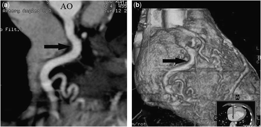
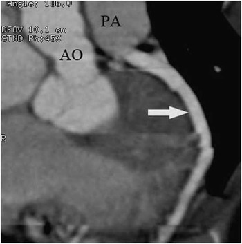
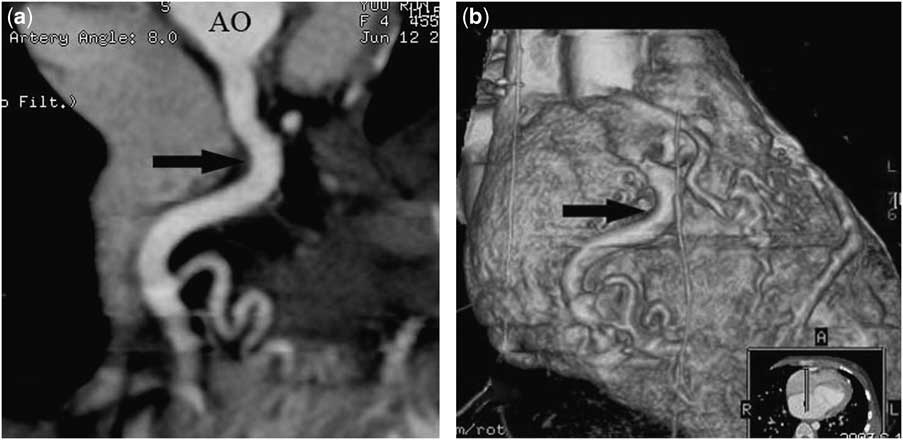
Computed tomography images including maximum intensity projection, curved reconstruction, and volume rendering show the left main coronary artery, which originated from the posterior aspect of the main pulmonary artery along with left anterior descending (Fig 1), and an enlarged tortuous right coronary artery, which originated from the right coronary sinus of the aortic root (Fig 2a and b), in keeping with the anomalous origin of the left coronary artery from the pulmonary artery.
Computed tomography angiogram demonstrates a left main coronary artery that originated from the posterior aspect of the main pulmonary artery along with left anterior descending (white arrow). PA = pulmonary artery; AO = aortic.

(a) Curved reconstruction and (b) volume rendering. The images demonstrate an enlarged tortuous right coronary artery (black arrow) that originated from the right coronary sinus of the aortic root. AO = aortic.