Introduction
Severe pulmonary valve regurgitation with or without right ventricular dysfunction following previous surgical repair of defects such as tetralogy of Fallot is a recognised indication for percutaneous pulmonary valve implantation. Reference Bonhoeffer, Boudjemline and Saliba1–Reference Eicken, Ewert and Hager3 Traditionally percutaneous pulmonary valve implantation has been limited to those with circumferential surgical right ventricular outflow tracts, such as those patients with surgically placed conduits or bioprosthetic valves. Reference Morgan, Prachasilchai and Promphan4 In patients with right ventricular outflow tracts reconstructed with a transannular patch or a dilated main pulmonary artery, the geometry and size of the right ventricular outflow tract can often preclude percutaneous pulmonary valve implantation with balloon expandable valves. Reference Morgan, Prachasilchai and Promphan4–Reference Hascoët, Bentham and Giugno7 More recently, self-expanding valves have been used. Reference Morgan, Prachasilchai and Promphan4,Reference Zablah and Morgan8–Reference Park, Kim and Lee14 The efficacy of percutaneous pulmonary valve implantation using the VenusP-valve has been demonstrated in the adult population, Reference Morgan, Prachasilchai and Promphan4,Reference Jalal, Valdeolmillos and Malekzadeh-Milani6,Reference Hascoët, Bentham and Giugno7 but a significant proportion of patients that require percutaneous pulmonary valve implantation in this setting are in the paediatric age group and for this subset, the utility of the VenusP-valve has not been demonstrated.
The aim of this study is to report the feasibility, safety and short- and medium-term outcomes of VenusP-valve™ implantation in the paediatric population.
Methods
Between June 2015 and February 2023, a retrospective review of patients under 18 years of age undergoing VenusP-valve implantation at a single institution was conducted.
In our institution, percutaneous pulmonary valve implantation is indicated in those patients who had undergone surgical repair of the right ventricular outflow tract using a transannular patch technique or replacement with a pulmonary homograft and who had severe pulmonary regurgitation on echocardiography, a pulmonary regurgitant fraction of > 30% on cardiac MRI, and an indexed right ventricular end-diastolic volume of > 150 ml/m2. A smaller right ventricular end-diastolic volume in the presence of severe pulmonary regurgitation was accepted in the presence of symptoms, such as shortness of breath on exertion. Reference Baumgartner, Bonhoeffer, De Groot and de Haan2,Reference Suradi and Hijazi16–Reference Stout, Daniels and Aboulhosn18 Patients were excluded from this review if they were over 18 years of age at the time of intervention. Other exclusions included patients in whom the geometry of the outflow tract precluded the implantation of the VenusP-valve™, such as those with a pyramidal shaped right ventricular outflow tract, significant right ventricular outflow tract or main pulmonary artery narrowing (MPA), or significant obstruction of the proximal branches of the pulmonary artery. Patients were also excluded if the coronary artery anatomy posed a risk of coronary compression after valve deployment.
Pre-procedure evaluation
All the patients were evaluated with transthoracic echocardiography for a baseline assessment of pulmonary regurgitation, right ventricular function, and dimensions, right ventricular outflow tract outflow gradient, tricuspid regurgitation, and the pulmonary artery branching and geometry. Patients also underwent an MRI to quantify right and left ventricular function and volumes, pulmonary regurgitant fraction, and to assess further the anatomy and geometry of the right ventricular outflow tract and pulmonary artery branches.
All the patients underwent complete haemodynamic and angiographic assessment by cardiac catheterisation, either as a stand-alone procedure 3–6 months before percutaneous pulmonary valve implantation or during the valve implant procedure itself. Right ventricular, main pulmonary artery, branch pulmonary arteries, and aortic pressures were measured. Angiography was performed with cranial angulation (either left / right anterior oblique depending on cross-sectional reconstruction predictions) and lateral projections for both the right ventricular outflow tract and the main pulmonary artery. Measurements were made of the maximum systolic diameter of the proximal right ventricular outflow tract, pulmonary valve annulus, proximal and distal main pulmonary artery, and the distance between these points. The midpoint of the main pulmonary artery and the narrowest portion were also measured.
Selective left coronary angiography was performed simultaneously with balloon sizing of the main pulmonary artery to assess for the possibility of coronary artery compression and for sizing of the right ventricular outflow tract.
VenusP-valve
The VenusP-valve™ (Venus Medtech, Shanghai, China) is a self-expanding Nitinol multilevel support frame containing a tri-leaflet porcine pericardial tissue valve. This is preserved in buffered glutaraldehyde to both cross-link the tissue and preserve flexibility and strength. The stent is covered along its entire length by porcine pericardial tissue apart from the distal portion of cells which are uncovered. Both ends of the stent are flared. The covered but flared proximal end allows conformation to the dilated right ventricular outflow tract. The flared distal portion acts as an anchor at the distal portion of the pulmonary artery bifurcation. There are radiopaque markers at the junctions of both the flares with the straight portion of the stent for visualisation. Reference Cao, Kenny and Zhou15 The straight section of the stent, with the valve sutured to it, is available in valve diameters from 28 mm to 36 mm (in 2 mm increments) and lengths of 25 and 30 mm, although was initially available in a wider selection of lengths of 20–35 mm in 5 mm increments. Delivery catheter systems are in 22 Fr for valve diameters of 28–30 mm and 24 Fr for valve diameter of 32–36 mm. Reference Cao, Kenny and Zhou15
Procedure
The technique of the valve implantation has been described in detail previously. Reference Morgan, Prachasilchai and Promphan4,Reference Cao, Kenny and Zhou15,Reference Garay, Pan, Zhang, Wang and Springmuller19 All the procedures were performed under general anaesthesia. Bilateral femoral vein access was obtained in addition to one femoral artery access. Heparin 100 U/kg was administered to achieve an activated clotting time of > 250 s. Intravenous antibiotics were given at the start of the procedure, according to the institutional protocol. If haemodynamic and angiographic assessment with balloon sizing had been performed more than three months prior to the percutaneous pulmonary valve implantation, these were repeated to ensure no significant changes had occurred. The selected valve diameter was 2–4 mm larger than the waist on the balloon in the right ventricular outflow tract. The valve was prepared according to the manufacturer’s instructions for use and was then crimped manually in ice-cold water and loaded onto the delivery system.
A 6 Fr JR guide catheter was passed to a distal pulmonary artery branch. Early experience in adult patients had led to a preference for the left pulmonary artery, however, as experience has increased, both pulmonary arteries have subsequently been used. A 260 cm 0.035’’ Lunderquist extra-stiff guidewire (Cook Medical, Bloomington, IN, USA) was then used to pass a Gore DrySeal sheath (Gore Medical, Flagstaff, ARI, USA) into a proximal branch pulmonary artery. The valve and delivery system were passed through the sheath to the proximal pulmonary artery branch. Check angiography was performed frequently, through a pigtail catheter in the right ventricular outflow tract/main pulmonary artery, as the distal flare portion of the valve was deployed. Subsequently, the mid-portion and the proximal flare were deployed before the final release of the valve (Figure 1).
(a)–(b): angiogram demonstrating dilated RVOT. (c)–(d): sizing of the RVOT and MPA with compliant balloon. (d)–(e): VenusP-valve™ in situ with no regurgitation or paravalvar leak. All angiograms in RAO 30 Cranial 30 and LAT.

Antibiotics were continued for 24 hours following the procedure and the patients were commenced on aspirin 5mg/kg OD lifelong (maximum 75 mg) and clopidogrel 1mg/kg OD (maximum 75 mg OD) for six months. All the patients underwent a post-procedural echocardiogram, ECG and CXR (antero-posterior and lateral projections) prior to discharge. Patients were reviewed at 8 weeks following the procedure and then at 6 months, when they underwent a cardiac MR, echocardiogram, and fluoroscopy of the valve.
Statistics
Continuous variables are presented as means and ranges. Categorical data are presented as counts and percentages. Comparison between both groups was made using the Wilcoxon signed-rank test. Statistical analysis was performed using Statistical Package for the Social Sciences (IBM SPSS Statistics version 27.0.1.0 SSPS Inc., Chicago, IL). Statistical significance was defined as a p-value < 0.05.
Results
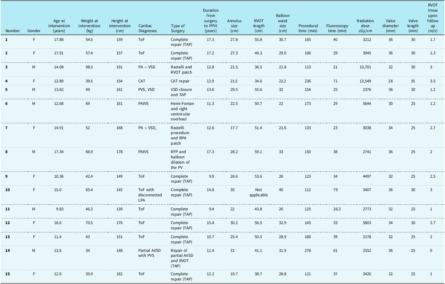
Between June 2015 and February 2023, 15 paediatric patients under 18 years of age underwent successful VenusP-valve implantation, of whom 9 were females and 6 males. During the same period, 34 adult patients underwent successful VenusP-valve implantation. All the patients had severe pulmonary regurgitation on echocardiogram or a regurgitant fraction of greater than 30% on MRI and met our institutions accepted criteria for pulmonary valve implantation. Table 1 demonstrates the patient anatomy, age at intervention, time from the initial surgery to VenusP-valve implantation and parameters measured at time of intervention and on follow up.
Patient anatomy, age at intervention, time from the initial surgery to venusP-valve implantation and parameters measured at time of intervention and on follow up. F = female, M = male, ToF =Tetralogy of fallot. PA = pulmonary atresia, VSD = ventricular septal defect, CAT = common arterial trunk, PVS = pulmonary valve stenosis, PAIVS = pulmonary atresia with intact ventricular septum, TAP = transannular patch. Pulmonary valve = PV, RFP = radiofrequency perforation

Mean age at valve implantation was 14.1 (range 9.8–17.9) years and was performed 13.2 (9.4 – 17.3) years after the initial surgery. Mean weight and height at valve implantation were 54.9 (34.0–98.5) kg and 159.9 (139–191) cm. The valve was deployed successfully in all the patients. The mean procedure and fluoroscopy times were 154.1 (106–278) minutes and 39.9 (21–79) minutes, respectively, and the radiation dose 4656 (2178–13549) cGy/cm. The valve diameter and length ranged between 28–36 mm and 25–35 mm, with some patients undergoing the procedure during the period with the previously available valve lengths. Only one patient had an implanted valve diameter < 30 mm. 26 French Gore DrySeal sheath was used in all but two cases, in whom a 24 French sheath was used.
Complications
There were four complications in four patients. One patient, with right pulmonary artery stenosis/hypoplasia, had jailing of the right pulmonary artery by the covered body of the VenusP-valve; the valve was manipulated successfully with a bioptome to uncover the right pulmonary artery and additional main pulmonary artery stenting was performed to maintain its patency and flow (Figure 2).
(a): angiogram RAO 30 Cr 30 demonstrating RVOT. (b): LAO 30 Cr 40 Mispositioned VenusP-valve with suspicion of jailing of the RPA. (c)–(d): angiography in LAO 30 Cr 40 confirming “jailing” of the RPA. (e)–(f): LAO 30 Cr 40 angiography demonstrating RPA stenting and post insertion dilatation of the RPA stent.

A second patient required surgical intervention to manipulate and suture the VenusP-valve more distally within the main pulmonary artery (Figure 3). This was following removal of the delivery system of the VenusP-valve, where the “nosecone” caught onto the distal struts of the valve, and the VenusP-valve migrated proximally within the main pulmonary artery and right ventricular outflow tract resulting in a paravalvar leak and unsatisfactory position. This required surgery, where the distal struts of the VenusP-valve were trimmed and pledgets were placed along the MPA to keep the valve in place.
(a)–(b): angiogram of VenusP-valve in satisfactory position post deployment. (c)–(d): angiogram of withdrawal of “nosecone” there is embolisation proximally of the device into the RVOT. (e)–(f): angiogram of attempted repositioning with CODA balloon (Cook medical, Bloomington, IN, USA). Angiograms in RAO 30 Cranial 30 and LAT. (g): final position of valve in PA CXR demonstrated cut distal struts.

A third patient experienced perforation of the right iliac vein. The femoral vein was found to be tortuous on angiography after some difficulty advancing the guidewire. However, a 12 Fr sheath was placed in the vessel with some resistance. Angiography demonstrated contrast extravasation. After attempting unsuccessful transluminal balloon tamponade to the vessel, an 8 Fr long sheath was passed distally to the point of extravasation and a covered stent was placed, with subsequent post dilation.
Finally, one patient had a fracture of the suture needle when performing a ’Z-suture’ in the groin for haemostasis, which required removal from the dermis.
There were no episodes of infection or post-procedural complications.
Follow-up
Mean follow-up duration was 3.4 (0.5– 8.1) years. To date, twelve patients have undergone follow-up MRI imaging. The right ventricular end-diastolic volume improved from a mean of 157.8 (140.0–197.0) ml/m2 to 117.6 (91 to 152.0) ml/m2 (p = 0.004). Pulmonary regurgitation fraction had reduced from a mean of 44.3 (31.0 – 60.0) % to 3.6 (0.0–15.0) % (p = 0.003). There was no stenosis noted in the right ventricular outflow tract. Two of the eight patients who had routine fluoroscopy between 6 and 12 months after the procedure had evidence of a single stent fracture, which has not been associated with any clinically important sequelae, such as stent integrity or valve dysfunction. There were no issues of dysrhythmia immediately after or during the follow-up period on serial 24-hour ECG monitoring. No patients developed subclinical thrombosis or micro-thrombosis on subsequent imaging.
Discussion
Experience with percutaneous pulmonary valve implantation worldwide had previously been limited to two balloon-expandable valves, the Melody Valve (Medtronic Inc., Minneapolis, MN) and the SAPIEN valve (Edwards Lifesciences, Irvine, CA). Reference Cao, Kenny and Zhou15 Both of these valves have been utilised in patients with circumferential surgical right ventricular outflow tracts, such as cardiac conduits. Reference Morgan, Prachasilchai and Promphan4 However, their use in patients with larger right ventricular outflow tracts, has been limited, as balloon-expandable stents are often not large enough in diameter to be implanted in the dilated regurgitant conduit-free right ventricular outflow tracts, which have been treated with transannular patches at the time of the surgical repair of the defects. More recently, the self-expanding VenusP-valve, and other self-expanding valves such as the Harmony transcatheter pulmonary valve (Medtronic Inc., Minneapolis MN), Pulsta transcatheter pulmonary valve (TaeWoong Medical Co., Gyeonggi-do, South Korea), or the Alterra Adaptive Prestent (Edwards Lifesciences, Irvine, California) in combination with SAPIEN 3 transcatheter heart valve (Edwards Lifesciences), have provided alternatives to surgical repair in the adult patients with suitable anatomical substrates, such as those with dilated right ventricular outflow tracts, that have traditionally not favoured a percutaneous approach. Reference Morgan, Prachasilchai and Promphan4,Reference Shahanavaz, Balzer and Babaliaros10,Reference Benson, Gillespie and Bergersen13,Reference Bergersen, Benson and Gillespie20
The Melody Valve has been used in the paediatric population in those above 15 kg for some time and in ages as young as 4 years of age. Reference Jalal, Valdeolmillos and Malekzadeh-Milani6,Reference Zablah and Morgan8,Reference Cools, Budts and Heying21 Its availability in smaller diameters (18–22 mm), and similarly the SAPIEN valve (20–29 mm), make it attractive for their paediatric use, with the SAPIEN valve being used in patients with weights of 25 kg and above. Reference Plessis, Hascoët and Baruteau22,Reference Kenny, Hijazi and Kar23 The delivery system profile is also more favourable for the paediatric patients with these valves. However, in paediatric patients with larger conduit-free right ventricular outflow tracts, especially those with diameters > 29 mm, balloon-expandable valves are limited by their largest diameter and are often not suitable. The introduction of the Alterra Adaptive Prestent goes some way to address this need and has been used in adolescents aged between 12 and 21 years of age but with weights as low as 56.5 kg. Reference Shahanavaz, Balzer and Babaliaros10 Self-expanding valves have widened the available percutaneous options in larger conduit-free patched outflow tracts, although their use in the paediatric cohort is not well documented. The Pulsta transcatheter pulmonary valve has been used in paediatric patients as young as 10 years of age and weights as low as 28.8 kg. Reference Lee, Kim and Kim11,Reference Park, Kim and Lee14 At the time of writing, data on the use of the Harmony transcatheter pulmonary valve has not been published in paediatric patients; however, there are reports suggesting its use in these patients. Reference Gillespie, McElhinney, Jones, Levi, Asnes and Gray24,Reference Zablah, Than, Browne, Rodriguez and Morgan25 Our group of 15 patients had a mean age and weight at valve implantation of 14.1 (range 9.8–17.9) years and 54.9 (34.0–98.5) kg, respectively, and therefore widens the use of self-expanding valves in paediatric patients.
Previous reports have focussed primarily on the early- and medium-term outcomes of VenusP-valves predominantly in the adult population. Reference Morgan, Prachasilchai and Promphan4,Reference Promphan, Prachasilchai, Siripornpitak, Qureshi and Layangool12,Reference Garay, Pan, Zhang, Wang and Springmuller19 Our study sought to focus on the current experience of the VenusP-valve and widening its use in the paediatric population, to assess whether medium-term outcomes are similar to adults, and importantly whether there are any increased complication risks given the potentially smaller size of patient. There was no increased complication rate in our patient cohort. An important consideration is how the valves behave with the growth of the younger patients. Because the valves are large sized, they seem to maintain function with the growth of the patients. It remains to be seen whether there is a progressive stenosis of the VenusP-valve in the longer term. The right ventricular outflow tract systolic velocity on Doppler during follow up was 1.92 (0–3.5) m/s. Two patients with the longest period (9 years) of follow-up had Doppler velocities of 3 and 3.5 m/s, which may indicate some progressive narrowing, although no intervention has been required. None of our patients experienced dysrhythmia either in the immediate post procedural period or during follow up. This is unlike the adult cohort, where patients have experienced ventricular dysrhythmias such as sustained ventricular tachycardia and fibrillation and albeit rare, is a concern of self-expanding valves. Reference Shahanavaz, Balzer and Babaliaros10,Reference Kramer, Schleiger, Duong and Berger26–Reference Barfuss, Samayoa and Etheridge29 The lower incidence of dysrhythmia may be related to the myocardium being under shorter conditions of volume loading, potentially less fibrosis and a weaker substrate for arrhythmia in a younger cohort of patients.
Percutaneous VenusP-valve implantation was performed in older children because the indications for pulmonary valve intervention in patients with dilated right ventricular outflow tracts are rarely met at a younger age, and the practicalities of needing large sheath sizes required for implanting the current device sizes and the equipment necessitates a larger weight and size of the patient. Despite this, our cohort of patients does include 10 children below 15 years of age and between 30 and 60 kg, including one child less than 10 years of age. Not surprisingly, the duration from initial surgery to valve implantation was shorter compared to the general cohort of patients. Technically, the procedure may be challenging but despite this, there were similar fluoroscopy and procedure times comparable to the adult patients, Reference Promphan, Prachasilchai, Siripornpitak, Qureshi and Layangool12 which may reflect our increasing experience.
All the paediatric patients who were accepted for VenusP-valve implantation had the procedure performed successfully. This is similar to the previous international experience reports of medium-term results of the VenusP-valve by Morgan et al, as well as other series using the Melody and Sapien valves, with success rates of around 97%. Reference Morgan, Prachasilchai and Promphan4,Reference Kenny, Hijazi and Kar23,Reference Zahn, Hellenbrand, Lock and McElhinney30
Our cohort of patients had one case of valve migration necessitating surgical repositioning. In another patient, the valve was deployed too distally, inadvertently jailing the right pulmonary artery; however, a satisfactory result was obtained after percutaneous manipulation and repositioning of the valve and subsequent stenting of the right pulmonary artery. There were two complications related to access in the form of femoral vein perforation and a local suturing complication. Of these, the only complication that may be attributed to patient size or age is that of femoral vein perforation. However, this may have been related to the vessel tortuosity rather than its absolute size. This gives a relatively high complication rate of 20% if the local suturing complication is excluded. This quite significantly contrasts to the findings of the large IMPACT registry in which complication rates were overall 2.4%, if only those below 18 were included, this figure is as low as 1.86%. Reference Stefanescu Schmidt, Armstrong and Aboulhosn31 Crucially, the IMPACT registry differs somewhat as it covers a much larger demographic and does not include self-expanding valves. Furthermore, only 25.0% of the total number of patients and 12.4% of those below the age of 18 had patched or native right ventricular outflow tracts. However, accounting for these patients only, complication rates are still only 3.4%. The reason for this difference is likely multifactorial. First, the valves included in the IMPACT registry are not self-expanding and are well established procedures in all age groups and so they are not on the same part of the “learning curve” as the VenusP-valve and those that implant it. Second, those that require a self-expanding valve, by their nature have large diameter right ventricular outflow tracts’ and so it may be argued are technically more challenging procedures with a higher complication rate. Finally, our cohort only includes 15 patients and so the weighting of any complications is high and needs to be considered. However, the complication rates seen in the IMPACT registry, and those in adult VenusP-valve insertion should act as target for larger and longer-term studies.
There have been no cases of infective endocarditis in our patients, although there have been cases of infective endocarditis in China, particularly in the first year after implant Reference Jin, Long and Zhang32
Stent fractures remain a concern for all the percutaneous valves. The incidence of Melody valve fractures has been reported at between 20% and 40%, depending on the time elapsed post-procedure. Reference Morgan, Prachasilchai and Promphan4,Reference Nordmeyer, Khambadkone and Coats33 In our study, four of our twelve paediatric patients had fluoroscopic evidence of single strut fracture. This is similar to the findings of Morgan et al. Reference Morgan, Prachasilchai and Promphan4 However, unlike the fractures in the Melody valve, the single strut fractures of the VenusP-valve have not had any impact on the valve function. The formation of hypo-attenuated leaflet thickening and hypo-attenuating material is an established finding in transcatheter aortic valve replacement and is present in 10–12% of cases and has been associated with long-term valve deterioration and mortality. Reference Akazawa, Higaki and Kashiwagi34–Reference Garcia, Fukui and Dworak36 In self-expanding pulmonary valves, the finding of subclinical and microthrombosis seems to be less prevalent and is currently limited to individual case reports for VenusP-Valve, although early failure has been described. Reference Pilati, Duong, Bordonaro, Secinaro and Butera37,Reference Iannaccone, Bautista-Rodriguez and Kacar38 Our patients did not demonstrate any evidence of subclinical or clinical thromboembolism despite the longest placed valves having increased gradients.
The use of the VenusP-valve in the paediatric patients is a novel application, but the patient numbers are small. However, it has demonstrated good functionality, and due to its flared distal and proximal ends, it maintains excellent stability in the dilated outflow tracts. In addition, remodelling of the right ventricle occurs after percutaneous pulmonary valve implantation, with a significant reduction in pulmonary regurgitation and right ventricular end-diastolic volumes.
The importance of pre-procedural evaluation of the right ventricular outflow tracts and branch pulmonary artery anatomy cannot be overstated for the successful planning for VenusP-valve implantation. Cardiac MRI provides the detailed indications for intervention; however, angiographic assessment with balloon interrogation is vital to assess the distensibility of the right ventricular outflow tracts for accurate measurements and valve sizing. Furthermore, whilst experience with self-expanding valves, such as the VenusP-valve, continues to evolve, those patients with sub-optimal anatomical geometry, such as branch pulmonary artery stenosis or a pyramidal shaped outflow tract, should be avoided in the paediatric cohort, until larger experience has been gained, to ensure safe application in children. Reference Husain, Praichasilchai, Gilbert, Qureshi and Morgan9
Limitations
This is a retrospective study in a small cohort of paediatric patients and so limited importance should be given to statistically significant results. Furthermore, follow up MRI volumes and fluoroscopic assessment of strut fractures was not available in three patients. Two of the patients were included in initial medium-term data on the VenusP-valve. Reference Morgan, Prachasilchai and Promphan4
Conclusion
This study shows the feasibility and safety of VenusP-valve implantation in the short- and medium term in the paediatric population, and merits further consideration. Remodelling of the right ventricle occurs with a decrease in right ventricular end-diastolic volume and pulmonary regurgitant fraction after implantation of the VenusP-valve.
Financial support
This research received no specific grant from any funding agency, commercial, or not-for-profit sectors. Guy’s & St Thomas’ NHS Foundation Trust received a grant for the CE Study of the Venus P-valve, in which some of these patients were included.
Competing interests
Prof Sir Shakeel Qureshi is a consultant, proctor, and Principal Investigator for Venus MedTech CE study.
Dr Matthew I Jones is a consultant & proctor for Venus MedTech Europe.
Ethical standard
This retrospective study was registered with our institutional Research and Development office. In accordance with the UK NHS National Research Ethics Service guidance, neither individual informed consent nor formal Research Ethics Committee review was required because the study was undertaken by the direct clinical team using information previously collected during routine care.