Introduction
Cowpea is the major legume grain crop in sub-Saharan Africa (SSA) and the main protein source for many rural people in this area of the world. However, the production of cowpea is limited by both abiotic and biotic constraints. Insect pests are the major biotic limiting factor for cowpea production in SSA (Singh and Allen, Reference Singh, Allen, Summerfield and Bunting1980; Singh et al., Reference Singh, Jackai, Dos Santos, Adalla and Singh1990). These insect pests include the legume pod borer Maruca vitrata Fab. (Lepidoptera: Crambidae), one of the most devastating insect pests of cowpea, which can cause typical yield losses ranging from 20 to 80% (Singh et al., Reference Singh, Jackai, Dos Santos, Adalla and Singh1990). In Burkina Faso, M. vitrata is endemic in the south-western region of the country (Ba et al., Reference Ba, Margam, Dabiré-Binso, Sanon, McNeil, Murdock and Pittendrigh2009; Baoua et al., Reference Baoua, Ba, Agunbiade, Margam, Dabiré-Binso, Sanon and Pittendrigh2011). Damage to cowpea, by M. vitrata, is due to larvae that feed on the tender parts of the stem, peduncles, flower buds, flowers and pods (Singh and Jackai, Reference Singh and Jackai1988).
The economic importance of this pest species has been the main rationale behind the work that has been performed on its biology (Gblagada, 1982; Karel, Reference Karel1985; Afun et al., Reference Afun, Jackai and Hodgson1991). The early-instar larvae feed mainly on flowers (Karel, Reference Karel1985) and a single larva can consume four to six flowers by the time it reaches the pupal stage (Gblagada, 1982). Up to 80% of cowpea flowers are infested by M. vitrata larvae in the field (Afun et al., Reference Afun, Jackai and Hodgson1991). However, little information is available on the feeding preferences of the larvae, as it pertains to the age of the cowpea flower and the preferred flower components. Thus, this study aims to identify which age and which component of cowpea flowers are preferred by M. vitrata larvae. Understanding these aforementioned parameters will be useful when engineering the Cry endotoxin of Bacillus thuringiensis (Bt) Berliner into cowpea (hereafter called Bt-cowpea), especially vis-à-vis which organs in the plant need the highest levels of Bt expression to maximize the impact on M. vitrata populations. Where (or if) promoters exist, or can be discovered/developed, this can drive higher levels of expression in these tissues, which might assure a more effective control of M. vitrata populations through transgenic Bt-cowpeas.
Materials and methods
Study sites
Field
Field trials were conducted at the research station of the Institut de l'Environnement et de Recherche Agricole (INERA) in Farako-Ba, Burkina Faso (latitude: 11 °11′N; longitude: 04 °18′W), during the 2011 rainy season. Burkina Faso has a unimodal rainfall pattern and a rainy season that lasts from June to October. A total rainfall of 831 mm was recorded in 2011 in Farako-Ba.
Laboratory
Laboratory bioassays were conducted in the Laboratory of Entomology of INERA, in Farako-Ba, within temperature ranges of 25–32 °C and 60–80% relative humidity.
Source of insects
M. vitrata larvae used in the bioassays were obtained from a mass rearing facility at the INERA in Farako-Ba. The insects were reared on a modified European corn-borer diet obtained commercially from Bio-Serv Company, USA (Bio-Serv product No. F9478B-M without corncob grits) and were supplemented with cowpea seed flour (KVX-61-1 INERA variety).
Field experiments on M. vitrata feeding preferences
For this experiment, a 1 ha plot of the KVx 404-8-1 (60 days) cowpea variety was planted in July 2011 with an intra-row spacing of 0.4 m and an inter-row spacing of 0.8 m. Mineral fertilizers (100 kg/NPK 15–15–15) were applied to the entire plot before planting. No pesticides were applied in the plot.
At flowering time, observations were made daily up until the pod maturation stage. Flowers and pods were randomly picked from the plot. Sampling included two types of flowers: the green flower (non-opened and non-fertilized) and the yellow, fecundated flowers. Sampling on pods included three types of pods according to Dabiré-Binso et al. (Reference Dabiré-Binso, Kini, Ba, Dabire and Fouabi2005): (1) newly formed pods without seeds (c. 3 days aged), (2) pods in the filling stage (c. 8 days old), and (3) mature pods (c. 14 days old). When available, each category of the plant material (flower and pods) was randomly sampled within the field and brought back into the laboratory for dissection. The yellow opened flowers and the pods were sampled according to visible signs of the presence of M. vitrata larvae (webbing or faeces or both). A total of 3000 non-opened, green flowers, 3000 yellow opened flowers and 700 pods were collected. The green flowers (non-opened and non-fertilized) were distributed into two batches before dissection: (1) flowers showing larval entrance holes, ‘perforated flowers’ and (2) flowers with no visible orifice ‘non-perforated flowers’. The organs were then spread on a laboratory bench to avoid the transfer of larvae onto other flowers. The flowers were dissected individually under a magnifying glass and the following parameters recorded: (i) number of larvae, (ii) larval stage, (iii) flower tissues consumed and (iv) number of green flowers with more than one M. vitrata larva.
Laboratory bioassays on the suitability of cowpea flower components for M. vitrata larval development
Continuous cowpea production of the KVx 404-8-1 variety, was maintained under irrigation during the dry season (April–June 2011) in a 500 m2 plot adjoining the laboratory. Healthy flowers were collected and dissected into six different components: (i) calyx, (ii) corolla as a whole, and then (iii) standard, (iv) wing and (v) keel separately and then (vi) reproductive organs. The corolla included the standard, wing and keel. The reproductive organs of the flower included the stamens and the pistil. These components were used as a food for bioassays with M. vitrata larvae. Neonate larvae were fed with each of the aforementioned cowpea flower components until they developed into pupae. For the flower components, the food was renewed daily. Larvae, in groups of 20, were placed in 250 cm3 capacity plastic boxes covered with a muslin cloth. An absorbent paper was placed at the bottom of each box to remove excess humidity contained in the flower components. Four batches of five boxes were used for each flower component, each batch representing one replicate. The larvae were supplied daily with 1.5 g of one given flower component, which remained the same component, until they developed into pupae. Every 24 h the larvae were transferred with a camel-hairbrush to new boxes, of the same size, with the same quantity of the same flower component. Dead insects were discarded. When the pupae were formed, soft pliers were used to carefully remove them from their envelope, and then the pupae were placed in the boxes until adult emergence. When the adults emerged they were placed in mating cages, in male and female pairs, in a mass rearing room (25 °C temperature and 80% relative humidity) until the females laid eggs.
The following parameters were measured to determine the impact of the larval diets: duration of (i) larval, (ii) prepupal and (iii) pupal stages, (iv) size of pupae, (v) weight of the pupae (weighed in batches of 50), (vi) adult emergence rate, (vii) fecundity and (viii) adult life span.
Statistical analysis
For each parameter, data were analysed by an ANOVA using SAS software version 9.2 (PROC GLM; SAS Institute, Reference SAS2001). When ANOVA F-values were significant, means were separated by the Student–Newman–Keuls test at the 5% level.
Results
Natural infestation of M. vitrata larvae in the field
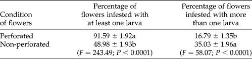
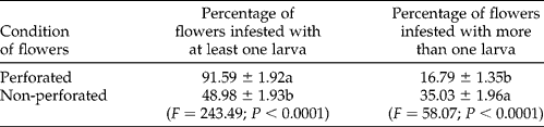
The perforated flowers, infested with at least one M. vitrata larva, were significantly higher than the non-perforated flowers (Table 1). However, the perforated flowers with at least two M. vitrata larvae were significantly lower than the non-perforated flowers (Table 1).
Maruca vitrata larval infestation (mean±SE) in green non-opened cowpea flowers
Mean values within a column followed by the same letters are not significantly different by the Student–Newman–Keuls test at the 5% level.
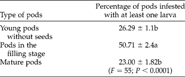
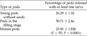
We observed M. vitrata larvae on all stages of the pods. However, the pods in the filling stage contained significantly more M. vitrata larvae than did the pods without seeds or mature pods (Table 2).
Maruca vitrata larval infestation (mean±SE) in cowpea pods
Mean values within a column followed by the same letters are not significantly different by the Student–Newman–Keuls test at the 5% level.
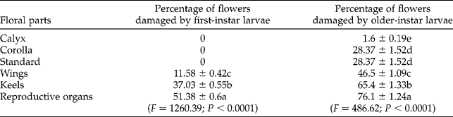
Upwards of 50% of the flowers, infested with first-instar M. vitrata larvae, had their reproductive organs damaged (Table 3). The first-instar larvae damaged 12 and 38% of the wings and the keels, respectively (Table 3). No damage, due to the first-instar larvae, could be observed on the standard, corolla and calyx of the flowers (Table 3). For subsequent M. vitrata larval instars there was a significant increase in damage to reproductive organs (Table 3).
Flower components (mean±SE) damaged by Maruca vitrata larvae
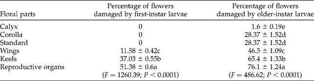
Mean values within a column followed by the same letters are not significantly different by the Student–Newman–Keuls test at the 5% level.
Development of M. vitrata larvae feeding on different flower components
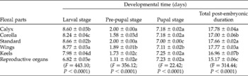
M. vitrata larvae were able to develop to adulthood when feeding on each of the individual cowpea flower components (Table 4). However, the larvae that fed on the reproductive organs of the flower had significantly shorter developmental times when compared with the larvae that fed on the other flower components.
Maruca vitrata larval development time (mean±SE) on different media

Mean values within a column followed by the same letters are not significantly different by the Student–Newman–Keuls test at the 5% level.
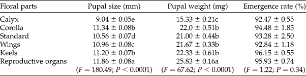
The component of the flower used as the diet, significantly influenced the size and weight of the pupae (Table 5), with the reproductive organs being the best diet for the insects. The pupae that emerged from the larvae, which were fed with the reproductive organs of the flower, were significantly greater in length, and were heavier, than the larvae that fed on the other flower components. The calyx was the least suitable substrate for insect development. However, there was no significant impact of the diet on the emergence of adults (Table 5).
Size and weight of Maruca vitrata pupae (mean±SE), and emergence rate of adults (mean±SE), depending on the medium upon which the larvae developed
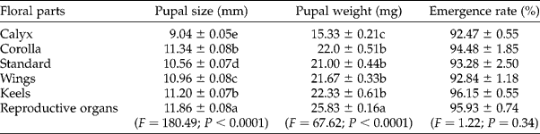
Mean values within a column followed by the same letters are not significantly different by the Student–Newman–Keuls test at the 5% level.
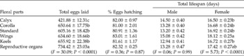
The females that emerged from the larvae, reared on the reproductive organs of the flower, laid significantly more eggs than the females that emerged from the larvae reared on the other flower components (Table 6). However, the eggs of all the females had the same percentage viability regardless of the medium upon which the larvae were raised (Table 6). The lifespan of females emerging from the flower's reproductive organs and wings was significantly longer than the females that emerged from the larvae raised on the other floral parts. The larval diet did not significantly affect the life span of the males (Table 6).
Fecundity of Maruca vitrata females, fertility of eggs, and longevity of M. vitrata adults reared (mean±SE), at the larval stage, on the different parts of the cowpea flower

Mean values within a column followed by the same letters are not significantly different by the Student–Newman–Keuls test at the 5% level.
Discussion
Our results demonstrate that M. vitrata larvae can develop on any single component of the cowpea flower up to adulthood. Regardless of the medium, we noticed a shorter total larval development duration when rearing M. vitrata larvae on cowpea whole flowers when compared with the findings from Naveen et al. (Reference Naveen, Naik, Manjunatha, Shivanna and Sridhar2009). Depending on the type of component upon which the larva fed, the biology of the insect was affected. Several studies have reported the quality of the food, in terms of nutrients, as a parameter affecting insect development (Pollet et al., Reference Pollet, Mauritz and Van Roon1978; Dabiré-Binso, 1980). In our case, when the insects were reared on the reproductive organs of the flowers (stamens and pistils), the larval development time was shortened. A reduction in insect developmental time due to the differences in feeding substrate has also been reported for the cowpea pod-sucking bug Clavigralla tomentosicollis Stål (Hemiptera: Coreidae) (Dabiré-Binso et al., Reference Dabiré-Binso, Kini, Ba, Dabire and Fouabi2005). As a consequence, the pupae from larvae that developed on the flower's reproductive organs were heavier than the pupae from larvae that developed on the other floral parts. Similar results have also been reported with M. vitrata pupae (Onyango and Ochieng-Odero, Reference Onyango and Ochieng-Odero1993). This is in accordance with Jackai and Singh (Reference Jackai and Singh1983) who reported that M. vitrata larvae feeding on more suitable substrates had higher weights when compared with the larvae reared on less suitable substrates. However, the medium did not much influence the emergence of adults, in contrast to what has been observed with M. vitrata larvae reared on artificial medium (Jackai and Raulston, Reference Jackai and Raulston1988).
The average lifespan of males was not affected by the rearing medium whereas females lived longer when originating from the flower's reproductive organs and wings. However, regardless of the type of the rearing medium, both male and female lifespans were longer than what had previously been observed by Naveen et al. (Reference Naveen, Naik, Manjunatha, Shivanna and Sridhar2009) and Huang and Peng (Reference Huang and Peng2001), but shorter than the findings of Chi et al. (Reference Chi, Sakamaki, Tsuda and Kusigemati2005). These differences are probably due to the rearing medium and environmental conditions (e.g. temperature). As regards fecundity, females from the larvae that developed on the flower's reproductive organs laid more eggs than the ones that derived from the other floral parts. The quality of the diet on female fecundity has previously been reported in the cowpea pod-sucking bug C. tomentosicollis system (Dabiré-Binso et al., Reference Dabiré-Binso, Ba, Sanon, Drabo and Foua Bi2010).
Our laboratory findings indicate that the reproductive organs of the flowers are much better for M. vitrata development: shorter developmental time for larvae, heavier weight for pupae, as well as increased longevity for females and higher fecundity. Our field data corroborate our laboratory findings. When dissecting the flowers that had visible signs of M. vitrata damage, we observed that the larvae fed preferentially on the reproductive organs. Taylor (Reference Taylor, Singh, Taylor and van Emden1978) reported similar observations. It is not known why M. vitrata larvae have this preference; however, the reproductive parts of the flowers may contain nutrients, water levels or plant secondary compounds, or a combination thereof, which is beneficial to the larvae.
However, the feeding preference of M. vitrata larvae in the field may also be related to a camouflage defence strategy of the larvae. They are thought to use several strategies to defend themselves against predators. This microhabitat feeding preference has been reported as an insect anti-predation strategy (Wellbornet and Robinson, Reference Wellbornet and Robinson1987; Pierce, Reference Pierce1988; Lima and Dill, Reference Lima and Dill1990). Thus, M. vitrata larvae may use the corolla as a shelter in an attempt to reduce predation. Within the flower, the pistils and stamens may offer a better shelter, to avoid predation, than the other parts of the flower. In fact, when feeding on this inner flower component, M. vitrata larvae are thereby covered by a series of three protective envelopes: the keel, the wing and the standard. The larvae are thus relatively shielded, securely embedded inside the flower.
In the field, we also noticed that M. vitrata larvae consumed green, non-opened cowpea flowers, either perforated or non-perforated. Previous oviposition behaviour studies have indicated that M. vitrata females deposited eggs on flower buds, vegetative buds, flowers and sometimes on leaf axils (Taylor, Reference Taylor1967, Reference Taylor, Singh, Taylor and van Emden1978). Thus, the presence of the larvae within flowers, with no visible entry orifice, indicates that the female has directly deposited eggs within the flower or flower buds. First-instar larvae were observed within cowpea flowers with sometimes more than one larva per flower. Since first-instar larvae are not highly mobile, they do not typically move from the flower where oviposition has occurred to a new flower. The presence of multiple larvae within a flower indicated that females might have deposited more than one egg in a flower bud or more than one female had deposited an egg on that flower. This is in accordance with previous studies, which indicated that eggs are deposited singly or in batches of two to six (Taylor, Reference Taylor1967, 1978). The young larvae were most often associated with young flowers as well as young pods, in keeping with previous observations (Singh and Jackai, Reference Singh and Jackai1988; Atachi and Gnanvossou, Reference Atachi and Gnanvossou1989).
Conclusion
Our findings indicate that M. vitrata larvae feed on all types of flowers regardless of their age and on all the components of the cowpea flower, with a preference for the reproductive organs of the flowers. Thus, for Bt-engineered cowpea, discovery and development of promoters, which would optimise the expression of Bt across all of these flower tissues without having negative impacts on cowpea productivity, would be highly desirable.
Acknowledgements
This study was made possible through support provided to the Dry Grains Pulses Collaborative Research Support Program (CRSP) by the Bureau for Economic Growth, Agriculture, and Trade, US Agency for International Development, under the terms of grant no. EDH-A-00-07-00005 to B.R.P., N.M.B. and C.L.D.-B. The opinions expressed herein are those of the authors and do not necessarily reflect the views of the US Agency for International Development or the US Government. We are also grateful to the African Agriculture Technology Foundation (AATF) for supporting the insect mass rearing facilities.