Introduction
Nematodes of the family Heterorhabditidae are obligate parasites of insects. They are considered as entomopathogenic nematodes (EPN), having a mutualistic association with bacteria of the family Enterobacteriacae, which are harboured in the intestine of the infective juveniles (IJs). The bacteria are lethal to their insect host, which makes them suitable for use as biocontrol agents. Numerous surveys for EPNs have been conducted worldwide, and many new species have been recovered (K.B. Nguyen et al., Reference Nguyen, Gozel, Koppenhofer and Adams2006; Malan et al., Reference Malan, Knoetze and Tiedt2012). Currently 19 species have been included in the family Heterorhabditidae (Nguyen, Reference Nguyen2015). In Pakistan, studies of entomopathogenic nematodes were initiated in 1996 (Shahina & Maqbool, Reference Shahina and Maqbool1996). More than 7000 soil samples were collected from different climatic regions of Pakistan to isolate EPNs that may be utilized effectively to control economically important agricultural insect pests. Surveys and studies of EPNs in Pakistan have revealed five new species, viz. Steinernema pakistanense Shahina, Anis, Reid, Rowe and Maqbool, Reference Shahina, Anis, Reid, Rowe and Maqbool2001; S. asiaticum Anis, Shahina, Reid and Rowe, Reference Anis, Shahina, Reid and Rowe2002; S. maqbooli Shahina, Tabassum, Mehreen and Salma, Reference Shahina, Tabassum, Mehreen and Salma2013; S. bifurcatum Shahina, Yan, Qiu, Han, Mehreen, Tabassum and Salma, Reference Shahina, Yan, Qiu, Han, Mehreen, Tabassum and Salma2014 and S. balochiense Shahina, Tabassum, Shaukat, Solangi, Mehreen and Salma, Reference Shahina, Tabassum, Shaukat, Solangi, Mehreen and Salma2015, which were described and illustrated, while seven known species S. abbasi Elawad, Ahmad and Reid, Reference Elawad, Ahmad and Reid1997; S. siamkayai Stock, Somsook and Reid, Reference Stock, Somsook and Reid1998; S. feltiae (Filipjev, Reference Filipjev1934) Wouts, Mráček, Gerdin and Bedding, Reference Wouts, Mracek, Gerdin and Bedding1982; S. carpocapsae (Weiser, Reference Weiser1955) Wouts, Mráček, Gerdin and Bedding, Reference Wouts, Mracek, Gerdin and Bedding1982; S. litorale Yoshida, Reference Yoshida2004; Heterorhabditis indica Poinar, Karunakar and David, Reference Poinar, Karunakar and David1992 and H. bacteriophora Poinar, Reference Poinar1976 were reported for the first time from Pakistan.
Molecular techniques, such as the polymerase chain reaction (PCR) and the analysis of sequence data, are considered as supplements to traditional identification and systematics of nematodes when dealing with taxonomic ambiguities, and they also help in resolving problems such as identification of species that are morphologically similar (Stock et al., Reference Stock, Griffin and Chaenari2004). Various gene regions have been used for the molecular characterization of Heterorhabditis species, such as the internal transcribed spacer (ITS) region of the ribosomal RNA gene (rRNA), the 18S ribosomal RNA gene region and the NADH dehydrogenase subunit 4 (ND4) gene of mitochondrial DNA (mtDNA) (Liu et al., Reference Liu, Berry and Blouin1999). The current study describes a new species of EPN, H. pakistanense n. sp. from Sindh, Pakistan, based on morphometrics, morphological and molecular evidence, which is compared and contrasted with the previously known species of the same genus. This new species is targeted for development as a biopesticide against insect pests in Pakistan.
Materials and methods
Collection and examination of nematodes
Heterorhabditis pakistanense n. sp. isolate Pak. S. H. 333 was recovered from soil using the insect baiting method (Bedding & Akhurst, Reference Bedding and Akhurst1975). Five last-instar Galleria mellonella larvae were placed in 250-ml plastic containers with moistened soil from samples, each being covered with a lid, turned upside down and kept at 27–30°C. Soil assays were checked daily, dead larvae were picked out, rinsed in distilled water and placed in modified White traps (Kaya & Stock, Reference Kaya, Stock and Lacey1997) until the nematode progeny emerged and migrated to the water reservoir.
Nematodes were cultured on G. mellonella at 25 ± 5°C according to Baliadi et al. (Reference Baliadi, Kondo and Yoshiga2009). The mortality of G. mellonella larvae was recorded after 24–48 h. Only IJs were collected from the White traps on the fourth day of emergence and used for identification. Hermaphroditic and amphimictic adults were collected 3 and 7 days after larval death, respectively, by dissecting the cadavers in Ringer solution. For morphological observations, IJs, first- (hermaphroditic females) and second-generation (amphimictic) males and females were killed by hot (85°C) water, then fixed in triethanolamine formaldehyde (TAF) (Courtney et al., Reference Courtney, Pooley and Miller1955) and processed in glycerine by the Seinhorst method (Seinhorst, Reference Seinhorst1959). Permanent slides were made using glass supports to avoid flattening of specimens. At least 25 specimens of each stage were selected at random and measured using a Nikon E400 light microscope (LM). Photomicrographs were also recorded with the Nikon E400 light microscope.
Scanning electron microscopy
For scanning electron microscopy (SEM), first-generation hermaphrodites, second-generation (amphimictic) males, females and IJs were fixed in TAF (ten of each). Each specimen was post-fixed with 2% glutaraldehyde solution overnight at 4°C and then washed with 0.1 m phosphate buffer. Nematodes were then dehydrated in a 30–100% (v/v) graded ethanol series for 20 min at each stage and finally washed three times in 100% ethanol. The nematodes were critical point dried with liquid CO2, mounted on SEM stubs and coated with gold before being examined with a JEOL JSM-840 scanning electron microscope.
Cross-hybridization
Cross-breeding tests were performed between H. pakistanense n. sp. and morphologically similar Pakistani isolates H. bacteriophora and H. indica using the modified hanging-drop method, as described by Kaya & Stock (Reference Kaya, Stock and Lacey1997). Twenty replicates were observed for each treatment and control. Sealed Petri dishes were incubated at 27–30°C and observations were recorded daily for reproductive compatibilities.
Molecular characterization and phylogenetic relationships
DNA was extracted from the mature first-generation single female using the method of Joyce et al. (Reference Joyce, Reid, Driver, Curran, Burnell, Ehlers and Masson1994). The forward primer 93 (5'-TTGAACCGGGTAAAGTCG-3') and the reverse primer 94 (5'-TTAGTTTCTTTTCCTCCGCT-3'), as described by Stock et al. (Reference Stock, Campbell and Nadler2001), were used for amplification and sequencing of the rDNA fragment containing the internal transcribed regions ITS1 and ITS2 as well as the 5.8S rRNA gene. The D2D3 expansion segment of the 28S rRNA gene was amplified using the forward primer D2F (5'-CCTTAG-TAACGGCGAGTGAAA-3') and the reverse primer 536 (5'-CAGCTATCCTGAGGAAAC-3') (K.B. Nguyen et al., Reference Nguyen, Gozel, Koppenhofer and Adams2006). For amplification of the mitochondrial 12S rDNA gene, the forward primer 505 (5'-GTTC-CAGAATAATCGGCTAGAC-3') and the reverse primer 506 (5'-TCTACTTTACTACAACTTACTCCCC-3'), as described by Nadler et al. (Reference Nadler, De Ley, Mundo-Ocampo, Smythe, Stock, Bumbarger, Adams, De Ley, Holovachov and Baldwin2006), were used. The PCR cycling conditions consisted of 4 min at 94°C; 35 cycles of 1 min at 94°C, 1.5 min at 55°C and 2 min at 72°C; followed by a final step of 10 min at 75°C (Nadler et al., Reference Nadler, De Ley, Mundo-Ocampo, Smythe, Stock, Bumbarger, Adams, De Ley, Holovachov and Baldwin2006). The PCR product was separated by electrophoresis on a 1.0% agarose gel and visualized on a UV-transilluminator.
PCR products were purified using the QIA quick Gel Extraction Kit (Qiagen, Valencia, California, USA) and cloned into pGemT easy vector System I (Promega, Madison, Wisconsin, USA). The plasmids were transformed into Escherichia coli strain JM109 competent cells (Promega) according to the manufacturer's protocols. Clones containing putative rRNA inserts were identified by blue/white colour selection. The plasmid preparations were extracted using the QIAquick Spin Miniprep Kit (Qiagen). From the plates with positive results, two clones for each EPN individual were sequenced using an ABI Prism 377 sequencer (PE Applied Biosystems, Foster City, California, USA) and dye terminator sequencing reagents at the Centralize Laboratory, University of Karachi, Karachi, Pakistan.
Sequences were compared to known sequences in the public databases by means of the Basic Local Alignment Search Tool (BLAST; http://www.blast.ncbi.nlm.nih.gov). Multiple alignments of the new sequence data, together with other sequences selected from National Center for Biotechnology Information (NCBI) databases, were performed using Clustal W2 software (Larkin et al., Reference Larkin, Blackshields, Brown, Chenna, McGettigan, McWilliam, Valentin, Wallace, Wilm, Lopez, Thompson, Gibson and Higgins2007). Phylogenetic analysis of the ITS, D2D3 regions and 12S rDNA gene, was performed by maximum parismony (MP) using PAUP* 4.0b8 (Swofford, Reference Swofford2001). Trees were evaluated statistically by bootstrap analysis based on 1000 resamplings of the dataset. The resulting trees were visualized by using MrEnt 2.0 (program distributed by A. Zuccon and D. Zuccon, 2010). Caenorhabditis elegans (EU131007) and Pellioditis typica (AF036946) were used as outgroups during calculation of the trees based on ITS rDNA sequence; C. elegans (X03680) was used as the outgroup for calculation of the trees based on D2D3 sequences.
Results and discussion
Morphology of Heterorhabditis pakistanense n. sp.
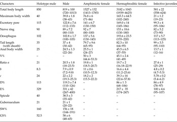
The morphometrics of H. pakistanense n. sp. are summarized in table 1.
Morphometrics (measurements in μm with mean values ± SD) of Heterorhabditis pakistanense n. sp. based on holotype and 25 paratypes of males, females and infective juveniles with ranges given in brackets.

V% = (distance of vulva from lips × 100/body length); a = body length/greatest width; b = body length/oesophagus length; c = body length/tail length; D% = (distance from anterior end to excretory pore divided by pharynx length) × 100; E% = (distance from anterior end to excretory pore divided by tail length) × 100; SW% = (spicules, length divided by anal body diameter) × 100; GS% = (gubernaculum length divided by spicules length) × 100; H, length of hyaline portion of tail.
Male
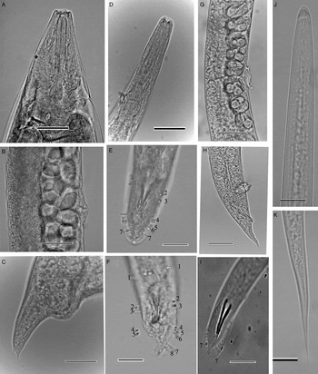
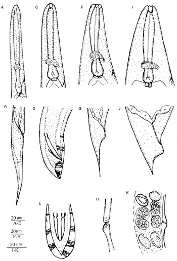
Body J-shaped when heat relaxed. Cuticle smooth under light microscopy but with fine striae visible under SEM. Lateral field inconspicuous. Head truncated, sometimes slightly swollen. Six labial papillae present. Cephalic papillae not observed. Amphidial opening conspicuous. Mouth opening funnel shaped, stoma short with barrel-shaped cheilorhabdions, other rhabdions indistinguishable. Pharynx with cylindrical corpus, metacorpus and isthmus distinct (fig. 1D, 2C). Basal bulb pyriform with reduced valve. Nerve ring surrounding isthmus just anterior to basal bulb. Excretory pore posterior to nerve ring or at the level of basal bulb (fig.1D, 2C). Testis monorchic, reflexed anteriorly. Vas deferens well developed. Spicule paired, separate, without rostrum. Lamina slightly curved ventrally. Gubernaculum slightly curved ventrally. Nine bursal papillae present on right side of tail, whereas on left side only six papillae are present. Number of genital papillae 1–6 (from anterior end on right side) are unchanged and typical for Heterorhabditis. The first pair of papillae is well anterior to cloaca, reaching bursal rim. Pairs 2 and 3 in a group anterior to cloaca, also reaching bursal rim. Pairs 4, 5 and 6 forming a group posterior to the cloaca, extending to the bursal rim. In 60% of specimens pairs 7–9 were missing on the left side of the tail, as per photograph (figs 1F and 2E), whereas in 40% of specimens the 8th and 9th pair of the terminal group were absent. Tail conoid, ventrally curved, slightly longer than anal body diameter with pointed tip. Bursa peloderan (figs 1E, F and 2E).
Photomicrographs of Heterorhabditis pakistanense n. sp. (A–C) Hermaphrodite, (A) pharyngeal region, (B) vulval region, (C) tail region showing mucron; (D–F, I) male, (D) pharyngeal region, (E) tail, (F, I) tail, showing papillae; (G, H) amphimictic female, (G) vulval region, (H) tail; (J, K) infective-stage juvenile, (J) pharyngeal region, (K) tail region. Scale bars: A–D, G = 20 μm; E, F, H, I = 10 μm; J, K = 20 μm.

Heterorhabditis pakistanense n. sp. (A, B) Infective stage juvenile, (A) pharyngeal region, (B) tail region; (C–E) male, (C) pharyngeal region; (D, E) tail, showing papillae; (F–H) amphimictic female, (F) pharyngeal region, (G) tail, (H) vulval region; (I–K) hermaphrodite, (I) pharyngeal region, (J) tail, (K) vulval region.

Hermaphroditic female
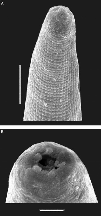
C-shaped after fixation, body robust, filled with many eggs, cuticle with five transverse striae. Head region truncated to slightly round (fig. 3B), tapering anteriorly, labial region with six prominent papillae (fig. 3B). Cephalic papillae conspicuous. Amphidial aperture small and pore-like, stoma shallow with refractile cheilorhabdions, other rhabdions fused. Posterior stoma funnel shaped, enclosed by anterior part of pharynx. Pharynx with cylindrical corpus but indistinguishable metacorpus and isthmus; basal bulb pyriform with valve (fig. 1A). Cardia present. Nerve ring distinct, located in the middle of the isthmus. Excretory pore located posterior to nerve ring mostly at the level of basal bulb or slightly posterior to it (figs 1A and 2I). Gonad didelphic, amphidelphic. Vulva located at mid body with transverse slit. Post-anal swelling well developed (figs 1C and 2J). Tail terminus conoid with prominent mucron (figs 1C and 2J). Phasmid inconspicuous.
SEM photographs of Heterorhabditis pakistanense n. sp. (A) Anterior region of ensheathed infective juvenile with tessellate pattern; (B) head of hermaphroditic female showing six labial papillae Scale bars: A = 5 μm; B = 2 μm.

Amphimictic female
Body C-shaped when heat relaxed. Cuticle with fine transverse striae. General morphology similar to that of hermaphroditic female but smaller in size. Vulva non-protuberant, without copulating plug located in the mid-body region. Post-anal swelling much smaller than that of hermaphroditic female. Tail conoid, about 2–3 times longer than anal body diameter, tapering to a finely pointed terminus.
Infective juvenile
Body long and slender, straight when heat relaxed, gradually tapering towards anterior and posterior ends, Cuticle of ensheathed juvenile with fine annulations. Cephalic region rounded not annulated, continuous with body contour. Anterior part of body (ensheathed juvenile) with tessellate pattern (fig. 3A). Posterior part with longitudinal ridges. Labial region with conspicuous dorsal tooth and amphidial aperture. Cephalic papillae absent. Stoma closed. Pharynx with cylindrical procorpus, narrow meta-corpus, slender isthmus and valvate basal bulb. Lateral field starting from posterior region of isthmus with two incisures, forming two ridges at mid body. Cardia present. Hemizonid usually 3–4 annules anterior to excretory pore. Excretory pore prominent, located at the anterior level of the basal bulb (fig. 2A). Nerve ring surrounding isthmus between posterior end of metacorpus and anterior end of basal bulb. Phasmid inconspicuous. Tail elongate filiform with pointed terminus.
Cross-hybridization
Progeny were not observed from cross-breeding treatments. Progeny were produced in the self-cross controls.
Taxonomic summary
Type host and material
The natural host of H pakistanense n. sp. is unknown, as the nematode was trapped by baiting soil around roots of Bermuda grass (Cynodon dactylon L.) from Malir, Karachi, Sindh, Pakistan. The holotype male (second generation) and numerous paratypes (first-generation hermaphrodites), the allotype male, female (second generation) and infective-stage juveniles are deposited in the Nematode Collection at the National Nematological Research Centre, University of Karachi, Pakistan.
Etymology
The species name reflects the country's name (Pakistan) and its first new species identification in the Heterorhabditis group.
Molecular and phylogenetic analysis
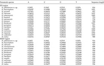
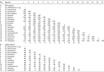
The ITS rDNA regions of H. pakistanense n. sp. (JX144740) are characterized by the sequence length of 816 base pairs and composition A = 0.2452, C = 0.1962, G = 0.2510 and T = 0.3076 (table 2). Pairwise distances between closely related species (table 3A) show that H. gerrardi and H. indica are the least divergent species from H. pakistanense, with 7 and 9 bp, respectively, while H. zealandica and H. megidis are the most divergent species, with 177 and 164 bp, respectively, when the ITS regions are considered.
Comparison of sequence length and base pair composition of ITS and D2D3 regions of Heterorhabditis pakistanense n. sp. with Heterorhabditis species.

Pairwise distances between the (A) ITS and (B) D2D3 regions of H. pakistanense n. sp. and other members of Heterorhabditis species.

The sequence length of the D2D3 expansion segment of the 28S rRNA gene of H. pakistanense n. sp. (KP096496) is characterized by 916 base pairs and its composition is A = 0.2452, C = 0.1962, G = 0.2510 and T = 0.3076 (table 2). Pairwise distances between closely related species (table 3B) show that H. pakistanense n. sp. differs least from H. indica (24 bp) and H. noenieputensis (25 bp) and most from H. megidis (68 bp) when the D2D3 regions are considered.
The 12S rDNA sequence obtained from H. pakistanense n. sp. (KM216180) is the first to be determined successfully for this gene region of the Heterorhabditis group and is characterized by a sequence length of 546 base pairs.
Previous studies have indicated that nematode mtDNA tends to be of less phylogenetic utility than nuclear genes (Nadler & Hudspeth, Reference Nadler and Hudspeth2000). The present study showed that ITS and D2D3 regions were more efficient and highly resolved in discriminating groups for identification of new species.
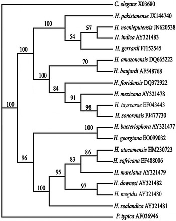
Phylogenetic relationships between 18 Heterorhabditis species inferred from ITS rRNA sequences, using the maximum parismony method (MP), are presented in fig. 4 (for MP tree length = 1139, CI = 0.8244, RI = 0.8657, RC = 0.7137, HI = 0.2699). In this tree, the indica and megidis groups are represented as two clades with several monophyletic groups included within them. In the first, the indica group, H. pakistanense n. sp., H. noenieputensis, H. indica and H. gerrardi form a monophylectic group with adequate bootstrap support (100%). Heterorhabditis amazonensis, H. baujardi, H. floridensis, H. mexicana, H. taysearae and H. sonorensis form a sister group within this clade with 100% bootstrap support. The second group, the megidis group, was formed by eight other species.
Phylogenetic relationships of Heterorhabditis pakistanense n. sp. with 18 Heterorhabditis spp. based on ITS rDNA sequences. Caenorhabditis elegans (X03680) and Pellioditis typica (AF036946) were used as outgroups. The percentage of replicate trees in which the associated taxa clustered together in the bootstrap test is shown next to the branches.

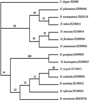
The phylogenetic relationships between 13 Heterorhabditis species inferred from D2D3 regions of the 28S rDNA gene sequences, using the maximum parismony method, are presented in fig. 5 (for MP tree length = 447, CI = 0.6805, RI = 0.7993, RC = 0.7027, HI = 0.1208). Heterorhabditis pakistanense n. sp., H. noenieputensis and H. indica comprise a monophyletic group with weak bootstrap support within the indica group.
Phylogenetic relationships of Heterorhabditis pakistanense n. sp. with 13 Heterorhabditis spp. based on D2D3 rDNA sequences. Caenorhabditis elegans (X03680) was used as the outgroup. The percentage of replicate trees in which the associated taxa clustered together in the bootstrap test is shown next to the branches.

We conclude that the molecular data presented here, together with morphological data from the previous sections, are sufficient to regard this isolate as a new species.
Diagnosis and relationships
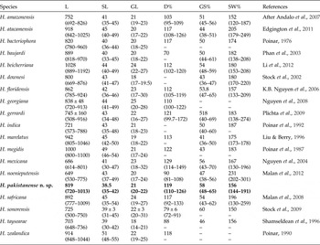
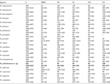
Heterorhabditis pakistanense n. sp. can be distinguished from other Heterorhabditis species by a combination of morphological and morphometric characteristics, and belongs to the indica-group on morphological (average length of IJ) and molecular bases. Male body length 819 μm (720–1013), cephalic papillae not observed, D% 119 (110–126), SW% 156 (144–191), GS% 58 (48–65) (table 4) and variations in the number of papillae of the terminal group of the bursa; 8th and 9th bursal papillae are sometimes absent on both sides; eight bursal papillae present on the right side vs six papillae present on the left. On the right side the arrangement of papillae is 1 + 2 + 3 + 2 whereas on the left side it is 1 + 2 + 3. The hermaphroditic female has a prominent post-anal swelling and a conoid tail 82 μm (64–95) long with pointed terminus. Infective juveniles have a medium-sized body 581 μm (558–624), long pharynx 117 μm (113–125), ensheathed tail 99 μm (95–110) and E% 99.9 (95–107) (table 5).
Comparative morphometrics of male of Heterorhabditis pakistanense n. sp. (in bold) and other related species. All measurements are in μm and in the form: mean (range).

L, total length; SL, spicule length; GL, gubernaculum length; GS% = (gubernaculum length divided by spicule length) × 100; SW% = (spicules, length divided by anal body diameter) × 100; D% = (distance from anterior end to excretory pore divided by pharynx length) × 100; –, data not available.
Comparative morphometrics of infective juveniles of Heterorhabditis pakistanense n. sp. (in bold) with other Heterorhabditis spp. All measurements are in μm and in the form: mean (range). References are the same as for table 4.

L, total length; MB, maximum body width; EP, excretory pore; TL, tail length (ensheathed); D% = (distance from anterior end to excretory pore divided by pharynx length) × 100; E% = (distance from anterior end to excretory pore divided by tail length) × 100; – data not available.
The indica group presently comprises eight species, viz. H. indica, H. amazonensis, H. baujardi Phan, Subbotin, Nguyen, and Moens, Reference Nguyen, Mrek and Webster2003, H. floridensis Nguyen, Gozel, Koppenhofer and Adams Reference Nguyen, Gozel, Koppenhofer and Adams2006, H. gerrardi Plichta, Joyce, Clark, Waterfield and Stock, Reference Plichta, Joyce, Clarke, Waterfield and Stock2009, H. mexicana Nguyen, Shapiro, Stuart, McCoy, James and Adams, Reference Nguyen, Shapiro, Stuart, McCoy, James and Adams2004, H. noenieputensis and H. taysearae Shamseldean, Abou El-Sooud, Abd-Elgawad and Saleh, Reference Shamseldean, Abou El-Sooud, Abd-Elgawad and Saleh1996. The new species closely resembles H. indica, H. gerrardi, H. amazonensis and H. noenieputensis, but the male can be distinguished from all species of Heterorhabditis by the absence of the 6th, 7th, 8th and 9th bursal papillae on the left side (fig. 1F).
Hermaphrodites of H. pakistanense n. sp. can be distinguished from all species of Heterorhabditis except H. downesi Stock, Griffin and Burnell, Reference Stock, Griffin and Burnell2002 by having a mucronate tail. In the phylogenetic analysis of sequence data, H. pakistanense n. sp. clustered with H. noenieputensis, H. gerrardi and H. indica (figs 4 and 5). However, it can be distinguished from these species by morphological and morphometric characters of IJs and adults. The males of H. pakistanense n. sp. differ from those of H. noenieputensis by having a higher D% (119 (110–126) vs 90 (81–108)) and a lower SW% (156 (144–191) vs 231 (202–301)). The IJs of H. pakistanense n. sp. also differ from those of H. noenieputensis by having a longer body length (581 μm (558–624) vs 528 μm (484–563)), longer pharynx (117 μm (113–125) vs 103 μm (99–108)) and longer tail with sheath (99 μm (95–110) vs 86 μm (78–95)).
Heterorhabditis pakistanense n. sp. differs from H. indica in the morphology of the male papillae (terminal pair of papillae absent vs present). The IJs of H. pakistanense n. sp. differ from those of H. indica by having a longer body length (581 μm (558–624) vs 528 μm (479–573)). Heterorhabditis pakistanense n. sp. can also be separated from H. gerrardi by the hermaphrodite females having a shorter tail (82 μm (64–95) vs 106 μm (90–195)). The males of H. pakistanense n. sp. differ from those of H. gerrardi by having longer body length (819 μm (720–1013) vs 745 μm (508–916)), shorter spicule length (38 μm (35–42) vs 43 μm (34–48)) and lower SW% (156 (144–191) vs 183 (138–274)).
Financial support
The present work was conducted under the Project No. PSF/ILP/S-KU/Bio (047) under 17th Protocol on Science and Technology Cooperation between Pakistan and China, entitled ‘Molecular characterization, mass production and formulation of entomopathogenic nematodes’, supported by the Pakistan Science Foundation.
Conflict of interest
None.