Introduction
Older adults with cardiovascular disease (CVD) often report experiencing cognitive difficulties in everyday life (Gunstad et al., Reference Gunstad, Cohen, Paul, Tate, Hoth and Poppas2006; Haley et al., Reference Haley, Hoth, Gunstad, Paul, Jefferson, Tate and Cohen2009; Humphreys et al., Reference Humphreys, Moser, Hynes, Reese and Haynes2007; Khatri et al., Reference Khatri, Babyak, Clancy, Davis, Croughwel, Newman and Blumenthal1999; McKhann et al., Reference McKhan, Selnes, Grega, Bailey, Pham, Baumgartner and Zeger2009; Newman et al., Reference Newman, Klinger, Ven, Smith, Harrison and Treasure1989; Selnes et al., Reference Selnes, Grega, Borowicz, Barry, Zeger and McKhan2004; Vingerhoets, de Soete, & Jannes, Reference Vingerhoets, de Soete and Jannes1995). Common self-reported complaints include forgetfulness, difficulties with sustained attention, verbal fluency, and short-term memory (Gunstad et al., Reference Gunstad, Cohen, Paul, Tate, Hoth and Poppas2006). The clinical significance of these complaints has been dismissed in cross-sectional and brief follow-up studies (<6 months) of CVD patients that found perceived cognitive problems to relate more closely to measures of psychological distress than to objective measures of cognitive function (Gunstad et al., Reference Gunstad, Cohen, Paul, Tate, Hoth and Poppas2006; Humphreys et al., Reference Humphreys, Moser, Hynes, Reese and Haynes2007; Khatri et al., Reference Khatri, Babyak, Clancy, Davis, Croughwel, Newman and Blumenthal1999; Vingerhoets et al., Reference Vingerhoets, de Soete and Jannes1995). However, reports from longitudinal studies with more extensive follow-up periods (≥12 months) have demonstrated that these increased levels of cognitive complaints among CVD patients relate to measurable cognitive decline over time, regardless of the level of reported depression (Haley et al., Reference Haley, Hoth, Gunstad, Paul, Jefferson, Tate and Cohen2009; McKhann et al., Reference McKhan, Selnes, Grega, Bailey, Pham, Baumgartner and Zeger2009; Selnes et al., Reference Selnes, Grega, Borowicz, Barry, Zeger and McKhan2004). These latter studies suggest that self-reported cognitive difficulties may reflect changes in intellectual functioning related to early stage neuropathology that has not yet expressed itself clinically and is therefore difficult to detect by cross-sectional studies. Support for this idea is provided by preliminary reports that show that cognitive complaints in older patients with CVD are associated with greater volume of white matter lesions a year before cognitive decline is documented on paper-and-pencil tests (Haley et al., Reference Haley, Hoth, Gunstad, Paul, Jefferson, Tate and Cohen2009).
In this work, we examine the question whether younger individuals at risk for CVD express cognitive complaints; and more importantly, if early self-reported cognitive difficulties relate to brain health while cognitive function is still intact. Sensitive markers of brain health would be invaluable for early detection of brain vulnerability and for testing the efficacy of early interventions to preserve cognition throughout the lifespan. Prevention of cognitive decline, on the other hand, is key to ensuring the successful aging of our growing population of elderly, as cognition is the most important determinants of quality of life and functional ability in older age (Gaugler, Yu, Krichbaum, & Wyman, Reference Gaugler, Yu, Krichbaum and Wyman2009).
In pursuit of the answer to these questions, we examined the relationship between self-reported complaints and blood oxygen level dependent (BOLD) response to a working memory task in cognitively normal adults at risk for CVD. We used functional magnetic resonance imaging (fMRI) to identify abnormal brain function because of its high sensitivity to a variety of disorders before clinical symptoms manifest (Bookheimer et al., Reference Bookheimer, Strojwas, Cohen, Saunders, Pericak-Vance, Mazziotta and Smal2000; Chang et al., Reference Chang, Speck, Miller, Braun, Jovicich, Koch and Ernst2001; Saykin et al., Reference Saykin, Flashman, Frutiger, Johnson, Mamourian, Moritz and Weaver1999; Sweet, Rao, Primeau, Durgerian, & Cohen, Reference Sweet, Rao, Primeau, Durgerian and Cohen2006; Sweet, Rao, Primeau, Mayer, & Cohen, Reference Sweet, Rao, Primeau, Mayer and Cohen2004). Coupled with well-targeted behavioral challenges that unmask early cognitive inefficiencies and compensation, fMRI can be invaluable for timely identification of early brain vulnerability. A middle-aged population was selected because midlife is a critical period of life when chronic diseases are first identified and many neurodegenerative processes are triggered (World Health Organization, 2005). Thus, midlife presents an ideal point for implementing early interventions (Center for Disease Control and Prevention, 2009). In view of consensus that vascular disease processes such as atherosclerosis are initiated long before clinical diagnosis of CVD (Singhal, Reference Singhal2009) and are related to cognitive decline, we hypothesized that middle-aged adults at risk for CVD will exhibit higher levels of cognitive complaints and concomitant lower BOLD response to a cognitive challenge. A verbal working memory task was chosen as the behavioral challenge because the task requires selective attention, executive ability and psychomotor speed. These abilities are typically affected early in vascular cognitive impairment and closely match the types cognitive complaints reported by CVD patients.
Method
The study was conducted in accordance with the Helsinki Declaration of 1975 and with approval from the local Institutional Review Board. All volunteers provided written informed consent before enrollment. Participants completed a medical history interview with a research assistant. Body weight in kilograms and height in centimeters were measured on a beam-balance scale for the subsequent calculations of body mass index (BMI). BMI was calculated by dividing weight in kilograms by height in meters squared. Participants also underwent a full neuropsychological evaluation and brain imaging on separate days, completing the study within one month.
Participants
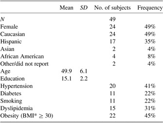
Right-handed (Oldfield, Reference Oldfield1971) participants between the ages of 40 and 60 were recruited through flyers and newspaper advertisements. Volunteers were excluded from participation if they had a history of neurological disease (i.e., large vessel stroke, seizure disorder, Parkinson's disease, clinically significant traumatic brain injury involving loss of consciousness for over 5 min, multiple sclerosis, or brain infection/meningitis), major psychiatric illness (e.g., schizophrenia, bipolar disorder), substance abuse (diagnosed abuse and/or previous hospitalization for substance abuse), or MRI contraindications. Fifty individuals enrolled in the study. The data of one participant were excluded from subsequent analyses due to low working memory task performance (41% accuracy), resulting in a final sample of 49. The mean (±SD) age of those participants was 49.9 ± 6.1 years (median age, 50 years). The mean education level was 15.1 ± 2.2 years. The mean full-scale IQ score was 114.4 ± 10.5, indicating high average global cognitive functioning according to published norms (Wechsler, Reference Wechsler1999). Enrollees identified themselves as follows: 49% Caucasian, 35% Hispanic, 8% African American, 4% Asian American, and 4% Other/Did Not Specify. CVD risk factors (hypertension, dyslipidemia, diabetes, and past history of smoking) were coded as present or absent based on participants’ answers to a medical history questionnaire and BMI was calculated based on measurements taken during one of the study visits (Table 1). These risk factors were then summed to create an overall CVD risk score (range, 0–5) to be included as a covariate in subsequent analyses.
Sample demographic and clinical characteristics

* Body mass index (BMI) = weight (kg)/ height2 (cm2).
Assessment of Subjective Cognitive Complaints
Participants completed a 39-item scale of perceived cognitive difficulties with attention, memory, perception, and psychomotor abilities (Cognitive Difficulties Scale, CDS; McNair & Kahn, Reference McNair and Kahn1983). The CDS has been used in both healthy and patient samples, including persons with CVD (Derouesné, Lacomblez, Thibault, & LePoncin, Reference Derouesné, Lacomblez, Thibault and LePoncin1999; Gunstad et al., Reference Gunstad, Cohen, Paul, Tate, Hoth and Poppas2006; Humphreys et al., Reference Humphreys, Moser, Hynes, Reese and Haynes2007; Khatri et al., Reference Khatri, Babyak, Clancy, Davis, Croughwel, Newman and Blumenthal1999; McNair & Kahn, Reference McNair and Kahn1983). Participants are asked to rate themselves using a 5-point Likert scale (from “never” to “most of the time”) on several statements describing everyday inefficiencies (e.g., “I find it hard to keep my mind on a task or a job.”), lapses of memory (e.g., “I have trouble recalling frequently used phone numbers”), and related functions that people often notice about themselves (e.g., “I have to do things very slowly to be sure I'm doing them right.”). Scores range between 0 and 156. Higher scores indicate a greater number of subjective complaints.
Objective Assessment of Cognitive Performance
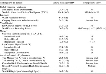
Participants completed a 2-hour assessment battery including standard clinical neuropsychological instruments with established reliability and validity (Lezak, Reference Lezak1995). The battery included measures of global cognitive functioning (Mini Mental Status Exam, MMSE (Folstein, Folstein, & McHugh, Reference Folstein, Folstein and McHugh1975); Wechsler Abbreviated Scale of Intelligence, WASI (Wechsler, Reference Wechsler1999), language (WASI Vocabulary Subtest; Category Fluency for Animals (Morris et al., Reference Morris, Heyman, Mohs, Hughes, van Belle, Fillenbaum and Clark1989), memory (California Verbal Learning Test-II, CVLT-II, Delis, Kramer, Kaplan & Ober, Reference Delis, Kramer, Kaplan and Ober2000; Rey Complex Figure Test recall, RCF, Lezak, Reference Lezak1995), attention-executive functioning (Controlled Oral Word Association Test, COWA (Eslinger, Damasio, & Benton, Reference Eslinger, Damasio and Benton1984); Trail Making Test A & B (Reitan, Reference Reitan1958); Wechsler Adult Intelligence Scale-III (WAIS-III) Digit Span Subtest), psychomotor speed (Grooved Pegboard (Klove, Reference Klove1963), and visual-spatial ability (RCF copy; WASI Matrix Reasoning Subtest). All tests were administered and scored by a trained research assistant using standard administration and scoring criteria.
Assessment of Depression
Current level of depressive symptoms was assessed using the Beck Depression Inventory-II (BDI-II) (Beck, Steer, & Brown, Reference Beck, Steer and Brown1996), a well-validated 21-item self-report questionnaire. Respondents are asked to endorse statements characterizing their mood. The maximum total score is 63, with scores greater than 14 suggesting clinically significant depressive symptoms.
Neuroimaging
A verbal working memory 2-Back task was chosen as the cognitive challenge in this study because the task is widely used and stable activation patterns are reported in a wide range of participants including patients with CVD (Haley, Sweet et al., Reference Haley, Sweet, Gunstad, Forman, Poppas, Paul and Cohen2007). Each imaging session included working memory task practice, T1-weighted imaging for anatomical reference, and two imaging runs of the working memory task. The 2-Back task was presented using E-Prime software (Psychology Software Tools, Inc., Pittsburgh, PA), back-projected onto a screen positioned at the participant's head, and viewed through a double-mirror attached to the head coil. Participants’ responses were collected using an MR-compatible response box.
MRI Data Acquisition
MRI data for each participant were acquired in a single session on a 3 Tesla GE Signa Excite MRI scanner equipped with a standard head coil. Structural imaging included a high-resolution Spoiled Gradient Echo (SPGR) sequence (256 × 256 matrix, field of view [FOV] = 24 × 24 cm2, 1 mm slice thickness, 0 gap) anatomical scan of the entire brain in the sagittal plane. Functional imaging was performed using a whole brain echo-planar imaging (EPI) sequence (repetition time [TR] = 3000 ms, echo time [TE] = 30 ms, FOV = 24 × 24 cm2, 64 × 64 matrix, 42 axial slices, 3 mm slice thickness, 0.3 mm gap).
Working memory paradigm
During this task, a series of individual consonants was presented visually for 500 ms each with a 2500-ms inter-stimulus interval. Consonants were arranged in random order from a list of all consonants except “L” due to ambiguity in the lowercase form. For each stimulus, participants were asked to determine if the letter on the screen was the same as, or different from, a previously presented letter. A 0-Back control condition was alternated with the 2-Back condition in a block design. Two imaging runs, each consisting of three blocks of the 0-Back, three blocks of the 2-Back, and three blocks of rest presented in alternating order and lasting approximately 6 minutes, were conducted. Rest preceded 0-Back, preceded 2-Back, always in the same order.
0-Back control condition
This task consisted of three blocks of 12 consonants of random case and order, 33% of which were targets. Participants responded “yes” when the upper or lowercase letter “H” appeared on the screen, or “no” if another letter appeared.
2-Back working memory condition
The experimental condition consisted of three blocks, each containing 15 consonants of random case and order, 33% of which were targets. A letter was considered a target if it was the same as the letter presented two stimuli earlier regardless of the case (e.g., s, G, S, d, V, D, v, etc.). Task performance was assessed by measuring accuracy rates and mean reaction time for all correct trials.
Rest
The participants were instructed to rest for 30 seconds while focusing their attention on a fixation cross that appeared in the middle of the screen.
fMRI data processing
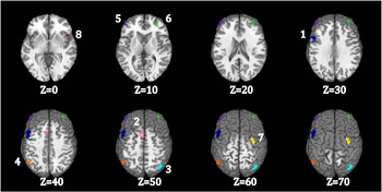
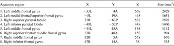
All EPI images were processed using Analysis of Functional NeuroImages (AFNI) software (Cox, Reference Cox1996). Each time series was spatially registered to the sixth volume of the session to reduce the effects of head movement. This AFNI 3-dimensional registration program also yields information on displacement and rotation for each volume, which was used later to further correct motion. All participants moved less than 1.5 mm per imaging run and no participants were excluded from the analyses due to excessive head motion. Data pre-processing also included adjustment for differences in adjacent slice timing due to interleaved slice acquisition, temporal smoothing and spatial blurring using a 4.5 mm kernel. Task-related brain activation was determined using within-subject voxel-wise multiple regression analyses including all task trials and the following parameters: separate regressors (reference waveforms) for rest, 0-Back and 2-Back, convolved with a gamma function, and covariates accounting for instruction screens and head movement. It is of note that motion correction for blocked designs is under some debate. Some have argued that the inclusion of motion covariates in a blocked N-Back design may serve to “steal” real variance away from the task regressors due to being highly correlated with the motor task response, thus lowering the power to detect real effects (Johnstone et al., Reference Johnstone, Ores Walsh, Greischar, Alexander, Fox, Davidson and Oakes2006). However, as this issue would serve to undermine our effect, rather than enhance it, and motion tends to increase with age, motion covariates were included in the analyses. Averaged task-related activation within a set of eight a priori regions of interest (ROIs) was examined in subsequent analyses. A separate data set was used to create empirically defined task-related ROIs for hypothesis testing to avoid circularity. The task used to create the ROIs was identical to the one used in the current study. The sample and the creation of the ROIs are described in detail in Haley, Sweet et al. (Reference Haley, Sweet, Gunstad, Forman, Poppas, Paul and Cohen2007). Briefly, the ROIs were created by automatically transforming the results from individual multiple regression analyses to standard stereotaxic space (Talairach & Tournoux, Reference Talairach and Tournoux1988) using linear interpolation. Individual t-statistics were thresholded at p < .05, corrected for multiple comparisons using the false discovery rate (FDR) supplied by AFNI. Voxels were included in the final mask if they were significantly active in over 90% of all participants. Finally, active voxels were defined as a cluster if they were contiguous and formed a volume of at least 200 μL. Eight cortical ROIs were identified using this process (Figure 1; Table 2). Anatomical designations were assigned to the ROIs according to the ROI center in Talairach coordinates. This empirically defined mask was then applied to the results from the individual multiple regression analyses in the current study sample transformed to standard stereotaxic space, using the fully automated 3dmaskave plugin in AFNI. This program allows one to compute the average over an ROI of all voxel values from an input dataset. Average unthresholded t-values within each ROI for each person were used as measures of task-related activation intensity in subsequent analyses for hypothesis testing. These eight a priori ROIs were used because they represented the most stable activation pattern in response to the task used in this study from a similar yet separate sample of participants.
Regions of Interest on Template Anatomy. 1 = Left Middle Frontal Gyrus; 2 = Left Medial Frontal/Superior Frontal Gyrus; 3 = Right Superior Parietal Lobule; 4 = Left Inferior Parietal Lobule; 5 = Left Middle Frontal Gyrus; 6 = Right Superior Frontal/ Middle Frontal Gyrus; 7 = Right Middle Frontal Gyrus; 8 = Right Inferior Frontal Gyrus.


Data Analyses
Neuropsychological measures were grouped into one of five cognitive domains: (1) global cognitive functioning, (2) language functions, (3) visual-spatial abilities, (4) memory functions, and (5) attention-executive-psychomotor functions. The following test scores were included in each domain, and raw total scores were used unless otherwise stated: (1) global: MMSE and WASI Full Scale IQ; (2) language: WASI Vocabulary Subtest and Category Fluency for Animals; (3) visual-spatial: RCF copy and WASI Matrix Reasoning Subtest; (4) memory: CVLT-II immediate recall, delayed recall, and recognition discrimination, RCF immediate recall, delayed recall, and recognition discrimination; (5) attention-executive-psychomotor functions: Trail Making A and B time to completion, COWAT, WAIS-III Digit Span Subtest, and Grooved Pegboard-Dominant Hand time to completion. Participants’ raw test scores were converted to Z-scores using the study sample mean and standard deviation. Timed test scores were multiplied by -1 so that higher scores indicate better performance. Five composite cognitive domain Z-scores were calculated for each participant by averaging the Z-scores of all tests within that domain.
The relationship between level of subjective cognitive complaints (CDS score) and 2-Back related brain activation within the a priori regions of interest was examined using hierarchical linear regression analyses. In the first step of the analysis, the relationship between CDS score and task-related brain activation was adjusted for the effects of age, education and reported depressive symptoms. The independent contribution of CDS score to variance in BOLD response to a working memory task was estimated in the second step. Covariates were chosen a priori based on their documented relationship with working memory-related brain activation and/or subjective cognitive complaints. Regression residuals were exported and examined for normality using Q-Q plots to ensure that regression assumptions were not being violated. All regression models used partial correlations. Relationships between subjective cognitive complaints, demographic variables, cardiovascular risk score, and cognitive performance were explored using nonparametric correlations. Data were analyzed using SPSS 16.0 computer software (SPSS Inc., Chicago, IL). A two-tailed alpha level of .05 was used as the criterion for statistical significance for all analyses. Multiple comparisons corrections were considered. However, in view of the preliminary nature of this study, the benefit of stringent multiple comparisons corrections in reducing Type I error was outweighed by the danger of missing an important effect that may be there, due to the inflation of Type II error associated with such corrections. This decision is supported by discussions of low statistical power and publication bias in behavioral studies due to stringent multiple comparisons corrections (Nakagawa, Reference Nakagawa2004).
Results
Descriptive Statistics
Demographic and clinical characteristics of the sample are reported in Table 1. Raw cognitive test scores and standard deviations are reported in Table 3. Descriptive statistical analyses revealed a cognitively normal, ethnically diverse, middle-aged sample, well representative of the population of the state of Texas based on the year 2000 US census data. On average, participants fulfilled criteria for two cardiovascular risk factors (e.g., obesity, hypertension, diabetes, dyslipidemia, or smoking) with a range between 0 and 4 (Table 1). Regression residuals were normally distributed and no transformations were performed.
Cognitive test scores

Subjective Cognitive Complaints in Relation to Demographic Variables and CVD Risk Score
Nonparametric correlation analyses revealed that increased report of cognitive difficulties was related to lower level of education (r = −0.31; p = .031), but not to age (r = 0.13; p = .379), sex (r = 0.06; p = .672), or CVD risk score (r = −0.003; p = .983).
Subjective Cognitive Complaints in Relation to Cognitive and Emotional Functioning
Consistent with previous cross-sectional studies of self-reported cognitive difficulties, cognitive complaints were not related to objective measures of cognitive function in any of the five domains: global (r = 0.01; p = .939), language (r = −0.21; p = .166), visuospatial (r = 0.02; p = .878) or attention-executive-psychomotor (r = −0.14; p = .358), although there was a trend toward weaker memory performance in the participants reporting higher level of cognitive difficulties (r = −0.30; p = .058). Self-reported symptoms of depression were not related to cognitive complaints (r = 0.11; 0.462) in this study, possibly due to the relatively low endorsement of depressive symptoms (mean BDI score = 6.06 ± 5.15).
Subjective Cognitive Complaints in Relation to BOLD Response to 2-Back and Cognition
The fully adjusted regression models successfully predicted mean 2-Back-related activation intensity in the right superior frontal/ middle frontal gyrus (F(4,44) = 3.26; p = .020) and the right inferior frontal gyrus (F(4,44) = 3.14; p = .024). Lower education was significantly associated with lower 2-Back-related intensity in the right superior frontal gyrus (ß = −0.34; p = .022). The independent effects of age and reported depressive symptoms did not account for any unique variance in 2-Back-related activation intensity.
Increased level of subjective cognitive complaints was significantly associated with attenuated 2-Back-related activation in the right superior frontal/ middle frontal gyrus (CDS ß = −0.39; p = .009) and in the right inferior frontal gyrus (CDS ß = −0.45; p = .003), independent of age, education, and self-reported depressive symptoms (Figure 2a & 2b). This relationship remained unchanged even after additional adjustment for CVD risk score (F(5,43) = 2.65; p = .035; CDS ß = −0.40, p = .008; F(5,43) = 2.48; p = .047; CDS ß = −0.46; p = .003). It is of note that the relationship between BOLD response and CVD risk, although in the expected direction, was relatively weak (r = −0.05; p = .731; r = −0.01; p = .951, respectively).
Partial regression plots of the relationship between reported cognitive dysfunction and working memory-related brain activation in the right superior frontal/ middle frontal gyrus (a) and the right inferior gyrus (b), adjusted for the effects of age, education and current level of depression.

2-Back Performance in Relation to BOLD Response, Demographic Variables, and Cognition
Mean 2-Back task accuracy (SD) was 81.46% (11.41). Mean reaction time (SD) for correct trials was 1110.19 ms (284.60). Neither task accuracy, nor reaction times were significantly related to self-reported cognitive difficulties (r = −0.20; p = .189; r = −0.06; p = .693). However, greater task accuracy was significantly related to better performance in the attention-executive-psychomotor domain (r = 0.493; p = .001). The 2-Back task performance was not related to any of the demographic variables (age, sex, education), reported depressive symptoms or CVD risk score, though there was a trend toward slower performance with older age (r = −0.25; p = .088). Higher 2-Back-related activation in the right superior frontal/ middle frontal gyrus was associated with trends toward better task accuracy (r = 0.28; p = .056) and faster reaction times (r = −0.13; p = .404).
Discussion
The present study found that increased report of cognitive difficulties by middle-aged adults at risk for CVD was associated with lower BOLD response to a working memory challenge despite intact global cognitive function. This effect was detected in functionally relevant brain regions where higher activation intensity relates to better working memory performance. The association was independent of age, education, and level of depression, and persisted after additional adjustments for level of CVD risk. Thus, an intriguing possibility is raised that subjective cognitive complaints by middle-aged individuals may indicate a unique and real aspect of early brain vulnerability that is difficult to detect with standard neuropsychological tests, or capture with traditional self-reported CVD risk indexes.
This finding is of special interest in view of discrepant reports in the aging literature regarding the value of subjective cognitive complaints in estimating current cognitive performance and predicting future cognitive trajectories. Cross-sectional and brief follow-up studies (<6 months) tend to dismiss self-reported cognitive difficulties as nothing more than a representation of current emotional state and personality (Gunstad et al., Reference Gunstad, Cohen, Paul, Tate, Hoth and Poppas2006; Humphreys et al., Reference Humphreys, Moser, Hynes, Reese and Haynes2007; Jungwirth et al., Reference Jungwirth, Fischer, Weissgram, Kirchmeyr, Bauer and Tragl2004; Khatri et al., Reference Khatri, Babyak, Clancy, Davis, Croughwel, Newman and Blumenthal1999; Lautenschlager, Flicker, Vasikaran, Leedman, & Almeida, Reference Lautenschlager, Flicker, Vasikaran, Leedman and Almeida2005; Minett, Da Silva, Ortiz, & Bertolucci, Reference Minet, Da Silva, Ortiz and Bertolucci2008). The majority of longitudinal studies with longer follow-up periods (1–8 years), on the other hand, paint a different picture. In those studies, a significant relationship is reported between memory complaints and objectively measured cognitive decline over time even after adjustment for symptoms of mood disturbance (Dufouil, Fuhrer, & Alpérovitch, Reference Dufouil, Fuhrer and Alpérovitch2005; Glodzik-Sobanska et al., Reference Glodzik-Sobanska, Reisberg, De Santi, Bab, Pirraglia, Rich and de Leon2007; Haley et al., Reference Haley, Hoth, Gunstad, Paul, Jefferson, Tate and Cohen2009; Jorm, Christensen, Korten, Jacomb, & Henderson, Reference Jorm, Christensen, Korten, Jacomb and Henderson2001; Kim et al., Reference Kim, Stewart, Kim, Yang, Shin and Yoon2006; McKhann et al., Reference McKhan, Selnes, Grega, Bailey, Pham, Baumgartner and Zeger2009; Selnes et al., Reference Selnes, Grega, Borowicz, Barry, Zeger and McKhan2004; Tobiansky, Blizard, Livingston, & Mann, Reference Tobiansky, Blizard, Livingston and Man1995). Self-reported cognitive difficulties therefore may reflect early changes in brain health and cognitive aging that are difficult to detect at a single time point using standard neuropsychological screening tools. The idea that cognitive complaints may reflect early neurodegenerative changes in the elderly is supported by findings of white matter lesions and significantly reduced gray matter volumes in non-depressed older adults reporting cognitive difficulties despite normal test performance (de Groot et al., Reference de Groot, de Leeuw, Oudkerk, Hofman, Jolles and Breteler2001; Dufouil et al., Reference Dufouil, Fuhrer and Alpérovitch2005; Minett, Dean, Firbank, English, & O'Brien, Reference Minet, Dean, Firbank, English and O'Brien2005; Stewart et al., Reference Stewart, Dufouil, Godin, Ritchie, Maillard, Delcroix and Tzourio2008).
In this study, we chose to examine the BOLD response to a cognitive challenge in relation to subjective cognitive complaints in middle-aged adults for several reasons. First, alterations in the BOLD response to cognitive challenges have been demonstrated in populations at risk for cognitive decline before significant impairment can be detected (Bookheimer et al., Reference Bookheimer, Strojwas, Cohen, Saunders, Pericak-Vance, Mazziotta and Smal2000; Chang et al., Reference Chang, Speck, Miller, Braun, Jovicich, Koch and Ernst2001; Sweet et al., Reference Sweet, Rao, Primeau, Mayer and Cohen2004). In addition, the BOLD response can be consistently measured in all subjects regardless of their age, whereas other measures of cerebrovascular health, such as white matter lesions, are relatively rare in individuals younger than 50 years of age (Sachdev, Chen, & Wen, Reference Sachdev, Chen and Wen2008). Finally, BOLD fMRI has been shown to be sensitive to subclinical changes in vascular health such as small decrements in peripheral endothelial function (Gonzales et al., Reference Gonzales, Tarumi, Tanaka, Sugawara, Swann-Sternberg, Goudarzi and Haley2010). Thus, the BOLD response to a cognitive challenge captured by fMRI may represent a unique early measure of brain health and vulnerability in younger populations. A working memory task was specifically chosen because it engages cognitive processes, such as executive function and attention, known to be especially susceptible to vascular cognitive impairment. We found that increased level of subjective cognitive complaints was associated with significantly lower task-related BOLD response in the right superior frontal/ middle frontal gyrus and right inferior frontal gyrus. One possible interpretation of the detected alterations in the BOLD response to working memory task in middle-aged adults with cognitive complaints is that self-reported cognitive difficulties reflect early vulnerability of the fronto-parietal executive system. The system is involved in regulating attention, switching attentional focus and task preparation (Ravizza, Delgado, Chein, Becker, Fiez, Reference Ravizza, Delgado, Chein, Becker and Fiez2004), and is affected early in the development of vascular cognitive impairment (Cohen et al., Reference Cohen, Poppas, Forman, Hoth, Haley, Gunstad and Gerhard-Herman2009; Forman et al., Reference Forman, Cohen, Hoth, Haley, Poppas, Moser and Gerhard-Herman2008; Haley, Forman, et al., Reference Haley, Forman, Poppas, Hoth, Gunstad, Jefferson and Cohen2007). This interpretation is supported by our present finding that greater task-related BOLD response in the right superior frontal gyrus was associated with trends toward better task performance and somewhat faster reaction times.
It is important to consider the direction of BOLD differences reported in this study. In the past, higher task-related BOLD response at comparable levels of performance in patients with multiple sclerosis, HIV, and genetic risk for Alzheimer's disease relative to healthy controls has been observed and interpreted as compensatory overactivation (Bookheimer et al., Reference Bookheimer, Strojwas, Cohen, Saunders, Pericak-Vance, Mazziotta and Smal2000; Chang et al., Reference Chang, Speck, Miller, Braun, Jovicich, Koch and Ernst2001; Sweet et al., Reference Sweet, Rao, Primeau, Durgerian and Cohen2006); however, the opposite trend (i.e., lower task-related activation at similar levels of performance) has been noted in patients at risk for cardiovascular and cerebrovascular disease (Gonzales et al., Reference Gonzales, Tarumi, Tanaka, Sugawara, Swann-Sternberg, Goudarzi and Haley2010; Haley et al., Reference Haley, Gunstad, Cohen, Jerskey, Mulligan and Sweet2008; Haley, Sweet et al., Reference Haley, Sweet, Gunstad, Forman, Poppas, Paul and Cohen2007; Irani et al., Reference Irani, Sweet, Haley, Gunstad, Jerskey, Mulligan and Cohen2009; Jennings, Muldoon, Price, Christie, & Meltzer, Reference Jennings, Muldoon, Price, Christie and Meltzer2008; Jennings et al., Reference Jennings, Muldoon, Ryan, Price, Greer, Sutton-Tyrrel and Meltzer2005). Based on these observations, we hypothesize that early in the vascular disease process, sub-clinical reductions in vascular responsivity and endothelial function may result in lower, yet sufficient, amounts of oxygen being delivered to activated neurons, thus producing a lower BOLD response to a cognitive challenge despite intact behavioral performance (Gonzales et al., Reference Gonzales, Tarumi, Tanaka, Sugawara, Swann-Sternberg, Goudarzi and Haley2010; Haley et al., Reference Haley, Gunstad, Cohen, Jerskey, Mulligan and Sweet2008). As vascular problems exacerbate, further declines in cerebrovascular reactivity and microvascular damage likely lead to vascular cognitive impairment. Support is provided by a growing body of literature documenting that peripheral cardiovascular dysfunction is related to poor cerebrovascular health and diminished cognitive function in older patients with cardiovascular disease (Cohen et al., Reference Cohen, Poppas, Forman, Hoth, Haley, Gunstad and Gerhard-Herman2009; Forman et al., Reference Forman, Cohen, Hoth, Haley, Poppas, Moser and Gerhard-Herman2008; Gunstad et al., Reference Gunstad, Cohen, Tate, Paul, Poppas, Hoth and Jefferson2005; Haley, Forman et al., Reference Haley, Forman, Poppas, Hoth, Gunstad, Jefferson and Cohen2007; Haley, Sweet et al., Reference Haley, Sweet, Gunstad, Forman, Poppas, Paul and Cohen2007; Hoth et al., Reference Hoth, Tate, Poppas, Forman, Gunstad, Moser and Cohen2007; Jefferson, Poppas, Paul, & Cohen, Reference Jefferson, Poppas, Paul and Cohen2007; Jefferson, Tate et al., Reference Jefferson, Tate, Poppas, Brickman, Paul, Gunstad and Cohen2007; Paul et al., Reference Paul, Gunstad, Poppas, Tate, Foreman, Brickman and Cohen2005).
Unfortunately, the cross-sectional nature of our study limits our ability to establish the time course of cognitive difficulties or infer future cognitive trajectories from the observed functional activation changes. It is possible that our results may reflect variability in brain function of normal or long-standing nature. Longitudinal studies that begin in mid-life could help validate the use of BOLD fMRI markers as indicators of long-term cognitive outcomes. The study was also limited to a relatively small sample of self-selected, non-depressed, and well-educated community volunteers. Ideally, the external validity of the results should be tested in larger, randomly selected community samples that represent the full range of education in the US population. Including participants with a wider range of BDI scores will allow for more in-depth analyses of the relationships between depression, cognitive variables and BOLD response to cognitive tasks. Our a priori decision not to use multiple comparisons corrections in this study could also be considered a limitation. It is of note, however, that the reported relationship between cognitive complaints and BOLD response to 2-back in the superior frontal/middle frontal gyrus would have been significant even if an adjusted Bonferroni alpha level of 0.02 was set as the criterion for statistical significance. Finally, additional assessments of white matter integrity may be useful in future studies to further elucidate the relationships detected in this study. While overt signs of microvascular damage in early adulthood are rare, small white matter lesions have been reported in randomly selected individuals in their 40s and 50s (Sachdev et al., Reference Sachdev, Chen and Wen2008). Therefore, assessments of white matter integrity may lend further insights into the cognitive complaints expressed by middle-aged individuals. Additional measures of BOLD response to non-cognitive tasks and hypercapnia, on the other hand, can help determine if the alterations in the cerebrovascular support for cognition reported in this study are specific to brain areas critical for cognitive function and/or related to global changes in cerebral perfusion, cerebrovascular reactivity or cerebrovascular coupling.
Summary and Conclusions
In summary, we present the first study to examine subjective cognitive complaints in relation to BOLD response to a cognitive challenge in middle-aged individuals at risk for CVD. Our results indicate that cognitively intact middle-aged individuals who self-report cognitive difficulties also demonstrate significantly lower BOLD response to a working memory task. These results were independent of age, education and current level of reported symptoms of depression. Thus, fMRI may be a helpful tool to identify early brain vulnerability associated with cognitive complaints. Future studies including BOLD response to cognitive and non-cognitive challenges as well as measures of cerebral perfusion and white matter integrity will be important. These approaches will help elucidate the pathophysiological mechanisms underlying the observed alterations in BOLD response to working memory among middle-aged individuals with self-reported cognitive difficulties.
Acknowledgments
This work was presented in part at the 36th Annual Meeting of the International Neuropsychological Society, Acapulco, Mexico. This work was supported by the American Heart Association (A.P.H., 09BGIA2060722); the American Federation for Aging Research (A.P.H., 8A0024); and the University of Texas at Austin. The authors have no conflicts of interest to report.