INTRODUCTION
The family Cirratulidae can be artificially divided into two groups, based on the number of tentacular filaments on the anterior segments; bitentaculate (with two tentacular filaments) and multitentaculate (with many tentacular filaments) (Blake, Reference Blake, Blake, Hilbig and Scott1996). The known multitentaculate genera are Cirratulus Lamarck, 1801, Cirriformia Hartman, 1936, Timarete Kinberg, 1866 and Protocirrineris Czerniavsky, 1881. They are characterized by having both capillary chaetae and spines on their parapodia, except for Protocirrineris which only has capillaries. Spines in these genera are generally short, stout and somewhat re-curved.
In the Mediterranean Sea, off Morocco, Fauvel (Reference Fauvel1928) described a new species, Cirratulus dollfusi, which has multiple branchiae and tentacular filaments together with long capillaries on parapodia. However, he did not mention the long finely tapering spines in noto- and neuropodia we observed on specimens of this species collected from the Sea of Marmara in 1992.
The present study focused on the re-description of C. dollfusi, based on the material collected in the Sea of Marmara and the type material, and the discussion of its taxonomic position in Cirratulidae
MATERIALS AND METHODS
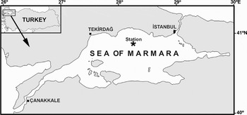
The specimens of Cirratulus dollfusi were collected on muddy substrate at 449 m in the Sea of Marmara by a bottom trawl (T-129) on 30 May 1992 during a survey aimed at studying demersal fish stocks along the Turkish coasts by RV ‘Koca Piri Reis’ (Figure 1). The specimens were fixed with 4% formaldehyde and transferred to the laboratory. The specimens were first rinsed with tap water and subsequently preserved in 70% ethanol. To make more visible morphological features such as scars of broken branchiae, the specimens were stained with ShirlastainA (brown stain). The biometric features of the specimens such as the total body length, the width at chaetiger 40 (the widest part) and length of chaetae were measured with an ocular micrometer.
Map of the investigated area with the location of sampling sites.

The holotype of Cirratulus dollfusi, deposited at the Muséum National d'Histoire Naturelle, France (MNHN) was examined and compared to the specimens collected from the Sea of Marmara.
Photographs of specimens were taken by a digital camera (Olympus, Camedia, C-7070) attached to stereo- and compound microscopes. Drawings of the specimens were made with the aid of a camera lucida.
Specimens of Cirratulus dollfusi were deposited at the Museum of the Faculty of Fisheries, Ege University (ESFM).
DIAGNOSIS
Body large, dorso-ventrally flattened. Prostomium broadly rounded anteriorly, without eyes. Tentacular filaments emerging on dorso-lateral area of chaetiger 1, arranged in 2 dense groups of circular pattern, with 25 filaments each. Branchial filaments are multiple in anterior end; they arise as 1–2 filaments from each of 4–8 raised notopodial lobes on parapodia of anterior and middle segments, with lobes reducing in size and fewer (holotype) or no branchiae posteriorly (our material). Two types of chaetae: long, fine capillaries, and straight, tapering spines.
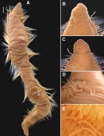
Fauvelicirratulus dollfusi. (A) Dorsal view of whole body (ESFM-POL/92-34); (B) dorsal view of anterior segments; (C) ventral view of anterior segments; (D) tentacular filaments; (E) branchiae on chaetigers 25 and 26. Scale bar A = 4.3 mm, B and C = 1.1 mm, D = 0.9 mm, E = 0.6 mm.

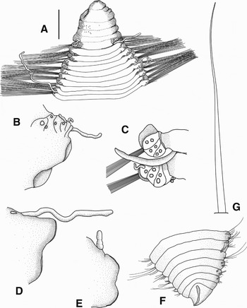
Fauvelicirratulus dollfusi. (A) Dorsal view of anterior segments (ESFM-POL/92-34); (B) lateral view of parapodium on chaetiger 8 (chaetae omitted); (C) dorsal view of parapodia on chaetigers 3 and 4; (D) posterior view of parapodium on chaetiger 30 (chaetae omitted); (E) posterior view of parapodium on chaetiger 119 (chaetae omitted); (F) dorsal view of posterior end and pygidium; (G) spine on neuropodium of chaetiger 100. Scale bar A = 1.8 cm, B, D and E = 0.8 mm, C = 0.6 mm, F = 0.74 mm, G = 270µm.

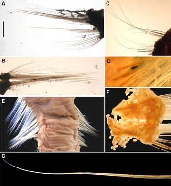
Fauvelicirratulus dollfusi. (A) Posterior view of parapodium on chaetiger 20 (ESFM-POL/92-34); (B) posterior view of parapodium on chaetiger 100; (C) posterior view of parapodium on chaetiger 162; (D) spines on neuropodium of chaetiger 100; (E) chaetigers in middle region of the body; (F) eggs within parapodium of chaetiger 70 (mean diameter 65.8 µm); (G) a spine on neuropodium of chaetiger 100. Scale bar A = 1 mm, B = 1.65 mm, C = 0.4 mm, D = 0.52 mm, E = 1.65 cm, F = 0.58 mm, G = 150 µm.

Cirratulus dollfusi Fauvel, Reference Fauvel1928: 9–10, figure 1h–k.
Ambo dollfusi; Fauvel, Reference Fauvel1936: 70, figure 8.
Timarete dollfusi; Rullier, Reference Rullier1965: 46; Intes & Le Lœuff, Reference Intes and Le Lœuff1975: 223.
MATERIAL EXAMINED
ESFM-POL/92-34, 5 specimens, 30 May 1992, Sea of Marmara, 40°53′N–28°15′E, 449 m, mud.
TYPE MATERIAL
Holotype of Cirratulus dollfusi, MNHN POLYTYPE 656, Morocco, Rabat.
DESCRIPTION
Largest specimen complete, 110 mm long, 13 mm wide for 182 chaetigers. Body dorsoventrally flattened, thickened throughout short, crowded segments, anteriorly narrow, widest around chaetiger 40, tapering posteriorly (Figure 2A). Body somewhat subrectangular in cross-section; ventral surface flat with trace of ventral nerve cord; dorsal surface rounded, particularly in middle region of body. Pygidium triangular; anal opening on dorsal surface (Figure 3F). Body colour rusty brown, with irregular black spots on dorsal and ventral surfaces of peristomium (Figure 2B). Black spots constituting a transverse band on posterior annulation of peristomium on ventral surface (Figure 2C). Chaetae yellowish.
Prostomium broadly rounded anteriorly, without eyes (Figures 2B, C, & 3A). Peristomium enlarged, massive, 6 times longer than prostomium, with at least 4 annulations (Figure 3A). First chaetiger and thoracic region wider than head. Tentacular filaments emerging on dorso-lateral area of chaetiger 1, arranged in 2 dense groups of circular pattern, with 25 filaments each (Figures 2D & 3A). Most of tentacular filaments missing, those present very small, thin, shorter than body width and branchiae. Branchiae present from chaetiger 1, to chaetiger 138. Each anterior chaetiger with many branchiae; anterior-most parapodium with 16 branchiae; 10 emerging on dorsal side of a rounded lobes just dorsal to notopodium, 6 located on posterior side of lobes (Figure 3B, C). Number of branchiae diminishing towards posterior end, with two present from approximately chaetiger 24 (Figures 2E & 3D) and one around chaetigers 40 to 138 (Figure 3E). Branchiae located on posterior surface of lobes generally longer and larger than others. Anterior branchiae longer than posterior ones. Lobes on notopodia reduced when number of branchiae decrease and are absent when when branchiae number 2 per parapodium.
Notopodia and neuropodia widely separated, somewhat highly developed in anterior region, poorly developed in posterior region (Figure 3B, D, E). Parapodia with long, thin capillaries and straight, tapering spines (Figure 4A–C). No short, stout, re-curved spines on parapodia. Capillary chaetae, yellowish, numerous on parapodia, maximally 2 mm long on chaetiger 1, 7.2 mm on chaetiger 25, 7.8 mm on chaetiger 50, 8 mm on chaetiger 110, 3 mm on chaetiger 150, 1.8 mm on chaetiger 170 (Figures 2A & 4E). After chaetiger 12 onwards, straight, tapering spines commencing both on noto- and neuropodia (Figures 3G & 4G). They are placed superiorly on notopodia and inferiorly on neuropodia. This type of capillary chaetae become larger on middle chaetigers, brownish; numbering 2 both on noto- and neuropodia in anterior region; numbering 2 on notopodia and 3–4 on neuropodia in middle region; numbering 2 both on noto- and neuropodia in posterior region; neuropodial chaetae larger and shorter than notopodial ones.
REPRODUCTION
The largest specimen of Fauvelicirratulus dollfusi has small eggs in its coelomic cavity (Figure 4F). Eggs spherical, with a diameter ranging from 58 µm to 80 µm (mean: 65.8 µm ±0.91 SE, N = 30). No sign of sexual maturity was observed.
REMARKS
Fauvelicirratulus dollfusi was originally described from Morocco and then subsequently reported from the Liberian coast and Côte d'Ivoire (Fauvel, Reference Fauvel1928; Intes & Le Lœuff, Reference Intes and Le Lœuff1975). The present study extends its distributional range to the Sea of Marmara.
The holotype is complete, 110 mm long, 15 mm wide (widest part) and has 170 chaetigers. The holotype and the specimens from the Sea of Marmara are closely similar with each other. Although Fauvel (Reference Fauvel1928) did not mention the straight, tapering spines in the original description, the holotype has this kind of chaeta emerging from notopodium and neuropodium of chaetiger 12. The only difference between the holotype and the Sea of Marmara specimens is that the holotype has more branchiae on anterior parapodia (20–24 per parapodium) than those of the Sea of Marmara specimens (maximally 18).
This genus is unique among cirratulids in having the combination of characters; tentacular filaments emerging only on chaetiger 1, and parapodia with straight, tapering spines. The type species of this genus has also multiple branchiae on anterior segments (maximum of 18 on each segment). However, the feature of having multiple branchiae on parapodium was also reported in the genus Timarete. The Californian species, Timarete perbranchiae (Chamberlin, 1918) has also 2–5 branchiae per segment (see Blake, Reference Blake, Blake, Hilbig and Scott1996). However, the genus Timarete mainly differs from the new genus Fauvelicirratulus in having tentacular cirri on more than one segment (only on chaetiger 1 in Fauvelicirratulus); stout and curved spines (long and tapering spines in Fauvelicirratulus); and branchiae moving dorsally in middle parapodia (always close to notopodia in Fauvelicirratulus).
Cirratulus hedgpethi described from Texas littoral zones by Hartman (Reference Hartman1951) shares some characters with F. dollfusi; body large, dorso-ventrally flattened; tentacular cirri small, spaghetti-like, placed on dorso-lateral parts of chaetiger 1; no stout curved spines on parapodia; chaetae two types both finely pointed at the tip. However, Hartman (Reference Hartman1951) did not mention the number and placement of branchiae on parapodia. To determine if C. hedgpethi belongs to Fauvelicirratulus, the type specimens of the species need to be re-examined.
Branchiae of F. dollfusi emerge on several lobes of anterior and middle parapodia. This character has not been reported for any multibranchiate cirratulids so far and seems to be unique for the genus. However, as these lobes can be easily overlooked, the other multibranchiate species need to be re-examined for the presence of these lobes.
KEY FOR THE MULTITENTACULATE CIRRATULID GENERA
1. Tentacular filaments arising only from chaetiger 1… … 2
– Tentacular filaments arising from 2 or more segments. . 3
2. Parapodia with stout, curved spines Cirratulus – Parapodia with straight, tapering spines
… .Fauvelicirratulus
3. Parapodia with capillary chaetae only… … … … … … Protocirrineris – Parapodia with capillary chaetae and spines 4
4. Branchiae placed next to notopodia throughout… … . Cirriformia – Branchiae placed far from notopodia on middle chaetigers Timarete