Introduction
During malignant progression of cancer cells, telomere length must be maintained for cell immortalisation. Telomerase, a ribonucleoprotein complex, plays a crucial role in cellular immortalisation and tumourigenesis via its effect on telomere elongation.Reference Marciniak and Guarente1, Reference Mennuni, Ugel, Mori, Cipriani, Iezzi and Pannellini2 The human telomerase complex consists of human telomerase reverse transcriptase, the catalytic subunit, and human telomerase-associated RNA, the template for telomeric repeat synthesis. Inhibition of telomerase activity limits cancer cell growth and leads to apoptosis. Therefore, telomerase-based therapy constitutes a novel approach to the treatment of many cancers.Reference Feldser and Greider3 Although there are many different ways to disrupt telomerase activity and function, human telomerase reverse transcriptase is essential for telomerase activity, and is thus a promising target for anti-cancer treatments.Reference Gillis, Schuller and Skordalakes4, Reference Zinn, Pruitt, Eguchi, Baylin and Herman5
However, some studies have demonstrated that telomere maintenance can occur in the absence of telomerase activity, by a mechanism termed ‘alternative lengthening of telomeres’.Reference Marciniak, Cavazos, Montellano, Chen, Guarente and Johnson6, Reference Nittis, Guittat and Stewart7 When assessing telomerase inhibitors as possible anti-cancer agents, we must determine whether telomerase inhibition selects for cancer cells that activate the alternative lengthening of telomeres mechanism.
Previously, we found that some laryngeal cancer cells maintained reproductive activity following RNA interference (RNAi) human telomerase reverse transcriptase based anti-telomerase therapy.Reference Chen, Tao, Hua, Xiao, Xu and Wang8, Reference Wang, Duan, Chen, Xiao, Cheng and Tao9 However, it is unclear if these cells survive because telomerase inhibition triggers the alternative lengthening of telomeres pathway.
This study aimed to reveal whether telomere maintenance mechanisms in laryngeal squamous carcinoma (Hep-2 line) cells change when telomerase is inhibited. Cells surviving the RNAi human telomerase reverse transcriptase treatment were carefully separated and collected. In these cells, we measured the activity of telomerase, quantified both human telomerase reverse transcriptase and human telomerase associated RNA, and assessed the formation of alternative lengthening of telomere specific promyelocytic leukaemia protein, to determine the telomere elongation mechanism.
Materials and methods
Cell lines and culture conditions
The human laryngeal cancer cell line Hep-2 was obtained from the American Type Culture Collection (Manassas, VA, USA) and grown in RPMI (RPMI-1640 was developed by Moore et. al. at Roswell Park Memorial Institute, hence the acronym RPMI) 1640 supplemented with 10 per cent Fetal Calf Serum (FCS) (Hyclone, Logan, Utah, USA) and 100 U/ml penicillin and streptomycin at 37°C in 5 per cent carbon dioxide.
RNAi plasmid construction and transfection
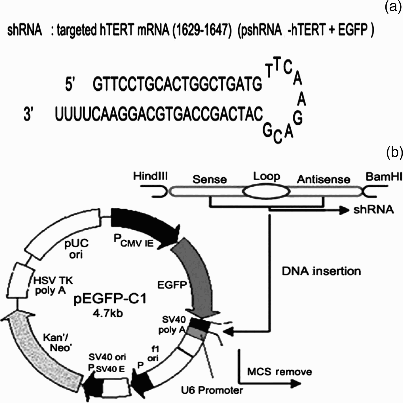
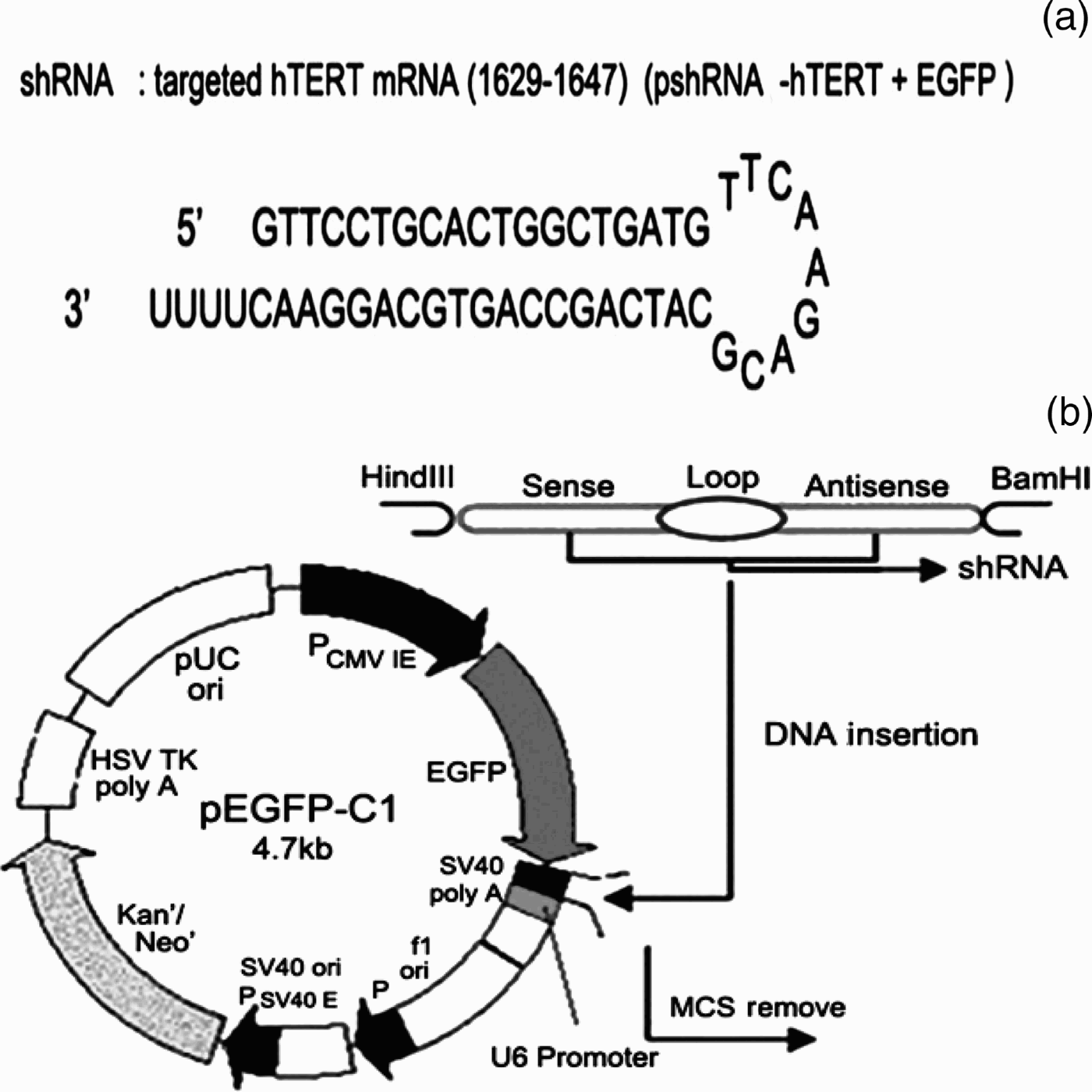
Short hairpin RNA was designed based on the complementary DNA sequence of human telomerase reverse transcriptase (GenBank AB085628). The short hairpin RNA encoded a 19-nucleotide target sequence as sense strand, followed by a spacer, the complementary antisense strand, and four consecutive thymines as a terminal signal (Figure 1a). The short hairpin RNA was subcloned into the pEGFP-C1 (the name of the Vector) vector with the human U6 promoter between the BamHI and HindIII restriction sites (Figure 1b). Plasmids were constructed by Wuhan Genesil Biotechnology Company (Wuhan, China). The Hep-2 cells were transfected with plasmids using Lipofectamine 2000 (Invitrogen, Carlsbad, California, USA), according to the manufacturer's protocol. The cells were then harvested 48 hours after transfection.
The Promoters (P) human telomerase reverse transcriptase (phTERT) Enhanced Green Fluorecence Protein (EGFP) reporter constructs. (a) Design of short hairpin RNA (shRNA) template. (b) Schematic diagram of the pEGFP vector. The shRNA encoding template was inserted between the BamHI and HindIII restriction sites downstream of the U6 promoter. The complete hTERT sequence of genome was inserted upstream of the EGFP gene. mRNA = messenger RNA; P = Promoters; CMV = Cytomegalovirus; IE = Cytomegalovirus Infection; SV40 = Simian vacuolating virus 40; poly A = Polynosinlc Acid; ori = Origin of replication; E = Enhancer; Kan = Kanamycin; Neo = Neomycin; HSV TK = Herpes Simplex Virus Thymidine Kinase; pUC = the name of the Plasmid; MCS = Multiple Cloning Site
Flow cytometry and cell sorting
The harvested cells were trypsinised, resulting in a single cell suspension that was analysed on a FACSCalibur flow cytometer (BD Biosciences, Franklin Lakes, New Jersey, USA). The Hep-2 cells that had been transfected with the plasmids expressed Green Fluorescent Protein (GFP). GFP positive cells were cultivated to determine their telomere elongation mechanism.
Measurement of telomerase activity
To determine telomerase activity, we used the telomeric repeat amplification protocol in the Trapeze Telomerase Detection Kit (Millipore, Billerica, MA, USA), according to the manufacturer's protocol. These experiments were performed in triplicate.
The cell extract was heated to 80°C for 10 minutes as a negative control, and cell extract from telomerase-active 293 cells was used as a positive control. Briefly, the telomeric repeated amplification protocol reaction involved the following: (1) primer elongation; (2) telomerase inactivation at 94°C for 5 minutes; and (3) amplification in 30 denaturation cycles at 94°C for 30 seconds, annealing at 50°C for 30 seconds, and polymerising at 72°C for 90 seconds, followed by a final 10 minutes at 72°C for balance. From each reverse transcription polymerase chain reaction, 5 µl was separated on a 10 per cent non-denaturing polyacrylamide gel, stained with Vistra Green (Amersham Biosciences, Aylesbury, UK) and visualised in a FluorImager 595 machine (Molecular Dynamics, Little Chalfont, UK). Semi-quantitative densitometric evaluation was performed using the ImageQuant 5.0 system (Molecular Dynamics).
Reverse transcription polymerase chain reaction
Total RNA was extracted from carcinoma cells using the TRIzol reagent (Invitrogen, La Jolla, California, USA). Complementary DNA (cDNA) was synthesised using the Thermoscript reverse transcription polymerase chain reaction kit (Invitrogen). For human telomerase reverse transcriptase, we used the forward primer 5′-GCC TGA GCT GTA CTT TGT CAA-3′ and the reverse primer 5′-CGC AAA CAG CTT GTT CTC CAT GTC-3′. For human telomerase associated RNA, we used the forward primer 5′-TCT AAC CCT AAC TGA GAA GGG CGT AG-3′ and the reverse primer 5′-CCA GCA GCT GAC ATT TTT TG-3′. Polymerase chain reaction products were separated on a 5 per cent non-denaturing polyacrylamide gel in a Protean-III electrophoresis chamber (Bio-Rad, Hercules, California, USA), stained with Vistra Green (Amersham Biosciences), visualised on a FluorImager 595 machine, and adapted for print with ImageQuant 5.0.
Western blotting
Proteins extracted from cells were analysed by sodium dodecyl sulphate polyacrylamide gel electrophoresis and transferred to nitrocellulous membrane. The human telomerase reverse transcriptase protein was detected with an anti-human telomerase reverse transcriptase polyclonal antibody (Santa Cruz Biotechnology, Santa Cruz, California, USA) at a 1:400 dilution. Polyclonal anti-actin (Santa Cruz Biotechnology) was used as an internal loading control.
Immunofluorescence
To determine if the residual cells had changed their telomere maintenance mechanisms to the alternative lengthening of telomeres pathway, we investigated whether the residual cells had raised levels of alternative lengthening of telomere specific promyelocytic leukaemia protein (a protein marker which characterises cells in which the alternative lengthening of telomeres mechanism is active), compared with the original cell line.Reference Venturini, Erdas, Costa, Gronchi, Pilotti and Zaffaroni10 To do this, we immunofluorescence-stained the cells using the N-19 anti-promyelocytic leukaemia body substrate (Santa Cruz Biotechnology) at a 1:200 dilution. The secondary antibody, Cy3-conjugated anti-rabbit (Sigma, St Louis, Missouri, USA), was used at a 1:300 dilution.
Results and analysis
Flow cytometry sorting for successfully transfected cells
To enable identification of successfully transfected cells, each RNAi plasmid also encoded GFP as a marker. By sorting for GFP positive cells by flow cytometry, we could accurately measure the gene silencing capabilities of RNAi in residual cells. Gene expression analysis was used as additional confirmation that the correct cells were measured for telomerase activity.
Telomerase activity
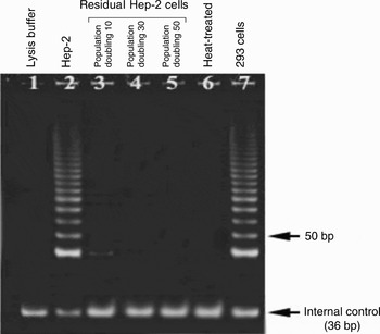
Telomerase activity in Hep-2 cells was significantly related to potential telomere maintenance mechanisms. To analyse telomerase status in the residual cells, we performed telomerase activity assays at cell doublings 10, 30, and 50 in culture. The inhibition of telomerase activity by a dominant-negative human telomerase reverse transcriptase was more robust in residual cells compared with the original cell line (Figure 2). Even after 50 cell passages, the residual cells continued to show telomerase inhibition, suggesting that no detectable increase in telomerase activity occurred during telomere shortening.
Telomerase activity in the original Hep-2 cell line and residual Hep-2 cells. Telomerase activity is indicated by a 6-bp ladder on a polyacrylamide gel. Lysis buffer containing no cell extract (lane one) and heat-treated samples (lane six) served as negative controls. Telomerase-positive 293 cells (lane seven) served as a positive control.

Expression of human telomerase reverse transcriptase and human telomerase associated RNA
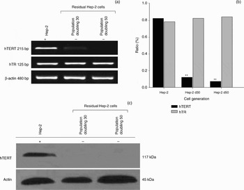
To determine the underlying mechanism regulating telomerase activity, we performed reverse transcription polymerase chain reaction and Western blot analyses for the telomerase catalytic subunit (i.e. human telomerase reverse transcriptase) and the telomerase RNA subunit (i.e. human telomerase associated RNA). Following RNAi treatment of Hep-2 cells, levels of human telomerase reverse transcriptase messenger RNA were significantly reduced (p < 0.01, chi-square test; Figure 3a and 3b). Similarly, in the residual Hep-2 cells, short hairpin RNA resulted in reduced human telomerase reverse transcriptase protein expression (Figure 3c). Telomerase activity decreased substantially in these cells, consistent with down-regulation of human telomerase reverse transcriptase expression. However, even after telomerase was inactivated, the cells continued to proliferate. After population doubling 50, the cells still exhibited no telomerase activity and only low levels of human telomerase reverse transcriptase expression, suggesting that they were using another mechanism to maintain telomere length. The original Hep-2 cell line and the residual Hep-2 cells showed no statistically significant difference in human telomerase associated RNA expression (p > 0.05, chi-square test; Figure 3b).
Expression of human telomerase reverse transcriptase (hTERT) and human telomerase associated RNA (hTR) in cell lines. No hTERT messenger RNA (mRNA) or protein expression was detected in residual Hep-2 cells, but hTR was expressed. (a) Reverse transcription polymerase chain reaction of hTERT and hTR mRNA. β-actin mRNA was amplified as the internal standard. (b) Ratio of hTERT and hTR to β-actin RNA levels in Hep-2 cells and residual Hep-2 cells. hTERT mRNA was significantly reduced in residual cells (p < 0.01; chi-square test), but there was no significant difference in hTR mRNA levels (p > 0.05). (c) Western blot for hTERT protein. Hep-2 d30 = residual Hep-2 population doubling 30; Hep-2 d50 = residual Hep-2 population doubling 50; += cells with telomerase expression; – = cells without telomerase expression

Detection of alternative lengthening of telomeres specific promyelocytic leukaemia protein
Promyelocytic leukaemia protein are one of the constituents of alternative lengthening of telomeres specific promyelocytic leukaemia protein; thus, detection of the latter was achieved by immunofluorescence staining for the former. Greatly raised levels of alternative lengthening of telomeres specific promyelocytic leukaemia protein were found in the residual Hep-2 cells (Figure 4), suggesting activation of the alternative lengthening of telomeres mechanism of telomere stabilisation. Combined with our telomerase activity analyses, these results suggest that the residual Hep-2 cells lacked telomerase but remained capable of proliferation via the alternative lengthening of telomeres mechanism for telomere stabilisation.
Immunofluorescence staining of promyelocytic leukaemia protein marker protein (arrows), indicating raised levels of alternative lengthening of telomeres specific promyelocytic leukaemia protein in the residual Hep-2 cells (b) compared to the original Hep-2 cell line (a).

Discussion
In this study, we showed that telomere elongation occurred even when telomerase activity was inhibited. In most cells, telomerase inhibition triggered apoptosis and induced cellular senescence; however, some cells continued to proliferate without detectable telomerase activity. Additionally, these cells had increased formation of alternative lengthening of telomeres specific promyelocytic leukaemia protein, a marker of the alternative lengthening of telomeres mechanism. These cells probably achieved telomere maintenance via the alternative lengthening of telomeres mechanism.
In most human malignancies, telomeres are maintained by the reactivation of telomerase. However, in a small but substantial fraction of human tumours, telomeres are maintained by a telomerase-independent telomere length maintenance mechanism referred to as alternative lengthening of telomeres.Reference Ohali, Avigad, Naumov, Goshen, Ash and Yaniv11, Reference Bollmann12 Although the nature of interaction between the telomerase mechanism and the alternative lengthening of telomeres mechanism is still not understood, it was widely believed that the alternative lengthening of telomeres pathway exists to enable survival following telomerase inhibition.Reference Bechter, Zou, Walker, Wright and Shay13 For example, when cells with an activated alternative lengthening of telomeres mechanism were fused with normal somatic cells or telomerase-positive cancer cells, alternative lengthening of telomeres was suppressed, indicating the presence of repressors that normally block this pathway.Reference Kumakura, Tsutsui, Yagisawa, Barrett and Tsutsui14 In another study, telomerase-positive immortal human fibroblasts showed sustained proliferation in culture after spontaneously inactivating telomerase.Reference Bechter, Zou, Walker, Wright and Shay13, Reference Kumakura, Tsutsui, Yagisawa, Barrett and Tsutsui14 However, these cells did not express alternative lengthening of telomeres specific promyelocytic leukaemia protein or a telomere length pattern typical of alternative lengthening of telomeres, and they reactivated telomerase upon treatment with the demethylating agent 5-aza-20-deoxycytidine. In contrast, other studies demonstrated that both the alternative lengthening of telomeres mechanism and the telomerase mechanism can coexist over a prolonged period in human cancer cells.Reference Grobelny, Kulp-McEliece and Broccoli15, Reference Perrem, Colgin, Neumann, Yeager and Reddel16
Our data indicate that an alternative lengthening of telomeres like telomere elongation mechanism is possible when telomerase is inhibited. However, a major obstacle to this theory is the fact the telomerase reactivated when the cells reached crisis. Previous studies have suggested that telomerase reactivation leads to human telomerase associated RNA expression.Reference Vulliamy, Marrone, Goldman, Dearlove, Bessler and Mason17 Unexpectedly, human telomerase associated RNA was still expressed in the residual cells, but these cells had minimally detectable telomerase levels. Thus, it is unlikely that telomerase activity in itself allowed the residual cells to survive. Human telomerase associated RNA expression in the absence of telomerase activity could have two explanations. Firstly, the telomere maintenance mechanism in human tumour cells could change from the telomerase mechanism to the alternative lengthening of telomeres mechanism following anti-telomerase therapy. Alternatively, these residual cells could have constituted the small fraction of cells already using the alternative lengthening of telomeres mechanism, which allowed them to survive the telomerase inhibition step. Distinguishing which of these explanations accounts for our results (or both) will require more research. Regardless of this, our results have direct implications for tumour treatment involving telomerase inhibitors.
• During malignant progression of cancer cells, telomere length must be maintained for cell immortalisation
• Telomerase, a ribonucleoprotein complex, plays a crucial role in cellular immortalisation and tumourigenesis
• This study investigated telomerase activity in laryngeal squamous carcinoma cells, and found that telomere elongation occurred even when telomerase activity was inhibited
• These results may have a major influence on the design of new cancer treatments
Although telomerase has been studied extensively as a cancer therapy target, there is currently a lack of suitable therapeutic targets in cells surviving via the alternative lengthening of telomeres mechanism. The alternative lengthening of telomeres pathway requires further exploration to characterise which tumours it involves, and to develop therapeutic targets that work in conjunction with current anti-telomerase treatments, in order to increase the efficacy of tumour therapy. Our study showed that telomere elongation via the alternative lengthening of telomeres pathway is present following telomerase inhibition. It remains unclear whether the residual cells were a small population that had already used this mechanism, or if they had initially been using telomerase but then switched to the alternative lengthening of telomeres mechanism following telomerase inhibition. Regardless of this, the precise nature of the alternative lengthening of telomeres mechanism in cancer cells remains unexplained. Elucidation of this telomere maintenance mechanism could be crucial for the development of an anti-cancer treatment encompassing all types of cancer.
Acknowledgement
This study was supported by grants number 30740012, 30672313 and 30872851 from the National Natural Science Foundation of China, Beijing.