Introduction
Retinopathy of prematurity (ROP), also called retrolental fibroplasia, is a disease of the eye affecting premature infants generally having received high-oxygen therapy due to the premature development of their lungs, which can attribute to the scarring and retinal detachment due to disorganized growth of retinal blood vessels, neural dysfunction, and microglial activation (Bashinsky, Reference Bashinsky2017; Higgins, Reference Higgins2019; Cai et al., Reference Cai, Therattil and Vajzovic2020). It is indicated that both preterm and low birth-weight babies are at high risk for ROP. As an iatrogenic condition, although ROP can be mild and self-resolved, it is still the leading cause of vision impairment and blindness worldwide (Hellstrom & Hard, Reference Hellstrom and Hard2019).
As a transcription factor, nuclear factor-erythroid 2-related factor 2 (Nrf2) could regulate the antioxidant process and confer cytoprotection against cellular stress (Li et al., Reference Li, Nasser, Masood, Adlat, Huang, Yang, Luo and Jiang2020; Madden & Itzhaki, Reference Madden and Itzhaki2020). Up-regulated Nrf2-Keap1-antioxidant element response (ARE) signaling pathway is testified to be the main cause of hyperoxia-induced lung damage associated with bronchopulmonary dysplasia (Chu et al., Reference Chu, Zhang, Gong, Zhou and Cai2020). In Nrf2 knockout mice, it is further testified that Nrf2 may protect the retina from hyperoxia-induced oxidative stress. While in this investigation, it is also indicated the concern of window time to produce Nrf2, which can regulate the anti-oxidant production and promote the survival of endothelial cells (Uno et al., Reference Uno, Prow, Bhutto, Yerrapureddy, McLeod, Yamamoto, Reddy and Lutty2010). Thus, appropriate intervention time is also important to ROP treatment.
As a naturally occurring quassinoid, brusatol is extracted from Brucea java, which is a unique inhibitor of Nrf-2 mediated signaling pathways with reduced Nrf2 protein transcription (Ren et al., Reference Ren, Villeneuve, Jiang, Wu, Lau, Toppin and Zhang2011; Turpaev et al., Reference Turpaev, Krizhanovskii, Wang, Sargsyan, Bergsten and Welsh2019). The role of brusatol in the peak time of ROP development is investigated in this study. The study suggests that brusatol is protective in Sprague–Dawley rats model of oxygen-induced retinopathy (OIR).
Methods and materials
Animals
Sprague–Dawley rats (160–180 g) purchased from Peking Vital River Laboratory Animal Ltd. (Beijing, China) were randomly classified into normoxia control, OIR, and OIR plus brusatol groups, which were placed in closed plexiglass containers and maintained the oxygen partial pressure between 80 ± 2% or normoxia from birth to postnatal day (P)11, and then cultured in the normoxia and treated with brusatol until P18. P18 represented a peak period of angiogenesis. All procedures performed were approved by the Ethics Committee of Cangzhou Central Hospital of Hebei Province and following the 8th edition of the National Institutes of Health Guide for the Care and Use of Laboratory Animals (2011).
Administration of brusatol
Brusatol ordered from Haihang Industry (Jinan, Shandong, China) was injected into the vitreous cavity of one eye with a 33-gauge syringe (Hamilton Company, USA) 1 mm behind nasal-side corneal limbus. The dose of brusatol (200 μg/5 μl) was referred based on a previous study (Turpaev et al., Reference Turpaev, Krizhanovskii, Wang, Sargsyan, Bergsten and Welsh2019).
Vessel morphology assessment
The number of vessels branching, percentage of avascular retinal territory, and clock hours of neovascular tufts were evaluated and measured with ImageJ software. Both peripheral nonvascularized and central vascularized areas were measured and the constitution was determined. The branching points of the widest vessels in each quadrant of the retina were evaluated as the branching of the vessels. The retinal plots were equally divided into 12 parts, the clock hours of neovascular tufts formation were estimated to indicate neovascularization.
Whole-mount staining
Retinal whole-mount preparation was performed as previously reported JOVE protocol (Ivanova et al., Reference Ivanova, Toychiev, Yee and Sagdullaev2013). The retina was fixedwith paraformaldehyde (4%) for 24 h and penetrated with Triton-100 (025%) for 2 h. Then the samples were incubated with GSA-IB4 or IbA1 solution (Santa Cruz Biotechnology Inc., Santa Cruz, CA) overnight at 4°C, and incubated with AlexaFluor 594-labeled second antibody. Afterward, the samples were further incubated with DAPI (Beijing Zhongshan Biotechnology Co. Ltd.) for 10 min, and examined under a Leica confocal microscope (A1R/A1, Barnack, Oskar, Germany). GSA-IB4-positive cells were recorded as microglial cells. For the analysis of microglia morphology, a total of 150 microglia were analyzed from six control rats, 240 microglia from six OIP rats, and 210 microglia from six OIR + Brusatol rats. Microglial activation was assessed based on the ratio of the somal area to the relevant dendritic area.
Quantitative real-time RT-PCR (qRT-PCR)
TRIzol reagent (Invitrogen, Carlsbad, CA) was utilized to extract total RNA from the retina, which was further reverse-transcribed with a High Capacity cDNA Reverse Transcription Kit (Applied Biosystems, Foster City, CA). To detect the relative expression of Nrf2, heme oxygenease-1 (HO-1), vascular endothelial growth factor receptor (VEGFR)-1, VEGFR-2, CD11b, IBA-1, TNF-α, and iNOS, SYBR Green master mix (Roche, Mannheim, Germany) was utilized to indicate the amount of amplified DNA. The reaction was set as follows: 95°C for 10 min, 40 cycles of 95°C for 15 s, and 60 $ {}^{\circ} $C for 1 min. Expression data were normalized to β-actin mRNA expression. Primer sequences were listed: β-actin, forward primer 5′-CTAAGGCCAACCGTGAAAAG-3′, reverse primer 5′-TACATGGCTGGGGTGTTGA-3′; Nrf2, 5′-AGATATACGCAGGAGAGGGAAGA-3′, reverse primer 5’-GTTTTCCAGCTTCCTTTTCCTACA-3′; HO-1, forward primer 5′-GGCTTTAAGCTGGTGATGGC-3′, reverse primer 5′-GGGTTCTGCTTGTTTCGCTC-3′; VEGFR-1, forward primer 5′-GGCCACCACTCAGGACTACT-3′, reverse primer 5′-GGCGCTTCCAAATcTCTAAC-3′; VEGFR-2, forward primer 5′-GCGACCCCAAATTCCATTATG-3′, reverse primer 5′-CTTCTGAGGCAAGGACCATCC-3′; CD11b, forward primer 5′-ATCGTGGAGAATAAGGTCA-3′, reverse primer 5′-CCCATCTGTTCATAGTCTTG-3′; IBA-1, forward primer 5’-TCTGAATGGCAATGGAGATA-3′, reverse primer 5′-GTTGGCTTCTGGTGTTCT-3′; TNF-α, forward primer 5′-TCTCATTCCTGCTCGTGG-3′, reverse primer 5′-CTCTGCTTGGTGGTTTGC-3′; iNOS, forward primer 5’-ATCCCGAAACGCTACACTT-3′, reverse primer 5′-AATCCACAACTCGCTCCA-3′.
Western blotting
RayBio® Lysis Buffer homogenized retina was separated by 10% sodium dodecyl sulfate-polyacrylamide gel electrophoresis and transferred to polyvinylidene fluoride (PVDF) membranes. After using 5% nonfat dry milk to inhibit nonspecific binding, the PVDF membranes were incubated with the primary antibodies specific for Nrf2, HO-1, glyceraldehyde-3-phosphate dehydrogenase (GAPDH), and IBA-1 (Santa Cruz Biotechnology Inc., Santa Cruz, CA) at a 1:1000 dilution at 4°C overnight. The PVDF membranes were then incubated with secondary antibody conjugated with peroxidase (Sigma-Aldrich, St. Louis, MO) at a 1:2000 dilution for 1 h at room temperature. The signal was developed with an ECL system (GE Healthcare Life Sciences, Chalfont, UK). The relative intensity of the interest bands was calculated by correcting with GAPDH.
Enzyme-linked immunosorbent assay (ELISA)
The retina was homogenized in RayBio® Lysis Buffer to prepare the tissue lysates, which was further assayed with GFAP and VEGF ELISA kits (eBioscience, San Diego, CA) according to the manufacturer’s instructions. The optical density was measured with SpectraMax M5 microplate reader at a wavelength of 450 nm.
Statistical analysis
Data were showed as the mean ± standard deviation and analyzed by Student’s t-test, or one-way ANOVA analysis with a Dunn’s post hoc test. P < 0.05 was considered to be statistically significant.
Results
Brusatol inhibits angiogenesis induced by OIR
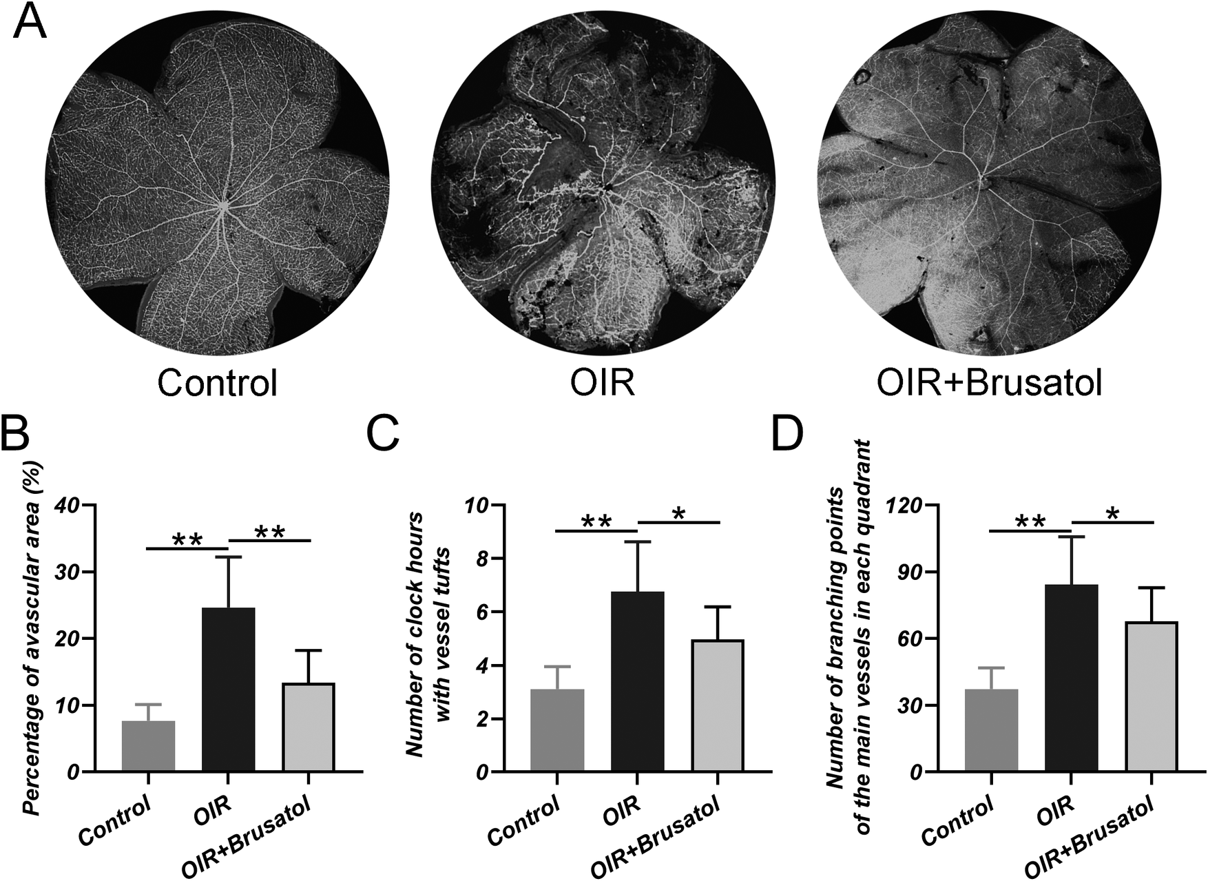
The effect of brusatol on the vessel extension and morphology in rats with OIR was first investigated. Fluorescein isolectin GS-IB4 staining was utilized to define the nonperfused peripheral retinal area, which indicated that brusatol treatment could reduce the extent of nonperfused area compared with OIR group (Fig. 1A). Intravitreal administration of brusatol could also decrease the percentage of avascular area (Fig. 1B), the number of clock hours with vessel tufts (Fig. 1C), and the number of branching points of the main vessels in each quadrant (Fig. 1D) when compared with OIR group. All of these indicated that brusatol treatment could inhibit the angiogenesis induced by hyperoxia.
Brusatol treatment alleviates angiogenesis in response to OIR. A, The nonperfused peripheral retinal area indicated by GS-IB4 whole mounted staining in different groups. B, Vaso-obliterated retinal area was quantified with the total retinal area. C, New vessel formation quantified as the number of clock hours with vessel tufts. D, The count of vessels branching points in each quadrant. N = 6 in each group. Values were represented in mean ± SD. *P < 0.05, **P < 0.01 between the indicated groups.

Brusatol alleviates microgliosis induced by OIR
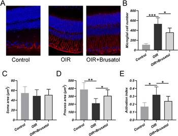
Whether brusatol could alleviate the neuronal dysfunction in OIR was further investigated. Microglia are implicated in the development of vascular and neuronal integrity maintenance in the central nervous system. The number of microglia increased significantly in the OIR retina compared with normoxia control, and brusatol treatment could decrease the number of over-growth microglia (Fig. 2A and 2B). Once activated, microglia can alter morphology from a stellate, highly ramified form to an amoeboid shape with process retraction (Caldeira et al., Reference Caldeira, Oliveira, Cunha, Vaz, Falcao, Fernandes and Brites2014; Avignone et al., Reference Avignone, Lepleux, Angibaud and Nagerl2015). As shown, soma size did not alter in OIR (Fig. 2C), while there was a reduction in the OIR process area compared with normoxia control (Fig. 2D). The ratio of somal to dendritic area was utilized as a microglial activation index to indicate microglial activation. The activation index increased in the OIR group compared with normoxia control rats, while such an increase could be diminished by brusatol administration (Fig. 2E). All of these suggest that OIR is associated with microglial activation or microgliosis, and brusatol treatment could prevent this activation response.
Alteration in the number and morphology of microglial in OIR retina after brusatol treatment. A, Representative IbA1 staining in different groups. B, The number of microglial cells comparison. Soma area (C), process area (D), and activation index (E) of microglia from different groups. N = 6 in each group. Values were represented in mean ± SD. *P < 0.05, **P < 0.01 between the indicated groups.

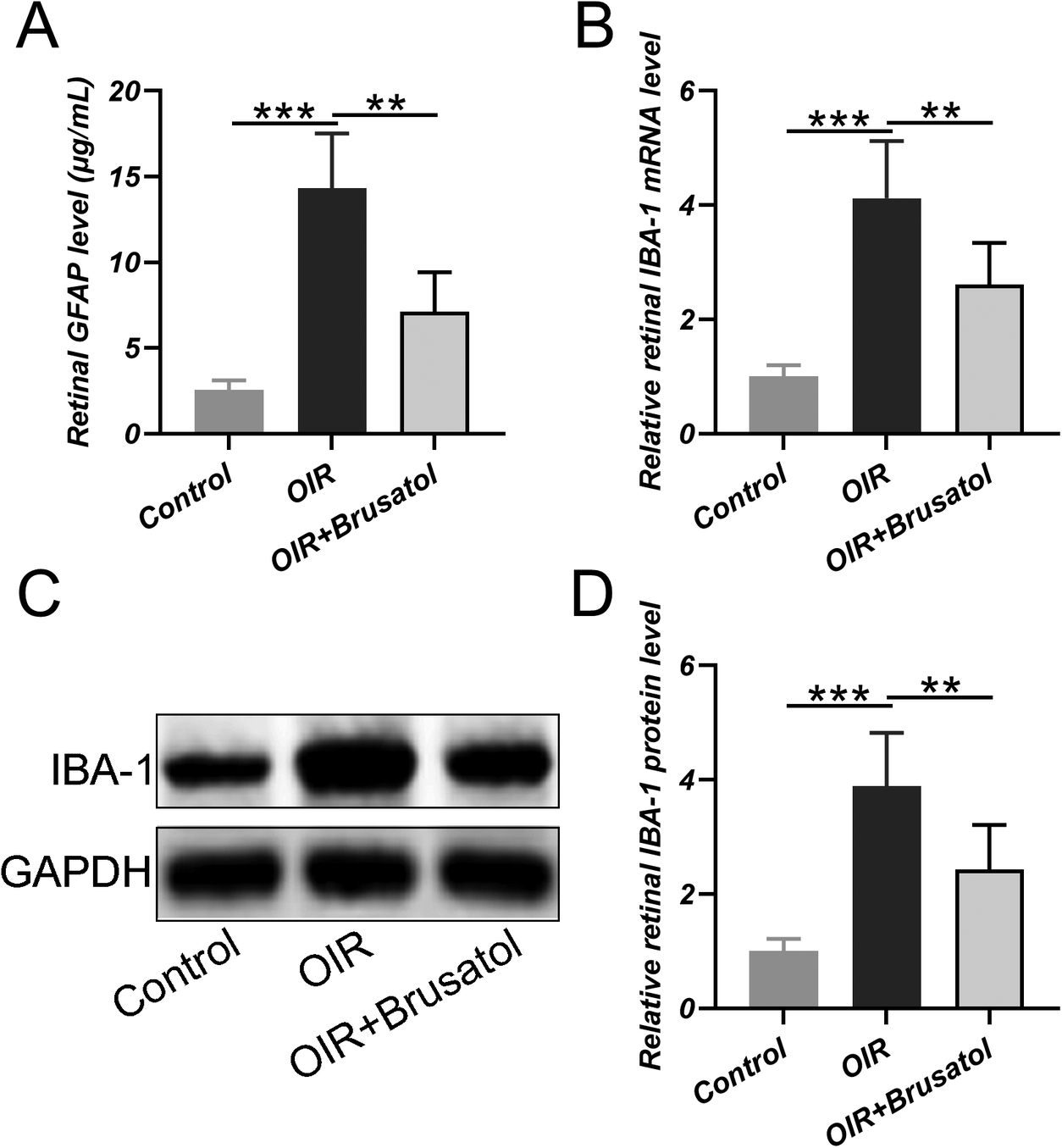
GFAP and IBA-1 expression were further assayed to indicate microglial activation. ELISA analysis indicated that GFAP could be up-regulated in the OIR group compared with normoxia control, which can be down-regulated by brusatol treatment (Fig. 3A). Both IBA-1 mRNA (Fig. 3B) and protein expression (Fig. 3C and 3D) also showed up-regulation in the OIR group, and such up-regulation could be inhibited by brusatol treatment.
Changes of GFAP and IBA-1 expressions in OIR retina in response to brusatol treatment. A, ELISA was used to test the levels of GFAP in retina from different groups. B, RT-PCR was utilized to measure the mRNA levels of IBA-1 in retina from different groups. C, Western blotting was used to measure the protein levels of IBA-1 in retina from different groups and relative expressions were normalized to control (D). N = 10 in each group. Values were represented in mean ± SD. **P < 0.01, ***P < 0.001 between the indicated groups.

Brusatol decreases angiogenesis and inflammation-related molecules
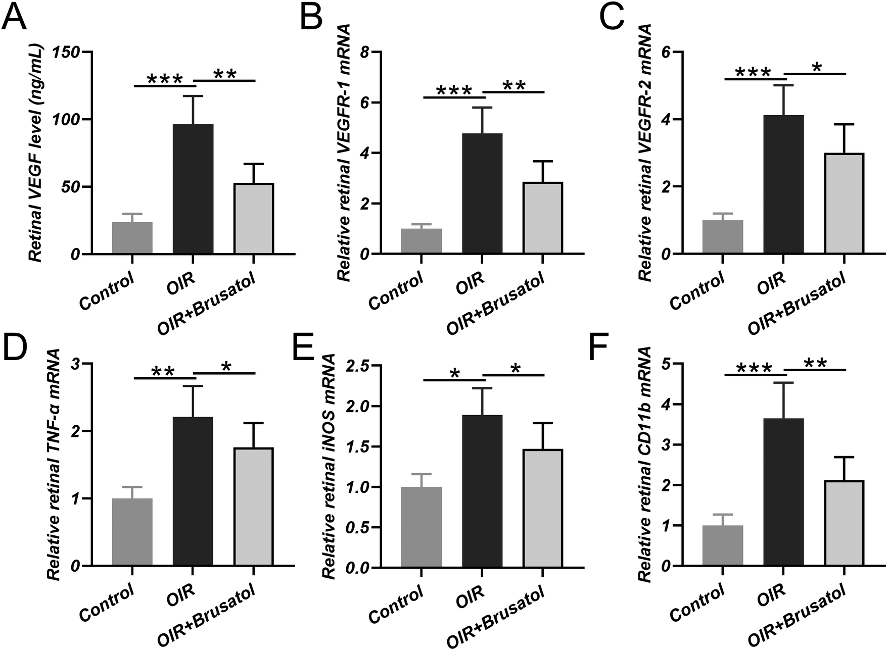
As expected, angiogenesis-related molecules, such as VEGF, were increased in the OIR group detected by the ELISA method (Fig. 4A). It was worth noting that other molecules related to angiogenesis, VEGFR1(Fig. 4B) and VEGFR1(Fig. 4C), inflammation-related molecules, such as TNF-α (Fig. 4D), INOS (Fig. 4E), and CD11b (Fig. 4F), were also up-regulated in the ORI group. At the same time, brusatol could decrease such angiogenesis and inflammation-related molecules expression.
Changes of mRNA and protein expressions of VEGF, VEGFR-1, VEGFR-2, CDllb, iNOS, and TNF-α in OIR retina in response to brusatol treatment. A, ELISA was used to test the VEGF level in the retina from different groups. The relative mRNA expression of VEGFR-1 (B), VEGFR-2 (C), TNF-α (D), iNOS (E), and CDllb (F) in retina from different groups. N = 10 in each group. Values were represented in mean ± SD. *P < 0.05, **P < 0.01, ***P < 0.001 between the indicated groups.

Brusatol decreases Nrf2 and HO-1 expression
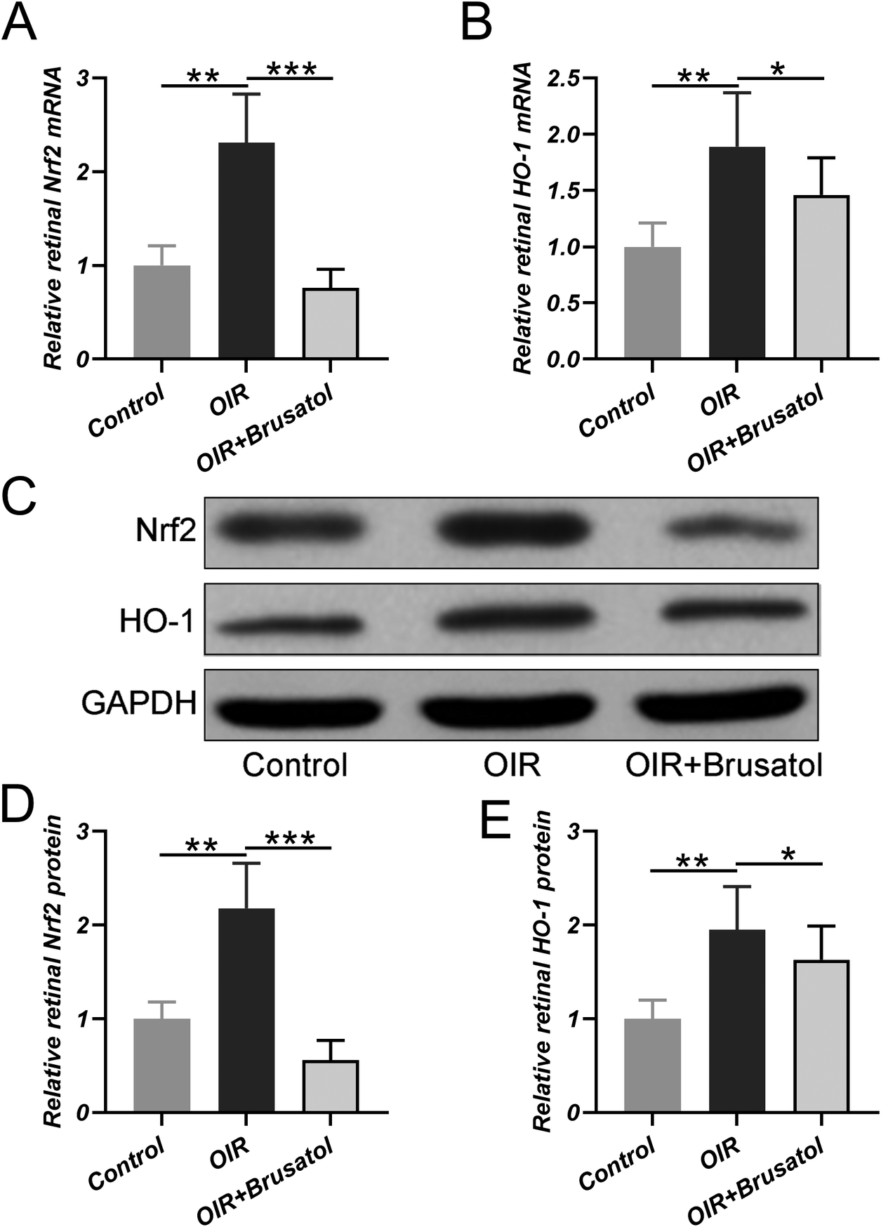
Hyperoxia could up-regulate both the mRNA expression of Nrf2 (Fig. 5A) and HO-1 (Fig. 5B) in the retina of OIR rats, and also induce the increased protein expression of Nrf2 (Fig. 5C and 5D) and HO-1 (Fig. 5C and 5E). As a heme-degrading enzyme, HO-1 could play a vital role in protecting against oxidative stress and inflammation-induced retina damage. While such up-regulation could be down-regulated by brusatol. All of these confirmed previous investigations that Nrf2 was a molecule target of brusatol (Ren et al., Reference Ren, Villeneuve, Jiang, Wu, Lau, Toppin and Zhang2011).
Changes of mRNA and protein expressions of Nrf2 and HO-1 in OIR retina after brusatol treatment. RT-PCR was used to measure the relative mRNA expression of Nrf2 (A) and HO-1 (B) in the retina from different groups. Western blotting was used to measure the protein levels of Nrf2 and HO-1 in the retina from different groups and relative expressions were normalized to control (D and E). N = 10 in each group. Values were represented in mean ± SD. *P < 0.05, **P < 0.01, ***P < 0.001 between the indicated groups.

Discussion
ROP progression can be divided into two phases. In the phase 1, acute hyperoxia and the subsequent maternal and fetal interaction loss can cause growth factors suppression and retinal vascularization arrest. Subsequently, during the phase 2, metabolically activated but poorly vascularized retina becomes hypoxic, which can promote growth factor-induced aberrant vascular proliferation, angiogenesis, and retinal detachment (Hellstrom et al., Reference Hellstrom, Smith and Dammann2013; Agarwal & Jalali, Reference Agarwal and Jalali2018).
In accordance with the previous report, ROP rats show increased vascularized retinal area and neuronal dysfunction(Chui et al., Reference Chui, Bissig, Berkowitz and Akula2012; Hansen et al., Reference Hansen, Moskowitz, Akula and Fulton2017), and this investigation testifies that brusatol has a retina-protective effect in a neonatal rats model of ROP through the inhibition of Nrf2. Nrf2 is testified to have a diverse role from anti-oxidant and anti-inflammation response to wound healing to tissue regeneration (Kahroba & Davatgaran-Taghipour, Reference Kahroba and Davatgaran-Taghipour2020; Kopacz et al., Reference Kopacz, Kloska, Forman, Jozkowicz and Grochot-Przeczek2020). Activated Nrf2 could be transferred into the nucleus, bind to ARE, and regulate basal and inducible antioxidants, detoxifications, and anti-inflammatory gene expression. The angiogenesis and inflammation-related molecules expression in the retina is counteracted by local brusatol treatment. The present findings further confirm the pathology nature of the Nrf2 in retinopathy.
Retinal microglial activation is correlated with the neovascularization process in both the hyperoxic and hypoxic phases of OIR (Fischer et al., Reference Fischer, Martin and Agostini2011; Kataoka et al., Reference Kataoka, Nishiguchi, Kaneko, van Rooijen, Kachi and Terasaki2011). Moreover, human tissue pathology analysis also indicates the association between microglia and retinal vascularization, and the number of microglial could affect the onset of neovascularization (Bodeutsch & Thanos, Reference Bodeutsch and Thanos2000). Down-regulated retinal microglial density induced by brusatol treatment is observed in this investigation, all of these suggest that brusatol could attenuate microglial mediated retinal neovascularization.
Laser photocoagulation and controlled oxygen administration are the main clinical treatment of ROP, which are performed to alleviate the disease progression (Jalali et al., Reference Jalali, Azad, Trehan, Dogra, Gopal and Narendran2010; Chen et al., Reference Chen, Stahl, Hellstrom and Smith2011; Sen et al., Reference Sen, Wu, Chandra, Vinekar, Manchegowda and Bhende2020). While in the most severe cases, intravitreal anti-VEGF therapy is utilized to diminish the disease progress (Hellstrom et al., Reference Hellstrom, Smith and Dammann2013). In this investigation, attenuated angiogenesis with down-regulated VEGFR1 and VEGFR2 expression is observed after brusatol injection, which indicates the possibility to perform further preclinical investigation.
In summary, although further detailed mechanism related to Nrf2 still needs to decipher, we have established the proof of concept that the intravitreal delivery of brusatol holds the potential to facilitate the clinical translation of the drug for the treatment of oxygen-induced ROP.
Conclusion
As an Nrf2 inhibitor, Brusatol is a promising compound to treat oxygen-induced ROP.
Conflict of interest
The authors declare no conflict of interest.
Supplementary Materials
To view supplementary material for this article, please visit http://dx.doi.org/10.1017/S095252382100002X.