Introduction
In building an embryo, complex mechanisms operate to increase, model and attribute specific traits to developing anlagen. Proliferation and apoptosis are the main sculptors acting in this process. In Xenopus laevis embryogenesis, apoptosis controls the absolute cell number and its occurrence is regulated in time and during development, starting from the beginning of gastrulation (Hensey & Gautier, Reference Hensey and Gautier1997). Bcl-2 and Bax balance and activation of downstream caspases are the main mechanisms involved in the control of apoptosis in this organism (Finkielstein et al., Reference Finkielstein, Lewellyn and Maller2001; Yeo & Gautier, Reference Yeo and Gautier2003). It was recently suggested that apoptosis may be further modulated during X. laevis development through p27BBP/eIF6 (β4 binding protein/eukaryotic initiation factor 6), which inhibits cell death by acting upstream of Bcl2 (De Marco et al., Reference De Marco, Iannone, Carotenuto, Biffo, Vitale and Campanella2010).
p27BBP/eIF6 is a highly conserved protein necessary for cell life (Sanvito et al., Reference Sanvito, Piatti, Villa, Bossi, Lucchini, Marchisio and Biffo1999; Gandin et al., Reference Gandin, Miluzio, Barbieri, Magri, Kiyokawa, Marchisio and Biffo2008). It regulates translation by preventing the interaction of 40S ribosomal subunits with 60S subunits through its binding to 60S ribosomes. In mammalian cells, upon PKC phosphorylation in serine 235, p27BBP/eIF6 is released from 60S to start translation (Ceci et al., Reference Ceci, Gaviraghi, Gorrini, Sala, Offenhauser, Marchisio and Biffo2003; Miluzio et al., Reference Miluzio, Beugnet, Volta and Biffo2009). This phosphorylative event may occur downstream of extra-cellular signalling such as insulin/growth factors indicating that p27BBP/eIF6 is a 60S-associated initiation factor able to modulate translation in vivo (Gandin et al., Reference Gandin, Miluzio, Barbieri, Magri, Kiyokawa, Marchisio and Biffo2008). The 235 SmR potential PKC phosphorylation site is also found in Xenopus p27BBP/eIF6 cDNA, in which a serine phosphorylated isoform has been reported as well (Carotenuto et al., Reference Carotenuto, Marco, Biffo, Wilding, Vaccaro, Marchisio, Capriglione, Russo and Campanella2005; Vaccaro et al., Reference Vaccaro, Cuccaro, De Marco and Campanella2006a,Reference Vaccaro, Cuccaro, De Marco and Campanellab).
Significantly, p27BBP/eIF6 appears to be selective in regulating translation of certain mRNAs, as in the case of β-catenin (Ji et al., Reference Ji, Shah, Soanes, Islam, Hoxter, Biffo, Heslip and Byers2008) and of a factor acting upstream of Bcl-2 (De Marco et al., Reference De Marco, Iannone, Carotenuto, Biffo, Vitale and Campanella2010). Moreover, its expression is variable among cell types (Donadini et al., Reference Donadini, Giodini, Sanvito, Marchisio and Biffo2001) and p27BBP/eIF6 mRNA and protein levels are high in rapidly proliferating cancer cells and low in cells committed to apoptosis (Sanvito et al., Reference Sanvito, Vivoli, Gambini, Santambrogio, Catena, Viale, Veglia, Donadini, Biffo and Marchisio2000).
During Xenopus embryogenesis, modulated expression of p27BBP/eIF6 occurs in developing anlagen in addition to a basal level of expression. In particular, p27BBP/eIF6 mRNA is abundant in the eye field and in the mid-hindbrain boundary, two regions with high proliferation levels suggesting a correlation between p27BBP/eIF6 and proliferative activity (Vaccaro et al., Reference Vaccaro, Cuccaro, De Marco and Campanella2006a,Reference Vaccaro, Cuccaro, De Marco and Campanellab). In Xenopus embryos, following p27BBP/eIF6 ectopic over-expression or down-regulation, cell proliferation appears unaffected (De Marco et al., Reference De Marco, Iannone, Carotenuto, Biffo, Vitale and Campanella2010), suggesting that under these experimental conditions, cell proliferation and apoptosis inhibition are uncoupled events. However, at present it is not known whether, during development of wild type embryos, the higher presence of p27BBP/eIF6 coincides with high activity of cell proliferation or low apoptosis levels, suggesting the existence of complex physiological co-ordination of such events.
In this paper we performed immunofluorescence with anti-p27BBP/eIF6-specific antibody, BrdU injection and TUNEL staining to investigate the correlation of p27BBP/eIF6 protein localization with proliferation and apoptosis levels in developing Xenopus embryos. Our data indicate that p27BBP/eIF6 is present throughout embryogenesis. However, the presence of this protein varies according to the sites where high proliferative activity occurs. Cell death appears variously associated to proliferative evidence and p27BBP/eIF6 localization. Indeed, at the beginning of embryogenesis a high concentration of p27BBP/eIF6 is correlated with high levels of both apoptosis and proliferation. Late in development, greater presence of p27BBP/eIF6 is always coincident with high mitotic activity whereas cell death decreases and is ubiquitously present.
Material and methods
Animals
Adult Xenopus laevis females were obtained from ‘Rettili’ Varese, Italy. They were kept and utilized at the Department of Structural and Functional Biology of the University of Naples, Federico II, according to the guidelines and policies dictated by the University Animal Welfare Office and in agreement with international rules.
To obtain eggs, X. laevis females were injected in the dorsal lymphatic sac with 500 units of Gonase (AMSA) in amphibian Ringer's solution (111 mM NaCl, 1.3 mM CaCl2, 2 mM KCl, 0.8 mM MgSO4, 25 mM HEPES, pH 7.8). Fertilized eggs and embryos were obtained by standard insemination methods (see De Marco et al., Reference De Marco, Iannone, Carotenuto, Biffo, Vitale and Campanella2010) and staged according to Nieuwkoop & Faber (Reference Nieuwkoop and Faber1967).
Immunofluorescence
X. laevis embryos were fixed in 4% formaldehyde at 4°C and stored in 100% MetOH at –20°C. For in toto immunofluorescence after incubation in hydrogen peroxide/methanol 1:2 (v/v) for about 2 days, the samples were incubated with rabbit anti-p27BBP/eIF6 antibody (Biffo et al., Reference Biffo, Sanvito, Costa, Preve, Pignatelli, Spinardi and Marchisio1997; Carotenuto et al., Reference Carotenuto, Marco, Biffo, Wilding, Vaccaro, Marchisio, Capriglione, Russo and Campanella2005) or polyclonal anti-pH3 antibody (UPSTATE) diluted 1:500 PBS/0.5%BSA/0.1% Triton X-100. The secondary antibody was goat anti-rabbit IgG BODIPY FL-conjugated (Molecular Probes). The embryos were then dehydrated in graded methanol and cleared in benzyl alcohol/benzyl benzoate 1:2 (v/v). Whole mounts were photographed with a Leica MZ 16F. For immunofluorescence on frozen section 10 μm-thick sections were obtained after embedding and freezing in Killik (Bio Optica). Non-specific background was blocked by incubating the sections for 30 min in normal goat serum 3% in PBS, 0.5% BSA, 0.1% Tween, prior to exposure O/N at 4°C to rabbit anti-p27BBP/eIF6 diluted 1:500 in PBS containing 0.5% BSA and 0.1% Tween. Staining was completed by incubating the samples with anti-rabbit goat IgG BODIPY FL-conjugated (Molecular Probes), followed by nuclei counter-staining with DAPI and mounting in PBS/glycerol (9:1, v/v). Sections were observed and photographed with a Leica CTR 6500 UV microscope equipped with the Leica application suite.
TUNEL staining
TUNEL staining to identify apoptotic cells was performed using ApopTag Peroxidase In Situ Apoptosis Detection Kit (Chemicon), in accordance with the manufacturer's instructions. The whole-mount TUNEL staining protocol was performed as described in Hensey & Gautier (Reference Hensey and Gautier1997). Detection and chromogenic reaction were carried out according to Harland (Reference Harland1991). The embryos were blocked in 20% goat serum, followed by incubation with anti-digoxigenin antibody coupled to alkaline phosphatase (Roche). Staining was developed using BM-purple (Roche). Embryos were viewed following dehydration in methanol. Whole mounts were photographed on a Leica MZ 16F.
BrdU experiments
Embryos were injected with BrdU (5-bromo-2′-deoxyuridine, Roche) in the gut, fixed 2 h later and cryostat sectioned. Preliminary experiments showed that cell division occurs in a few minutes (see also Viczian et al., Reference Viczian, Vignali, Zuber, Barsacchi and Harris2003). Sections were washed with 2 N HCl for 45 min and then neutralized with several PBS washes. The mouse anti-BrdU antibody (Sigma) was added at 1:500 dilution and the sections were incubated at 4°C overnight. Staining was completed by incubation with BODIPY FL goat anti-mouse IgG (Invitrogen, Molecular Probes).
Results
Stage 14
p27BBP/eIF6 localization was studied by in toto immunofluorescence in stage 14 embryos. The immunostaining was highly concentrated in the neural plate along the anteroposterior axis of the embryo and in the anteriormost region in territories fated to develop eyes and part of the encephalon (Fig. 1a,b). Apoptosis was detected by in toto TUNEL-staining. Labelled cells were found in the dorsal region where they appear to mark the neural plate, being highly concentrated at its anterior edge (Fig. 1c). We performed immunostaining with an antibody against phosphorylated histone H3, which specifically recognizes mitotic chromosomes. High levels of cell proliferation were present in whole embryos and were mostly localized in the developing neural plate (Fig. 1d). The immunostaining was specific, as indicated by the absence of a signal, when the primary antibody was absent (Fig. 1e). Therefore, at the beginning of embryogenesis, high levels of p27BBP/eIF6, proliferation and apoptosis are coincident.
In toto immunofluorescence with anti-p27BBP/eIF6 antibody of stage 14 embryo, dorsal (a) and lateral (b) view. Staining is relevant in the neural plate (arrow) and in the anteriormost region indicated by the arrowhead. (c) Apoptosis detected by in toto TUNEL assay at stage 14 embryo. A strong signal is present in the neural plate (arrow) with the most intense staining corresponding to the anteriormost portion (arrowhead). (d) immunofluorescence with anti-pH3 antibody. At the early neurula stage numerous mitotic cells are present in the whole embryo. The strongest staining is localized in the developing neuroepithelium (arrow for posteriormost and arrowhead for anteriormost neuroepithelium). (e) In toto immunofluorescence using only secondary antibody BODIPY-conjugates anti-rabbit goat IgG. Staining is absent.

Stage 24
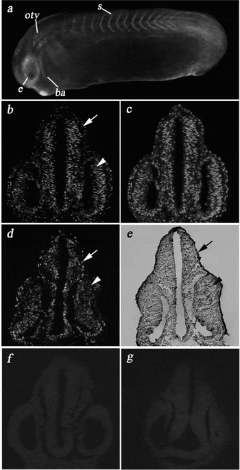
At tailbud stage p27BBP/eIF6 in toto immunofluorescence was detected throughout the developing brain, otic and optic vesicles, branchial arches and somites (Fig. 2a). In particular, the pattern of p27BBP/eIF6 distribution was studied during brain and eye formation. At stage 24, the neural tube is fully formed and differentiated into forebrain, midbrain and hindbrain. The optic vesicle has evaginated from the prosencephalon and makes contact with the overlying epidermis. The retina consists of a single population of neuroepithelial cells (Nieuwkoop & Faber, Reference Nieuwkoop and Faber1967; Holt et al., Reference Holt, Bertsch, Ellis and Harris1988; Eagleson et al., Reference Eagleson, Ferreiro and Harris1995). Immunofluorescence was performed, as well, on embryo frozen sections. Extensive p27BBP/eIF6 labelling was observed in all brain regions and in the undifferentiated cells of the optic stalk, presumptive retinal pigment epithelium and neural retina (Fig. 2b,c). BrdU and TUNEL experiments were performed on serial section of stage 24 embryos. BrdU labelling showed mitotic cell throughout the brain and eye. Indeed, at this stage most of the cells are still actively proliferating. However, TUNEL assay showed diffuse staining in the developing brain and eye and no tissue-specific localizations were found (Fig. 2e). The immunostaining was specific (Fig. 2f,g). Therefore while the greater presence of p27BBP/eIF6 and proliferation are coincident, apoptosis diminishes in the brain and eyes and does not appear to have specific localizations.
(a) At stage 24, in toto immunofluorescence shows the presence of p27BBP/eIF6 in somites (s), branchial arches (ba) and in the anlagen of optic (e) and otic (otv) vesicles. (b) immunofluorescence with anti-p27BBP/eIF6 antibody on a section of a stage 24 embryo. The protein is abundant in developing brain (arrow) and retina (arrowhead). (c) DAPI staining of section (b). BrdU immunolocalization (d) and TUNEL assay (e) was performed in serial cross-sections of stage 24 embryos. High proliferation rates are present in developing brain (arrow) and eye (arrowhead), while apoptosis levels are reduced compared with earlier stages of neurulation both in brain and in retina (e). Immuno-fluorescence controls using only anti-rabbit goat IgG BODIPY-conjugates as control for anti-p27BBP/eIF6 immunofluorescence (f) and anti-mouse goat IgG BODIPY-conjugates as control for anti-BrdU immunofluorescence (g). Staining is absent.

Stage 32
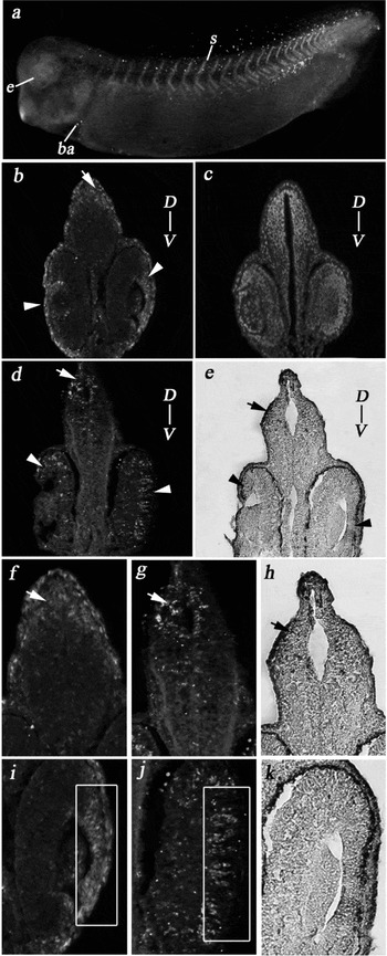
The general pattern of the brain and connected sensorial placodes is established at about stage 28 (Nieuwkoop & Faber, Reference Nieuwkoop and Faber1967). At stage 32, the eye cup is formed and cells are being generated in the central retina while some cells continue to divide in the ciliary marginal zone (CMZ), the most peripheral region of the retina (Straznicky & Gaze, Reference Straznicky and Gaze1971; Wetts et al., Reference Wetts, Serbedzija and Fraser1989). High levels of p27BBP/eIF6 were expressed in the brain, otic vesicle, optic cup as well as in branchial arches and somites (Fig. 3a). In particular, anti-p27BBP/eIF6 immunostaining detected high concentrations of the protein in more restricted regions than in earlier stages. Indeed the stain was segregated in the dorsal region of the brain and in the CMZ (Fig. 3b,f,i). BrdU and TUNEL experiments on serial sections of stage 32 embryos showed that higher proliferation and apoptosis rates were found in the dorsal with respect to the ventral region of the brain (Fig. 3d,e,h,j). In the eye, many cells were post-mitotic and the majority of the BrdU immunostaining was found in the CMZ, while TUNEL-positive nuclei were detected in the whole developing retina including the CMZ (Fig. 3d,e,j,k). Taken together, these data show that in the brain the levels of p27BBP/eIF6, proliferation and apoptosis are nearly overlapping. In contrast, in the eye the TUNEL reaction is found in the whole retina and does not appear specifically concentrated in the same sites where strong localization of both anti-BRDU and anti- p27BBP/eIF6 immunostaining was found.
p27BBP/eIF6 localization in stage 32 embryos. (a) A strong signal is present in somites (s), branchial arches (ba) and developing eye (e). (b) Immunofluorescence on sections shows that p27BBP/eIF6 label is more abundant in dorsal region of the brain (arrow) and in ciliary marginal zone of the eye (arrowhead). (c) DAPI staining of section (b). Immunostaining with anti-BrdU antibody (d) and TUNEL assay (e) on serial sections of a stage 32 embryo show that proliferation and apoptosis are overlapping in the brain. In contrast, in the eye anti-BRDU immunostaining is restricted to CMZ, while TUNEL staining is diffuse in the whole retina. (f) Higher magnification of the brain showed in b. Most anti-p27BBP/eIF6 immunostaining is next to the ventricle in dorsal region of the brain (arrow). (g, h) Details of parts (d) and (e) respectively show that BrdU and TUNEL assays stain the same sites (arrows) strongly labelled by anti-p27BBP/eIF6 antibody. (i) Higher magnification of eye showed in section (b). High levels of p27BBP/eIF6 are present in CMZ (square). (j) detail of section (d) shows a strong localization of mitotic cells in CMZ (square) overlapping with p27BBP/eIF6 immunostaining. (k) detail of section (e) shows that apoptotic cells are diffuse in all developing retina including CMZ. D-V, dorsoventral axis.

Stage 45
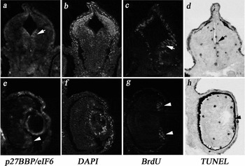
In the stage 45 brain, p27BBP/eIF6 immunofluorescence was abundant in the subependymal layer (Fig. 4a), cell division and apoptosis were localized as well in the subependymal layer as revealed by intense BrdU and TUNEL signals close to the ventricle (Fig. 4c,d). At the same stage, the retina is fully laminated, with three distinct nuclear layers separated by two plexiform layers where retinal cell types can be distinguished by morphological and cytological criteria (Straznicky & Gaze, Reference Straznicky and Gaze1971). In the mature retina p27BBP/eIF6 was localized in the CMZ (Fig. 4e), where high proliferative activity was demonstrated by the presence of BrdU staining (Fig. 4g). TUNEL signal was detected in all layers of the retina including the CMZ (Fig. 4h). Therefore, at stage 45, higher p27BBP/eIF6 presence, proliferation and apoptosis are fully overlapping in the brain. However, in the eye high levels of p27BBP/eIF6 and mitosis are restricted to the CMZ while cell death is widespread and not layer-specific as in previous stages.
p27BBP/eIF6 (a); DAPI staining (b); BrdU immunofluorescence (c); and TUNEL assay (d) in brain of stage 45 embryo. Most p27BBP/eIF6, BrdU and TUNEL signals are close to the brain ventricle (arrow). In the fully laminated retina p27BBP/eIF6 (e) and BrdU (g) staining is restricted to CMZ (arrowhead), whilst the TUNEL signal (h) is present in the whole retina. (f) DAPI staining.

Discussion
In adult eIF6+/− mice, while the eIF6 level is 50% lower in all tissues, the cell proliferation rate is largely unchanged except for a reduction in the liver, fat cells and cultured fibroblasts (Gandin et al., Reference Gandin, Miluzio, Barbieri, Magri, Kiyokawa, Marchisio and Biffo2008). In contrast, in p27BBP/eIF6 morphants of Xenopus laevis, cell proliferation levels are unchanged with respect to controls (De Marco et al., Reference De Marco, Iannone, Carotenuto, Biffo, Vitale and Campanella2010). It should be observed that the conditions of down-regulation experiments are not fully comparable in the mouse and the frog. Indeed, in eIF6+/− mice, p27BBP/eIF6 haploidism is genetically fixed and leads to chronic depletion of eIF6 in contrast to Xenopus p27BBP/eIF6 morphants where depletion is acute. Moreover, different organs and time-points were analyzed (Gandin et al., Reference Gandin, Miluzio, Barbieri, Magri, Kiyokawa, Marchisio and Biffo2008; De Marco et al., Reference De Marco, Iannone, Carotenuto, Biffo, Vitale and Campanella2010). In the opposite condition, i.e., when p27BBP/eIF6 is over-expressed, the data available for Xenopus embryogenesis indicate that any increase in cell proliferation occurs in p27BBP/eIF6 over-expressors. Yet in these experimental conditions, the induced high levels of p27BBP/eIF6 expression were found to reduce endogenous apoptosis, acting upstream of Bcl2 (De Marco et al., Reference De Marco, Iannone, Carotenuto, Biffo, Vitale and Campanella2010).
In the present paper we studied p27BBP/eIF6 protein localization in wild-type embryos and compared it to proliferation and apoptosis, in light of the recent data on the role of p27BBP/eIF6 as an anti-apoptosis factor and taking into consideration that in natural conditions high levels of p27BBP/eIF6 expression occur in developing anlagen of Xenopus laevis (Vaccaro et al., Reference Vaccaro, Cuccaro, De Marco and Campanella2006a,Reference Vaccaro, Cuccaro, De Marco and Campanellab). We focused on brain and eye development because they are excellent models for studies of cellular proliferation and differentiation. Indeed, these organs have reproducible stereotypic pattern of cell division and apoptosis required for their morphogenesis (Eagleson et al., Reference Eagleson, Ferreiro and Harris1995; Hensey & Gautier, Reference Hensey and Gautier1998; Yeo & Gautier, Reference Yeo and Gautier2003).
Immunofluorescence data showed that high levels of p27BBP/eIF6 protein are present in the neural tube and the presumptive eye field from the start of neurulation. Similarly, high proliferation levels were observed in the majority of cells of stage 14 embryos and these rates are greatest in antero-dorsal areas i.e. coincident with the localization of p27BBP/eIF6. In these embryos, dorsal embryonic regions are also heavily labelled with TUNEL assay, in agreement with previous studies where a possible interaction was suggested between apoptosis and proliferation in regulating the physiological number of cells within the developing neuroectoderm (Yeo & Gautier, Reference Yeo and Gautier2003).
At stage 24, high levels of proliferation are present in the same regions where p27BBP/eIF6 major expression is observed, while cell death was considerably reduced compared with earlier stages of neurulation. This finding may be because, at this stage, p27BBP/eIF6 activity as a modulator of apoptosis becomes evident.
In later stages of development (stages 32–45) when the brain acquires most of its form and structure (Nieuwkoop & Faber, Reference Nieuwkoop and Faber1967), the cell proliferation rate and p27BBP/eIF6 presence are restricted to specific areas of the brain. Indeed, the BrdU-detected mitoses localize in selected areas such as the sub-ependymal layer of CNS and the CMZ of the eye, a perpetually self-renewing proliferative neuroepithelium at the perimeter of the retina in amphibians and fish (Straznicky & Gaze, Reference Straznicky and Gaze1971; Johns, Reference Johns1977; Reh & Kljavin, Reference Reh and Kljavin1989; Wetts et al., Reference Wetts, Serbedzija and Fraser1989). Apoptosis is similarly localized, but it is more widespread and not restricted to the same areas highly reactive with anti-p27BBP/eIF6 and anti-BrdU antibodies. We surmise that high levels of p27BBP/eIF6 are required for fine regulation of apoptosis which appears to play a pivotal role in the morphogenesis of the brain (Hensey & Gautier, Reference Hensey and Gautier1998). Indeed, apoptosis regulates primary neurogenesis at the level of neuronal determination (Yeo et al., Reference Yeo and Gautier2003).
In conclusion this study shows that high levels of p27BBP/eIF6 are present in highly proliferative regions, where apoptosis is also physiologically required for normal development. We hypothesize that p27BBP/eIF6 finely regulates the development machinery in stages of neural differentiation and organogenesis by acting through its ability to switch the apoptotic Bcl-2-Bax balance to cell survival.
Acknowledgements
This work was funded by a Grant of the Italian Ministry of University and Research (P.R.I.N. 1997) to C.C. and by a Grant of the ‘Compagnia di San Paolo’ (Integration of spatial and biochemical clues during brain development) to C.C.