Case presentation
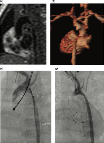
A 5-day-old term, 3 kg female was born at our institution. Heart murmur was detected and aortic coarctation was diagnosed. Through lateral thoracotomy, an end-to-end anastomosis was performed. The aortic wall was unusually hard and fluorescence in situ hybridization examination confirmed the diagnosis of Williams syndrome. The infant returned at the age of 2 months with severe recoarctation, peak echocardiographic gradient 130 mmHg (mean = 79 mmHg), with diastolic tail. On MRI and cardiac catheterisation, long-segment stenosis was appreciated (Fig 1a to c). Maximum diameter at the restenosis site measured 1.7 mm. Gadolinium contrast dropout was noted on three-dimensional MRI angiography due to extreme stenosis. Surgical consultation was against reoperation at this vessel diameter, hence angioplasty was suggested. The lesion was successfully ballooned with a Traveler 3.5 × 20 mm balloon (Abbott), upgraded to a non-compliant NC Euphora RX 5 × 15 mm (Medtronic) and finally a 6-mm Tyshak balloon (NuMED) (Fig 1d). Post-angioplasty, right axillary artery pressure dropped from 194/77 to 97/62 mmHg. Haemodynamic gradient dropped from 100 mmHg (ascending aortic pressure –AoA 194/77 mmHg, descending aorta –AoD 94/62 mmHg) to 39 mmHg (AoA 86/30/54 mmHg, AoD 47/30 mmHg). Peak echocardiographic gradient fell from 130 to 70 mmHg max (mean = 35 mmHg).
(a) Black blood sagittal MRI section, showing severe recoarctation, (b) three-dimensional reconstruction with gadolinium dropout at the site of the coarctation due to the severity of the stenosis, (c) angiographic view of the coarctation showing the narrowest point estimated at 1.7 mm, (d) initial balloon angioplasty – final result.

The lesion gradually restenosed within the next 3 months. On catheterisation, long-segment restenosis was noted more distally to the original lesion. Peak echocardiographic gradient measured 120 mmHg (mean = 75 mmHg), with prominent diastolic tail. The stenosis was re-ballooned with a non-compliant NC Euphora RX, 5 × 15 mm balloon (Medtronic), followed by a 6-mm Tyshak balloon. Haemodynamic gradient dropped from 99 mmHg (AoA 170/78 mmHg, AoD 71/45 mmHg) to 28 mmHg (AoA 110/40 mmHg to AoD 82/42 mmHg). Peak echocardiographic gradient fell from 120 to 46–50 mmHg (mean = 25 mmHg).
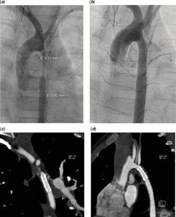
The patient remained clinically well for 1 year. However, subsequent monthly reviews revealed gradual long-segment restenosis. Double antihypertensive therapy was required. Peak echocardiographic gradient measured 105 mmHg (mean = 52 mmHg), with diastolic tail. Catheterisation was performed again at the age of 18 month. Both femoral arteries were found completely occluded with collateral network, hence access was obtained via a 4-Fr sheath placed in the right axillary artery. Repeat angioplasty was deemed unsuitable. Therefore, a 5 × 15 mm Resolute Onyx™ drug-eluting stent was implanted (Medtronic). The affected lesion was completely covered (Fig 2a and b). Baseline gradient of 80 mmHg (AoA 150/75 mmHg, AoD 70/48 mmHg) fell to 15 mmHg (AoA 92/52 mmHg to AoD 77/50 mmHg). Peak echocardiographic gradient fell from 105 to 45 mmHg (mean = 22 mmHg). Following this intervention, the patient was started on aspirin and clopidogrel which she has continued to this date (aka 19 months since the procedure and 18 months after the CT angiography). Aspirin will be continued indefinitely. Clopidogrel was adjusted for weight according to somatic growth up to 1- year post-stent implantation and has been left at the same dose since. It will empirically be discontinued at 2 years post-catheterisation. The patient has remained clinically well and free from further interventions. Echocardiographic max Doppler gradient has remained stable at 50 mmHg (mean = 20–25 mmHg) (Supplementary material Fig S1). CT angiography 18 months post-implantation showed good stent apposition without evidence of intimal proliferation (Fig 2c and d). The patient’s femoral pulses are well palpable and four-limb blood pressures measure an upper–lower peak gradient of 20 mmHg.
Implantation of a 5 × 15 mm Resolute Onyx™ drug-eluting sten (Medtronic) with an excellent angiographic result: (a) pre-stenting and (b) post-stenting, (c–d) CT angiography at 18 months post-implantation revealed no intimal in-stent growth.

Discussion
Williams syndrome results from a microdeletion in chromosome 7 (q11.23 region). This deletion involves several genes, including the ELN elastin-encoding gene. Structural cardiac and vascular disease occurs in about 80% of patients, most commonly supravalvar aortic stenosis and branch pulmonary artery stenosis.Reference Mannarino and Collins1 Due to the elastin deficiency, coarctation of the aorta, mid-aortic syndrome, as well as coronary, renal and mesenteric artery stenosis have also been described.Reference Mannarino and Collins1 Coarctation in these patients may manifest as discrete stenosis or a long-segment narrowing.Reference Collins, Kaplan and Rome2 Patch implantation is successful in most cases of Williams infants with long-segment coarctation.Reference Mannarino, Keizman, Pasotti, Codazzi, De Sando and Giamberti3 In our patient, vessel diameter was only 1.7 mm, which was considered to be too narrow for effective patch enlargement. Long-segment stenosis, rapid recoarctation following balloon angioplasty, as well as the fact that balloon angioplasty may trigger intimal proliferation further, prompted us to stent the lesion. Cohen et al recently reported successful use of drug-eluting balloons in a Williams patient with branch pulmonary artery in-stent stenosis.Reference Cohen, Glickstein and Crystal4 Angioplasty with a drug-eluting balloon was performed for in-stent restenosis and to remodel the distal pulmonary vessels bilaterally. This resulted in immediate improvement of the in-stent restenosis and resultant decrease in the right ventricular pressure. The antiproliferative effects of drug-eluting balloons may be of benefit in Williams arteriopathy. We chose a drug-eluting stent, based on the same hypothesis that the drug might minimise the risk of in-stent restenosis.
Drug-eluting balloons and drug-eluting stents have been tried in adults for both coronary lesions and peripheral femoropopliteal artery stenosis.Reference Scheinert, Duda and Zeller5,Reference Palmerini, Benedetto and Biondi-Zoccai6 In recent years, their use in paediatric cardiac interventions has anecdotally increased. Lee et al recently reported on nine newborns who underwent stent implantation in the arterial duct with a sirolimus-eluting single stents.Reference Lee, Seto, Benson and Chaturvedi7 The Zotarolimus Resolute Onyx™ stent is a new generation drug-eluting stent that delivers a synthetic analogue of Sirolimus, which inhibits the m-TOR activity leading to inhibition of cell cycle progression from the G1 to the S phase, thus minimising neointimal hyperplasia and the risk of in-stent restenosis.Reference Wessely, Schomig and Kastrati8,Reference Burke, Kuntz and Schwartz9 We chose a Zotarolimus drug-eluting stent based on its known safety profile. Indeed, based on available Zotarolimus pharmacokinetic data, systemic safety margins of ≥78-fold have been established for the Resolute Onyx™ stent at 380 μg due to the extended elution of Zotarolimus from the BioLinx® polymer, meaning that we delivered 78 × less drug than what it would be considered unsafe.Reference Burke, Kuntz and Schwartz9
At 18-month follow-up our patient has not developed recoarctation, arterial hypertension, or other significant adverse effects. Most recent echocardiography and CT angiography revealed that the stent is of good diameter with no collapse and laminar pulsatile forward flow. Further surgery will most likely be required when the patient is of a suitable age and weight to undergo a more definitive surgical procedure, although even this may be avoided, if further dilation and potentially cautious intentional stent fracture manages to deal with growth-related restenosis.Reference Khan, Qureshi and Justino10
Conclusion
Drug-eluting stents may be used in recoarctation patients with abnormal vascular wall to decrease neointimal proliferation and prevent rapid restenosis. Due to the small available diameters of such stents of up to 5 mm, it is anticipated that re-interventions for stent dilation and/or stent fracture or a reoperation may be required in the future. Therefore, such procedures may not be definitive but offer palliation in patients when imminent surgery carries a high risk with low success rate.
Supplementary Material
To view supplementary material for this article, please visit https://doi.org/10.1017/S1047951119003202
Financial Support
This research received no specific grant from any funding agency, commercial or not-for-profit sectors.
Conflicts of Interest
None.