INTRODUCTION
The striped dolphin, Stenella coeruleoalba (Meyen, 1833) is a small pelagic dolphin distributed across the warm-temperate to tropical waters of the world (Archer & Perrin, Reference Archer and Perrin1999). It is the most abundant dolphin species in the Mediterranean Sea (Notarbartolo di Sciara & Demma, Reference Notarbartolo di Sciara and Demma1994), but is not considered resident in the Croatian part of the Adriatic Sea. Bottlenose dolphin (Tursiops truncatus) is the only resident cetacean species in the Adriatic; several other cetacean species occur there occasionally (Gomeričić et al., Reference Gomercic, Curas, Lucic, Gomercic, Huber, Ckrtic, Cukrovic, Galov and Vukovic2002; Bearzi et al., Reference Bearzi, Holcer and Notarbartolo Di Sciara2004). Strandings of striped dolphins along the Italian coast have been regularly reported. Seven specimens were stranded in the northern, 21 in the central and 148 in the southern part of Italian coast of the Adriatic between years 1986 and 1996 (Podestà & Bortolotto, Reference Podestà and Bortolotto2001). The first stranding of the striped dolphin in the Croatian part of the Adriatic was reported in 1991 (Gomerčić et al., Reference Gomerčić, Huber, Gomerčić, Gomerčić, Škrtić, Vuković and Gomerčić1994) and the only other sighting of the species in the same area was reported in 1996 (Bearzi et al., Reference Bearzi, Fortuna and Notarbartolo di Sciara1998).
Genetic variability is thought to be essential to the long-term persistence and adaptability of populations. With co-dominant inheritance and high degree of polymorphism, microsatellite DNA markers have proved highly informative for population genetic studies (Bruford & Wayne, Reference Bruford and Wayne1993). The mitochondrial DNA (mtDNA) control region is commonly variable on the intraspecific level and is suitable for studies of genetic variability, phylogeography, assignment to management units and forensics (Kohn & Wayne, Reference Kohn and Wayne1997). Genetic markers have been used in many cetacean species to describe genetic diversity and identify genetic differentiation of populations (e.g. Bérubé et al., Reference Bérubé, Aguilar, Dendanto, Larsen, Notarbartolo Di Sciara, Sears, Sigurjónsson, Urban and Palsbøll1998; Natoli et al., Reference Natoli, Peddemors and Hoelzel2004). It has been shown by mtDNA restriction analysis that two different striped dolphin populations exist in European waters, corresponding to the Atlantic Ocean and Mediterranean Sea, with limited gene flow between them (García-Martínez et al., Reference García-Martínez, Moya, Raga and Latorre1999). Later studies based on microsatellite analysis corroborated this finding (Valsecchi et al., Reference Valsecchi, Amos, Raga, Podestà and Sherwin2004; Bourret et al., Reference Bourret, Macé and Crouau-Roy2007; Gaspari et al., Reference Gaspari, Azzellino, Airoldi and Hoelzel2007). Although Valsecchi et al. (Reference Valsecchi, Amos, Raga, Podestà and Sherwin2004) found little evidence for population structure within the Mediterranean, Gaspari et al.'s results (2007) revealed small but significant differences between putative populations within the Mediterranean Sea.
Here we summarize stranding records of striped dolphins from the Croatian coast of the Adriatic from 1999–2007, and present data on the genetic diversity of these animals based on a fragment of the mtDNA control region and 12 microsatellite markers. Genetic results represent a limited, but potentially valuable contribution to molecular genetic investigations of the Mediterranean population of striped dolphins.
MATERIALS AND METHODS
During our studies of marine mammals, we collected striped dolphins found dead in the Croatian part of the Adriatic Sea in the last eight years. Post mortem examination which included muscle tissue sampling for genetic analyses was either performed at the site of finding or at the Department of Anatomy, Histology, and Embryology, Faculty of Veterinary Medicine, University of Zagreb, Croatia. Age was estimated using annual growth layer groups (GLGs) from extracted teeth and based on modified methods described in Slooten (Reference Slooten1991). It is assumed that one GLG corresponds to one year in the striped dolphin (Calzada et al., Reference Calzada, Lockyer and Aguilar1994).
Physical maturity was determined based on fusion of vertebrae epiphyses. Stomach content was recorded. Total genomic DNA was isolated for tissue samples using JETQUICK Tissue DNA Spin Kit (GENOMED GmbH, Germany). The samples were genotyped at 12 dinucleotide microsatellite loci: EV1Pm, EV14Pm derived from Physeter macrocephalus, EV37Mn, EV94Mn from Megaptera novaeangliae (Valsecchi & Amos, Reference Valsecchi and Amos1996) and D08, D14, D18, D22, D28, TexVet3, TexVet5, TexVet7 from Tursiops truncatus (Shinohara et al., Reference Shinohara, Domingo-Roura and Takenaka1997; Rooney et al., Reference Rooney, Merritt and Derr1999). Six microsatellite loci were tested for the first time in striped dolphin (Table 2). For amplification three primer pairs were multiplexed in one polymerase chain reaction (PCR) using the QIAGEN Multiplex PCR Kit (QIAGEN GmbH, Hilden, Germany). PCRs were carried out in a 8-μl volume containing 80–120 ng of genomic DNA, 1 × QIAGEN Multiplex PCR Master Mix (consisting of QIAGEN Multiplex PCR buffer with a final concentration of 3 mM MgCl2, dNTP mix, and HotStar Taq DNA polymerase), 0.2 μM of locus-specific fluorescent-labelled forward primer (fluorescent dyes were FAM, JOE and TAMRA) and non-labelled reverse primer. PCR cycling profile was 15 minutes at 95°C; then 30 cycles of 30 seconds at 94°C, 90 seconds at 55°C, 60 seconds at 72°C; then 30 minutes at 60°C. The PCR products were run on an ABI PRISM, 310 Genetic Analyzer (Applied Biosystems) according to the manufacturer's instructions. GeneScan Analysis Software 3.1 and Genotyper 2.5.2. (both Applied Biosystems) software were used to determine the allele sizes, using GeneScan ROX 350 standard. Allelic diversity (number of alleles), expected (He) and observed (Ho) heterozygosities were obtained using Genetix 4.05. (Belkhir et al., Reference Belkhir, Borsa, Chikhi, Raufaste and Bonhomme1996–2004). Deviations from Hardy–Weinberg equilibrium (HWE) were evaluated for all loci by calculating probabilities with the program GENEPOP 3.3 (Raymond & Rousset, Reference Raymond and Rousset1995) using complete enumeration for loci with up to four alleles and a Markov chain method for loci with more than four alleles.
A mtDNA control region was amplified by PCR using universal primers MTCRf (5′-TTCCCCGGTCTTGTAAACC-3′) and MTCRr (5′-ATTTTCAGTGTCTTGCTTT-3′) after Hoelzel & Green (Reference Hoelzel, Green and Hoelzel1998). PCRs were carried out in a 30-μl volume containing 150–250 ng of genomic DNA, 1 × QIAGEN Multiplex PCR Master Mix (consisting of QIAGEN Multiplex PCR buffer with a final concentration of 3 mM MgCl2, dNTP mix, and HotStar Taq DNA polymerase) and 0.2 μM of each primer. The PCR cycling profile was 15 minutes at 95°C, 36 cycles of 30 seconds at 94°C, 90 seconds at 61°C and 90 seconds at 72°C, followed by 20 minutes at 72°C.
PCR products were purified using Wizard® SV Gel and PCR Clean-Up System (Promega, USA) and sequenced directly using the ABI dye-terminator method. Sequence alignment was performed using ClustalW (Thompson et al., Reference Thompson, Higgins and Gibson1994), implemented in BioEdit software (Hall, Reference Hall1999). We eventually analysed an aligned sequence comprising 882 base pairs (bp). Both haplotype (h) and nucleotide (π) diversities (±SE) were estimated according to Nei (Reference Nei1987), using the program ARLEQUIN 3.1. (Excoffier et al., Reference Excoffier, Laval and Schneider2005). We compared our haplotypes to the haplotypes submitted in the GenBank by Mace et al. (Accession numbers AM 498667–AM498740, unpublished).
RESULTS
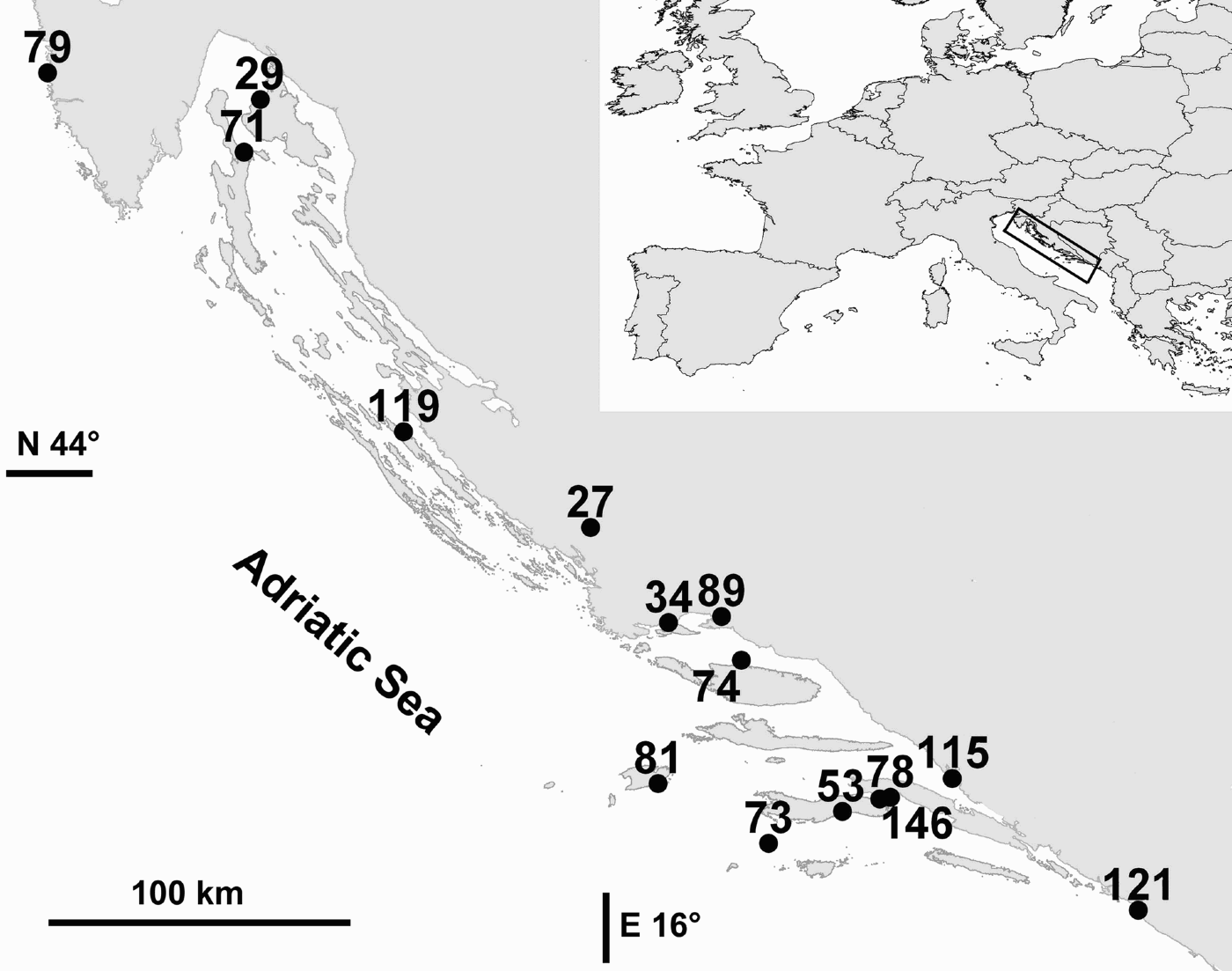
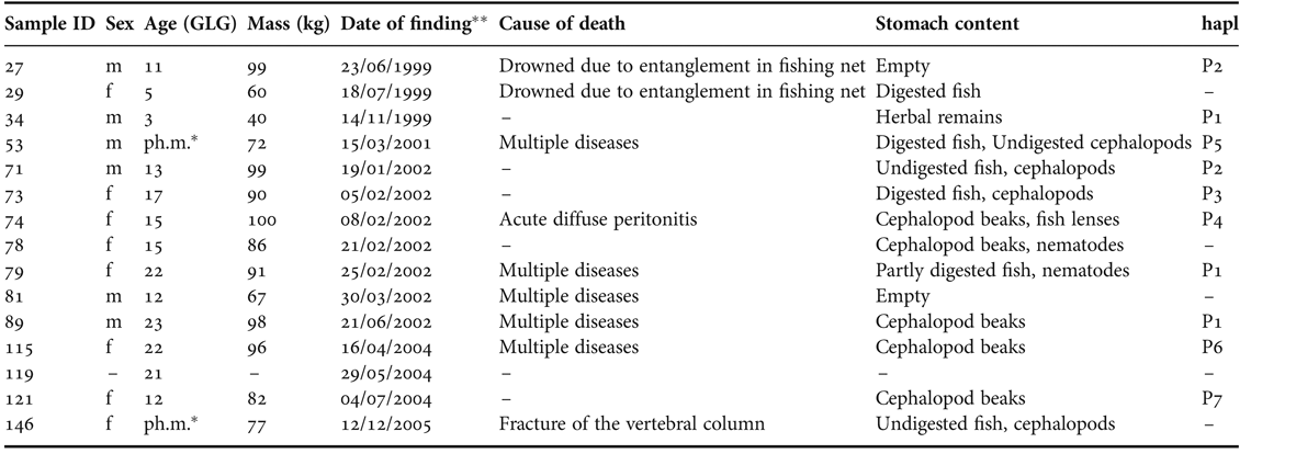
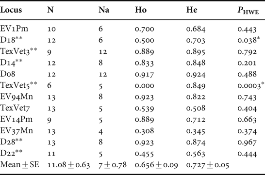
Fifteen striped dolphins, six males and eight females were found dead in Croatian part of the Adriatic Sea in the last eight years (Table 1). No data on specimen 119 apart from estimated age were obtained due to heavy decomposition of the body (Table 1). A gradient in distribution is evident, as five animals were found on the northern part versus ten animals found on the southern part of the coast (Figure 1). Two animals were sub-adults, others were adults. Two animals had empty stomachs, one had herbal remains while others had cephalopods and/or fish in various stages of digestion in their stomachs. Post mortem examination revealed cause of death in nine animals (Table 1). Although tested on a small number of samples, all microsatellite loci are highly polymorphic (Table 2). Four to twelve alleles per locus were found and allelic diversity across loci was 7±0.78. The mean expected heterozygosity was 0.727±0.05 and ranged from 0.345–0.924, while mean observed heterozygosity was 0.656±0.09 and ranged from 0–0.923. Significant deviation from HWE was observed at loci D18 and TexVet5. In addition, locus TexVet5 showed complete heterozygote absence. The 882 bp fragment at the 5′-end of the mtDNA control region was sequenced in a total of ten animals. Seven unique haplotypes were identified showing 22 polymorphic sites (Table 3). Sequences were deposited into GenBank (Accession numbers provided in Table 3). Twenty-one transitions, one transversion and no insertions/deletions were observed. Distribution of haplotypes is shown in Table 1. The haplotype diversity (h) was 0.911±0.077 and nucleotide diversity (π) was 0.006±0.003. The pairwise distances between haplotype P4 and all other haplotypes ranged between 19 and 21 nucleotides, whereas all other pairwise distances ranged between 1 and 3 nucleotides. Comparison of 630 bp long fragments with GenBank sequences revealed potential matches between haplotypes (Table 3). Haplotypes P1, P2, P4 and P5 match striped dolphin sequences from the Mediterranean coast of France. We identified sequence match between our haplotype P6 and the sequences from France, Spain and Italy (haplotype med1_B1). For haplotype P3 we did not identify any potential match among sequences deposited in GenBank.
Locations of findings of dead striped dolphins in the Croatian part of the Adriatic Sea in the last eight years (1999–2007).

Sex, estimated age, mass, date of finding, cause of death, stomach content and mitochondrial control region haplotype distribution (hapl) of striped dolphin found dead in Croatian part of the Adriatic Sea in the period 1999–2007.

*, ph.m. – physically mature; **, day/month/year.
Number of genotyped individuals (N), number of alleles per locus (Na), expected heterozygosity (He), observed heterozygosity (Ho), probability of the data under the assumption of the null hypothesis of Hardy–Weinberg equilibrium (PHWE). Six microsatellite loci tested for the first time in this species are marked.

*, significant PHWE-values; **, loci tested for the first time in striped dolphin species.
Seven mitochondrial control region haplotypes. Twenty-two polymorphic sites are identified within the 882 bp sequence where (•) represents identity with haplotype P1. The frequency (f) of occurrence of each haplotype, GenBank Accession numbers and potential matches with the GenBank sequences (Mace et al., unpublished) are provided. The match with GenBank sequences is based on a 630 bp long fragments.

DISCUSSION
A greater number of striped dolphin findings along the southern than the northern Croatian part is congruent with findings along the Italian coast of the Adriatic (Podestà & Bortolotto, Reference Podestà and Bortolotto2001). Since the striped dolphin is resident in the Mediterranean Sea, the gradient of its occurrence in the Adriatic reflects the distance from the main body of the Mediterranean. It could also reflect the bathymetry of the Adriatic basin, the striped dolphin being a deep-water species (Bearzi et al., Reference Bearzi, Holcer and Notarbartolo Di Sciara2004). Dates of dead animal findings do not reveal mass mortality of striped dolphins on the Croatian coast of the Adriatic Sea, but individual, sporadic deaths brought about by different causes (Table 1). Not all mitochondrial control region fragments and microsatellite loci were successfully amplified, probably due to DNA degradation in samples taken from carcasses that were not fresh. Significant deviation from HWE at loci D18 and TexVet5, combined with complete heterozygote absence at locus TexVet5 is either likely to indicate locus-specific genotyping problems due to null alleles or may be a consequence of small sample size. Further investigation on a larger sample size should be performed to reveal the usefulness of these two loci. The investigated group showed a high level of genetic variation, measured as both expected heterozygosity and haplotype diversity. The average expected heterozygosity of 0.723 is very similar to He value of 0.731 of the ‘Adriatic’ population estimated by Gaspari et al. (Reference Gaspari, Azzellino, Airoldi and Hoelzel2007). The expected heterozygosity of striped dolphins reported here is higher than the expected heterozygosities of bottlenose dolphins from the same geographical area, estimated at 0.69 (Galov et al., Reference Galov, Lauc, Gomerčić, Kocijan, Đuras Gomerčić, Bašić, Gomerčić, Besendorfer and Klobučar2006). High microsatellite variability detected within the investigated group of striped dolphins suggests high genetic diversity within the probable Mediterranean source population. That conclusion is consistent with the estimate of Bourret et al. (Reference Bourret, Macé and Crouau-Roy2007) of the He value for the Mediterranean population of striped dolphins of 0.756. The mtDNA haplotype analysis revealed a high number of unique haplotypes with respect to the number of samples. Seven unique haplotypes in a total of 10 animals were found, compared to just five unique haplotypes in a total of 25 bottlenose dolphin samples from the same geographical area (Galov, Reference Galov2007). This suggests striped dolphins investigated here might be a part of a larger, more diverse source population. Haplotype diversity (0.911) of the striped dolphin is also higher than the haplotype diversity (0.557) of bottlenose dolphin but, interestingly, nucleotide diversity is considerably lower (0.006) than that of bottlenose dolphins (0.022) from the same area (Galov, Reference Galov2007). Haplotype analysis also revealed that haplotype P4 differs substantially from other haplotypes. Nevertheless, it matches three other sequences found on the Mediterranean coast of France (Accession numbers AM498672, AM498728 and AM498729; Mace et al., unpublished). We did not find genetic differentiation between striped dolphin haplotypes found in the Croatian part of the Adriatic Sea and the rest of the Mediterranean, since six haplotypes (P1, P2, P4, P5, P6 and P7) match with sequences of striped dolphins found in the Mediterranean as far as France and Spain (Mace et al., unpublished). Only one haplotype (P3) is unique, but it differs from other haplotypes by a maximum of three nucleotides (P4 excluded). Although the matches are somewhat equivocal because the sequences submitted by Mace et al. are 630 bp long, only three out of 22 variable sites lie outside the span of Mace et al.'s sequences, and should not contribute substantially to genetic differentiation.
From 1999 till the end of 2006 fifteen carcasses of striped dolphins were found, compared to 99 carcasses of the bottlenose dolphin (H. Gomerčić, unpublished data). This ratio and the lack of genetic differentiation between the striped dolphin mtDNA control region haplotypes found in the Croatian part of the Adriatic Sea and the rest of the Mediterranean support a notion of the striped dolphin being a non-resident species in the Croatian part of the Adriatic Sea.
ACKNOWLEDGEMENTS
The work was supported by grants from the Ministry of Science, Education and Sports of the Republic of Croatia (Project No. 0053317) and Gesellschaft zur Rettung der Delphine e.V., Munich, Germany, and was carried out according to the annual permission of the State Institute for Nature Protection, Ministry of Culture, Republic of Croatia. We also want to thank Helena Ćetković from ‘DNA service’, Ruđer Bošković Institute, Zagreb, Croatia, for sequencing.