Introduction
Triclabendazole (TCBZ) remains the most significant drug for the control of fasciolosis, because of its high activity against juvenile stages (Fairweather, Reference Fairweather2005). Its widespread use has inevitably led to the development of resistance. Despite many unsubstantiated reports of TCBZ resistance, it is a genuine problem in many areas of the world: Australia, Europe and South America (Fairweather, Reference Fairweather2011a, Reference Fairweatherb). Understanding how resistance to TCBZ has developed in the fluke is important: for the design of treatment strategies to deal with the problem; for the identification of markers of resistance; and for the development of assays for the diagnosis of resistance.
Resistance of a parasite to an anthelmintic drug can arise in a number of ways. Modification of the target molecule, which will decrease drug binding, is a specific mechanism. However, this does not appear to be the case for Fasciola hepatica, because no differences in α- and β-tubulin isotypes were detected between TCBZ-susceptible (TCBZ-S) and TCBZ-resistant (TCBZ-R) isolates (Ryan et al. Reference Ryan, Hoey, Trudgett, Fairweather, Fuchs, Robinson, Chambers, Timson, Ryan, Feltwell, Ivens, Bentley and Johnston2008). In searching for alternative, non-specific mechanisms, one possibility is a potential change in the drug metabolizing capacity of the fluke. It is known that F. hepatica can metabolize TCBZ to its TCBZ.SO and TCBZ.SO2 metabolites (Solana et al. Reference Solana, Rodriguez and Lanusse2001; Mottier et al. Reference Mottier, Virkel, Solana, Alvarez, Salles and Lanusse2004; Robinson et al. Reference Robinson, Lawson, Trudgett, Hoey and Fairweather2004; Alvarez et al. Reference Alvarez, Solana, Mottier, Virkel, Fairweather and Lanusse2005). Also, that the metabolism is greater in TCBZ-R than TCBZ-S isolates (Robinson et al. Reference Robinson, Lawson, Trudgett, Hoey and Fairweather2004; Alvarez et al. Reference Alvarez, Solana, Mottier, Virkel, Fairweather and Lanusse2005). Evidence has been advanced for the existence in trematode parasites of Phase I xenobiotic detoxification systems, involving flavin monooxygenase (FMO) and cytochrome P450 (hereafter referred to as CYP) enzyme systems (Solana et al. Reference Solana, Rodriguez and Lanusse2001, Reference Solana, Scarcella, Virkel, Ceriani, Rodríguez and Lanusse2009; Saeed et al. Reference Saeed, Mostafa, O'Connor, Rafferty and Doenhoff2002; Cvilink et al. Reference Cvilink, Lamka and Skálová2009a, Reference Cvilink, Szotáková, Křižová, Lamka and Skálováb; Bártíková et al. Reference Bártíková, Vokřál, Skálová, Lamka and Szotáková2010; Pakharukova et al. Reference Pakharukova, Ershov, Vorontsova, Katokhin, Merkulova and Mordvinov2012). Specific inhibitors of the mammalian forms of the enzymes have been used for several in vitro and in vivo experiments on the liver fluke. The results have shown that inhibition of these enzyme pathways leads to an increased susceptibility of TCBZ-R flukes to TCBZ (Alvarez et al. Reference Alvarez, Solana, Mottier, Virkel, Fairweather and Lanusse2005; Devine et al. Reference Devine, Brennan, Lanusse, Alvarez, Trudgett, Hoey and Fairweather2009, Reference Devine, Brennan, Lanusse, Alvarez, Trudgett, Hoey and Fairweather2010a, Reference Devine, Brennan, Lanusse, Alvarez, Trudgett, Hoey and Fairweatherb, Reference Devine, Brennan, Lanusse, Alvarez, Trudgett, Hoey and Fairweatherc, Reference Devine, Brennan, Lanusse, Alvarez, Trudgett, Hoey and Fairweather2011a, Reference Devine, Brennan, Lanusse, Alvarez, Trudgett, Hoey and Fairweatherb, Reference Devine, Brennan, Lanusse, Alvarez, Trudgett, Hoey and Fairweatherc, Reference Devine, Brennan, Lanusse, Alvarez, Trudgett, Hoey and Fairweatherd, Reference Devine, Brennan, Lanusse, Alvarez, Trudgett, Hoey and Fairweather2012).
Another possible non-specific resistance mechanism involves altered drug uptake and removal, which will reduce the amount of drug reaching its intracellular target. This process involves drug transporter molecules, among which the main focus of research has been on P-glycoproteins (Pgps). Enhanced activity of Pgp-linked efflux pumps has been associated with anthelmintic resistance in nematodes and schistosomes (see Discussion for references). For F. hepatica, it has been shown that the uptake of TCBZ and its sulphoxide metabolite (TCBZ.SO) by TCBZ-R flukes is lower than in TCBZ-S flukes (Alvarez et al. Reference Alvarez, Solana, Mottier, Virkel, Fairweather and Lanusse2005; Mottier et al. Reference Mottier, Alvarez, Fairweather and Lanusse2006b). Again, it has been possible to reverse the susceptibility of TCBZ-R isolates by co-incubation with Pgp inhibitors (Mottier et al. Reference Mottier, Alvarez, Fairweather and Lanusse2006b). These two phenomena (decreased uptake and altered susceptibility) were specific to TCBZ, as they were not seen with the related benzimidazole compound, albendazole (Mottier et al. Reference Mottier, Alvarez, Fairweather and Lanusse2006b).
The aim of the present investigation was to gain further insight into the role of efflux pumps in resistance to TCBZ, by carrying out a morphological study to examine the sensitivity of a TCBZ-R isolate to TCBZ as a result of Pgp inhibition. The isolate used was the Oberon isolate, which was used by Devine and colleagues for the series of metabolic inhibition studies mentioned above. The Pgp inhibitor chosen was R(+)-verapamil (R(+)-VPL). Verapamil (VPL) belongs to the phenylalkylamine class of calcium channel antagonist: specifically, it binds to L-type calcium channels, thereby blocking calcium transport in smooth and cardiac muscle (Guerrero and Martin, Reference Guerrero and Martin1984; Hockerman et al. Reference Hockerman, Peterson, Johnson and Catterall1997; Striessnig et al. Reference Striessnig, Grabner, Mitterdorfer, Hering, Sinnegger and Glossman1998; Triggle, Reference Triggle2006). It is widely used for the treatment of cardiovascular disease, as a racemic mixture of R- and S-enantiomers. Verapamil is also recognized as an inhibitor of Pgp-mediated drug efflux (Pereira et al. Reference Pereira, Teodori, Dei, Gualtieri and Garnier-Suillerot1995; Toffoli et al. Reference Toffoli, Simone, Corona, Raschack, Cappelletto, Gigante and Boiocchi1995; Tiberghien and Loor, Reference Tiberghien and Loor1996; Choi et al. Reference Choi, Lee, Kim, Choi, Park, Shin, Yoo, Jung and Lee1998; Biscardi et al. Reference Biscardi, Teodori, Caporale, Budriesi, Balestri, Scappini, Gavazzi and Grossi2006). Multidrug resistance, linked to over-expression of drug transporters such as Pgp, is a major problem in the effective treatment of many diseases, particularly cancers. Verapamil and other efflux pump inhibitors have been utilized in one approach to overcome this problem (Tan et al. Reference Tan, Piwnica-Worms and Ratner2000; Biscardi et al. Reference Biscardi, Teodori, Caporale, Budriesi, Balestri, Scappini, Gavazzi and Grossi2006; Nobili et al. Reference Nobili, Landini, Giglioni and Mini2006; Perez-Tomas, Reference Perez-Tomas2006). A similar strategy has been suggested to overcome resistance to anthelmintic drugs (for references, see Discussion). The R(+) isomer of VPL was used in the present investigation as it is more specific for Pgp than racemic VPL and is less active as a calcium channel blocker (Echizen et al. Reference Echizen, Brecht, Niedergesass, Vogelgesang and Eichelbaum1985; Ye and Van Dyke, Reference Ye and Van Dyke1988; Varma et al. Reference Varma, Ashkraj, Dey and Panchagnula2003). In the context of the current study that was important, because racemic VPL is known to induce paralysis of the fluke (Holmes, Reference Holmes1983; Chapter 6, figs 6.5 and 6.7). The study was carried out in vitro and changes to the flukes were assessed by scanning electron microscopy (SEM). The focus is on the tegument, as it is the main route of entry for TCBZ compounds into the fluke (Mottier et al. Reference Mottier, Alvarez, Ceballos and Lanusse2006a; Toner et al. Reference Toner, McConvery, Brennan, Meaney and Fairweather2009, Reference Toner, Brennan, McConvery, Meaney and Fairweather2010) and carries out many important functions for the parasite (Fairweather et al. Reference Fairweather, Threadgold, Hanna and Dalton1999).
materials and methods
Two laboratory-maintained F. hepatica isolates were used in these experiments. Adult male Sprague–Dawley rats were infected with 20 metacercarial cysts of either the Oberon TCBZ-R or Cullompton TCBZ-S isolate. The Oberon isolate was originally obtained from a farm where TCBZ resistance was suspected, in Oberon, New South Wales, Australia in 1999. TCBZ has been shown to have limited activity against this isolate both in vitro and in vivo. Significantly, the Oberon isolate was used for the series of experiments carried out by Devine et al. (Reference Devine, Brennan, Lanusse, Alvarez, Trudgett, Hoey and Fairweather2009, Reference Devine, Brennan, Lanusse, Alvarez, Trudgett, Hoey and Fairweather2010a, Reference Devine, Brennan, Lanusse, Alvarez, Trudgett, Hoey and Fairweatherb, Reference Devine, Brennan, Lanusse, Alvarez, Trudgett, Hoey and Fairweatherc, Reference Devine, Brennan, Lanusse, Alvarez, Trudgett, Hoey and Fairweather2011a, Reference Devine, Brennan, Lanusse, Alvarez, Trudgett, Hoey and Fairweatherb, Reference Devine, Brennan, Lanusse, Alvarez, Trudgett, Hoey and Fairweatherc, Reference Devine, Brennan, Lanusse, Alvarez, Trudgett, Hoey and Fairweatherd, Reference Devine, Brennan, Lanusse, Alvarez, Trudgett, Hoey and Fairweather2012) in relation to the role of altered drug metabolism in resistance to TCBZ. The Cullompton isolate was originally obtained as a field isolate from an abattoir in Cullompton, Devon, UK in 1998. It has been shown to be susceptible to triclabendazole both in vitro and in vivo. The reader is referred to the review on fluke provenance by Fairweather (Reference Fairweather2011c) for details of previous experiments carried out with these isolates.
Experimental design
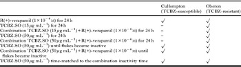
Adult flukes were recovered from the bile duct of rats under sterile conditions in a laminar flow cabinet and washed in several changes of warm (37 °C), sterile NCTC 135 culture medium (pH 7·4) containing antibiotics (penicillin, 50 IU mL−1; streptomycin, 50 μg mL−1) (NCTC was obtained from Flow Laboratories, Thame, Oxfordshire, UK; antibiotics were obtained from Sigma-Aldrich Co. Ltd., Poole, Dorset, UK). Between 6 and 8 flukes, taken from at least 3 individual rats, were placed randomly into each of the treatment groups shown in Table 1. Treatment groups are described below.
Treatment groups

Verapamil treatment
Flukes were placed in NCTC culture medium containing R(+)-VPL at a concentration of 1×10⁻⁴m and incubated at 37 °C for 24 h. R(+)-VPL was obtained from Sigma-Aldrich Co. Ltd., Poole, Dorset, UK. A stock solution of R(+)-VPL was initially prepared at a concentration of 1×10⁻²m in distilled water.
TCBZ.SO treatments
Flukes were incubated at 37 °C in NCTC culture medium containing TCBZ.SO at a concentration of either 15 μg mL−1 or 50 μg mL−1. The drug was initially prepared as a stock solution in dimethyl sulphoxide (DMSO) and added to the culture medium to give a final solvent concentration of 0·1% (v/v). The concentration of 15 μg mL−1 corresponds to the maximum blood level (13·3 μg mL−1) of TCBZ.SO reached in vivo following a therapeutic dose of 10 mg kg−1 TCBZ in sheep (Hennessy et al. Reference Hennessy, Lacey, Steel and Prichard1987); it has been used for several morphological studies on F. hepatica (for references, see Fairweather, Reference Fairweather2011c). A concentration of 50 μg mL−1 is the highest concentration used in vitro (Coles, Reference Coles1986) and has been used in previous morphological studies on F. hepatica involving TCBZ.SO. At this concentration, it has limited impact on the morphology of TCBZ-R flukes and does not cause any diminution of tubulin staining over a 24-h period (Robinson et al. Reference Robinson, Trudgett, Hoey and Fairweather2002; Walker et al. Reference Walker, McKinstry, Boray, Brennan, Trudgett, Hoey, Fletcher and Fairweather2004). The two concentrations were chosen so that comparisons with the existing body of data could be made and to determine whether any potentiation of drug sensitivity demonstrated is manifest at both high and low concentrations of TCBZ.SO. Flukes were incubated for different time periods, depending on the particular experiment, but no experiment was allowed to go beyond 24 h.
Combination treatments
Flukes were first pre-incubated in NCTC culture medium containing 1×10⁻⁴m R(+)-VPL for 2 h at 37 °C, then transferred to a fresh culture medium containing 1×10⁻⁴m R(+)-VPL plus TCBZ.SO at a concentration of either 15 μg mL−1 or 50 μg mL−1 for 24 h at 37 °C.
Inactivity experiments
The flukes in each treatment were observed at 1-h intervals under a light microscope and their motility noted. When all flukes had ceased to move, that time was taken as the time to inactivity. The experiment was stopped at this point and the flukes were recovered and processed for SEM.
Time-match experiments
These experiments involved incubation in TCBZ.SO for a similar time to that recorded in the combination inactivity experiments. The flukes were initially incubated in NCTC culture medium for 2 h, then transferred to a fresh culture medium containing TCBZ.SO at the relevant concentration for the required time period.
Controls
Controls were prepared by incubating whole flukes in NCTC culture medium containing 0·1% (v/v) DMSO for 24 h at 37 °C. Controls at 0 h were also prepared.
Tissue preparation for SEM
Material was taken for SEM analysis from each of the groups in Table 1. After treatment, flukes were washed in several changes of 0·1 m sodium cacodylate buffer (pH 7·4) containing 3% (w/v) sucrose. Flukes were lightly flat-fixed under a coverslip for 30 min in 4% (w/v) glutaraldehyde in 0·1 m sodium cacodylate buffer (pH 7·4) containing 3% (w/v) sucrose and then transferred to fresh fixative (as above) and free-fixed for a further 4 h. Specimens were subsequently washed overnight at 4 °C in 0·1 m sodium cacodylate buffer (pH 7·4) containing 3% (w/v) sucrose, post-fixed in 1% osmium tetroxide for 1 h and again washed in buffer overnight. The flukes were then dehydrated in a series of alcohols, dried in hexamethyldisalazine, fixed onto aluminium stubs and coated with gold-palladium in a Polaron E.5000 sputter-coating unit. The specimens were viewed in an FEI Quanta 200 scanning electron microscope operating at 10 keV.
results
Visual observations
Time to inactivity results are presented in Table 2. Following incubation in R(+)-VPL (1×10⁻⁴m) for 24 h, flukes of both isolates were alive and very active. After treatment for 24 h with TCBZ.SO at a concentration of 15 μg mL−1, all the flukes of the two isolates were alive and reasonably active. The Cullompton flukes remained motile at the end of a 24-h incubation in the combination treatment, although the level of activity was slightly less than that of the TCBZ.SO-treated flukes. In contrast, of the 12 Oberon flukes examined, 6 were motionless and the other 6 were very sluggish in their movement. At the higher TCBZ.SO concentration of 50 μg mL−1, both the Cullompton and Oberon flukes were inactive at the end of a 24-h incubation in either TCBZ.SO on its own or in combination with R(+)-VPL. Of the time-matched specimens, the Cullompton flukes were extremely sluggish and barely moving; the Oberon flukes were motile, but at a reduced level of movement. The control flukes of both isolates were highly active at the end of the 24 h incubation period.
Results of the time to inactivity experiments

Scanning electron microscope observations
Controls
The surface architecture of the control specimens appeared to be normal. For images of normal morphology, the reader is referred to the paper by McConville et al. (Reference McConville, Brennan, Flanagan, Hanna, Edgar, Castillo, Hernández-Campos and Fairweather2009: figs 1–6).
Oberon isolate treated with R(+)-VPL (1×10⁻⁴m, 24 h)
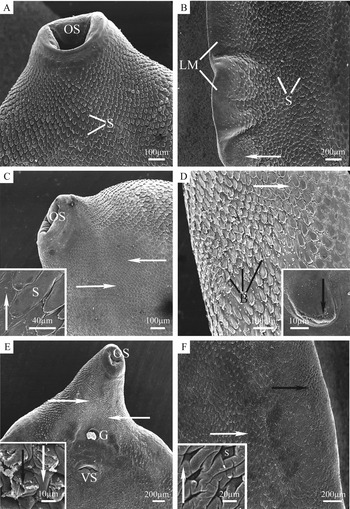
The majority of flukes showed little change from normal morphology. Spines were seen to project clearly from the tegumental surface and there was no change to the surface of the oral sucker (Fig. 1A), ventral sucker or the gonopore. However, some indications of disruption were seen in a number of the flukes examined. This consisted of small isolated patches of swelling of the tegument between the spines in the midbody and tail regions (Fig. 1B).
Scanning electron micrographs (SEMs) of Fasciola hepatica (Oberon isolate) which had been treated for 24 h in vitro with either verapamil (1×10⁻⁴m) (A, B); TCBZ.SO (15 μg mL−1) (C, D); or a combination of verapamil (1×10⁻⁴m) and TCBZ.SO (15 μg mL−1) (E,F). (A) Oral cone. Spines (S) can be seen projecting from the surface. OS, oral sucker; (B) Midbody region. An area of mild swelling (arrow) can be seen towards the lateral margin (LM). Spines (S) cover the rest of the surface; (C) Oral cone. Widespread, relatively severe swelling (arrows) can be seen behind the oral sucker (OS). Inset shows swelling (arrow) of the tegument between the spines (S); (D) Midbody region. Swelling of the tegument can be seen between the spines (arrow) and blebs (B) are present on and between the spines. Inset shows the beginning of sloughing from the tip of the spine (arrow); (E) Oral cone. Severe swelling and blebbing (arrows) of the tegumental syncytium are evident. G, gonopore; OS, oral sucker; VS, ventral sucker. Inset shows swelling between the spines (white arrow) and sloughing of the tegumental syncytium (black arrow) from the tip of a spine; (F) Midbody region. Severe swelling of the tegument (white arrow) can be seen towards the lateral margin. In areas, this swelling totally engulfs the spines (black arrow). Inset shows a higher-power SEM of swollen tegument (arrow) and a submerged spine (S).

Oberon isolate treated with TCBZ.SO (15 μg mL−1, 24 h)
In the oral cone region, swelling was present to some extent on all the specimens, ranging from small isolated areas of mild swelling to widespread, moderately severe, swelling which covered almost half of the oral cone (Fig. 1C).
In the midbody region, the disruption was concentrated towards the lateral margins. Swelling of the tegumental syncytium between the spines was the main form of disruption observed and was present to varying degrees of severity. Blebbing was also observed in the midbody region in approximately half of the specimens examined and was generally found in association with swollen tegument (Fig. 1D). In the most severely affected specimens, swelling of the tegument covering the spines had occurred. Disruption to the tegument at the tips of some of the spines was evident, and this was accompanied by limited loss of the apical plasma membrane (Fig. 1D inset).
In the tail region, swelling of the tegument between the spines was observed in most specimens, but was less extensive than that seen in the midbody region. However, swelling of the tegument covering the spines was more widespread. No blebbing of the surface was observed in the tail region.
Oberon isolate treated with a combination of R(+)-VPL (1×10⁻⁴m) and TCBZ.SO (15 μg mL−1) for 24 h
In the oral cone region, some degree of tegumental swelling was observed in all specimens. In more severe cases, this swelling covered more than half of the region (Fig. 1E) and, in the most seriously affected areas, the swollen tegument had engulfed the spines. Blebbing of the tegumental surface was also present in approximately half of the specimens observed and was associated (both in location and severity) with swollen tegument. In a small number of specimens, generally in areas where severe swelling and blebbing had occurred, sloughing of the tegumental syncytium from the tips of the spines was observed (Fig. 1E inset), although no spine loss had occurred.
In the midbody region, the disruption was very similar to that seen in the oral cone region. Swelling of the tegument was most severe towards the lateral margins, where swollen tegument had engulfed the spines in a number of specimens (Fig. 1F). Blebbing was seen in less than half of the specimens and was most severe in areas that were swollen. Towards the midline of the flukes, swelling of the tegument between the spines was less severe, although swelling of the tegument covering the spines was more widespread. Sloughing of the tegument at the tips of the spines was seen in the most severely disrupted areas at the lateral margins, but again no spine loss had occurred.
Less disruption was seen in the tail region than in the other regions and was more evenly distributed, with swelling predominating. Little or no blebbing and no tegumental sloughing were seen in this region.
Oberon isolate treated with TCBZ.SO (50 μg mL−1, 24 h)
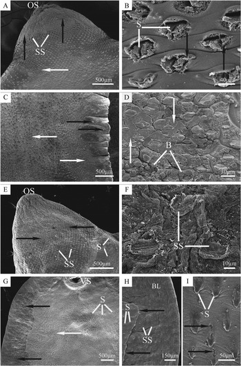
In the oral cone region, widespread disruption was observed in all specimens. In the most severe cases (but only a few specimens), complete sloughing of the tegumental syncytium had occurred over large areas of the oral cone (Fig. 2A). In the majority of flukes, however, sloughing of the tegument around the spine tips was observed (Fig. 2B) and, in some cases, this had also resulted in the loss of the spines. Some swelling of the tegumental syncytium between the spines was seen in all specimens. In those specimens where tegumental sloughing was not observed, there was widespread swelling that was severe in localized patches. Blebbing of the tegumental surface was present only in those specimens that had severe swelling and was restricted to these areas.
Scanning electron micrographs (SEMs) of Fasciola hepatica (Oberon isolate) which had been treated in vitro for 24 h with either TCBZ.SO (50 μg mL−1) (A–D) or a combination of TCBZ.SO (50 μg mL−1) and verapamil (1×10⁻⁴m) (E–I). (A) Oral cone. An area of tegumental sloughing (black arrows) can be seen behind the oral sucker (OS) on the dorsal surface. Spine sockets (SS) can be seen in the disrupted tegument posterior to the sloughed area. On the shoulders of the fluke, the tegument is swollen (white arrow); (B) High-power SEM of the oral cone showing the tegumental syncytium sloughing (arrows) away from the spines (S); (C) Midbody region. Swelling and blebbing (white arrows) can be seen over large areas of the midbody. Very small patches of tegumental sloughing (black arrows) are also visible at the lateral margin; (D) Higher-power SEM of the midbody region, showing severe swelling (arrows) and blebbing (B) of the tegumental surface; (E) Oral cone, dorsal surface. The tegumental syncytium has been completely sloughed away over most of this region, exposing the underlying basal lamina (black arrows) and empty spine sockets (SS). Posterior to this area of sloughing, spines (S) remain in the intact tegument. OS, oral sucker; (F) Higher-power SEM of the oral cone showing empty spine sockets (SS) on the exposed basal lamina; (G) Midbody region. Extensive sloughing (black arrows) can be seen along the lateral margin. Swelling and blebbing (white arrow) of the tegumental surface can be seen in large patches on the tegument that remains. S, spines; VS, ventral sucker; (H) A higher-power SEM showing the boundary (black arrows) between the sloughed and intact areas shown in Fig. 2G. Exposed basal lamina (BL), pitted with empty spine sockets (SS), can be seen in the sloughed area, while spines (S) remain on the adjacent area of intact tegumental syncytium; (I) Higher-power SEM of tegument from the midline in the midbody region. The beginning of tegumental sloughing (black arrows) can be seen on and between the spines (S).

Less severe disruption was seen in the midbody region and was focused on the lateral margins, with widespread, moderately severe, swelling in all specimens. In a limited number of specimens, small patches of tegumental sloughing were observed at the lateral margins (Fig. 2C). In contrast, towards the midline of the flukes, swelling and blebbing were only seen in localized patches (Fig. 2D). Blebbing of the tegumental surface was associated with the most badly swollen areas and so followed the same pattern.
The least severe disruption was seen in the tail region. Areas of mild swelling were seen along the margin in all specimens and this swelling was widespread in half of the specimens. No blebbing or sloughing was evident on any of the specimens.
Oberon isolate treated with a combination of R(+)-VPL (1×10⁻⁴m) and TCBZ.SO (50 μg mL−1) for 24 h
The oral cone region showed very severe disruption: in every specimen tegumental sloughing was seen and was so severe as to expose the underlying basal lamina and empty spine sockets (Fig. 2E and F). This sloughing was also extensive. In half of the flukes examined, total sloughing had occurred over the entire area between the oral sucker and the gonopore and in the other half the basal lamina was exposed over the entire oral cone region (Fig. 2E). In the specimens where the tegument remained over part of the surface, it was extremely disrupted.
Very severe disruption was seen in the midbody region, but it was less extensive than that seen in the oral cone region. Tegumental sloughing was seen in all specimens, but was confined to the lateral margins, exposing the basal lamina along the entire length of this region (Fig. 2G). Disruption decreased towards the midline of the flukes. Spine loss from otherwise intact tegument occurred close to the boundary between intact and sloughed tegument (Fig. 2H). Closer to the midline, sloughing was confined to the spine tips and occasionally to the apical plasma membrane between the spines (Fig. 2I). Along the midline itself, severe patches of swelling and blebbing were seen in most specimens (Fig. 2G).
In the tail region, sloughing occurred in the majority of specimens, around the margin of the fluke in the most severe cases, leaving intact tegument present only along the midline well above the excretory pore. However, most specimens only bore patches of tegumental sloughing at the lateral margins, the tegument covering up to two-thirds of the surface in this region. Swelling and blebbing occurred where the tegument remained.
Oberon isolate treated with TCBZ.SO (50 μg mL−1), time-matched to combination treatment time (13 h); and Oberon isolate treated with TCBZ.SO (50 μg mL−1) until flukes became inactive (15 h)
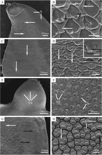
The results of these two treatments were similar and so will be described together. The oral cone region was the most disrupted region. Severe and widespread swelling of the interspine tegument was seen in all of the specimens, accompanied by mild blebbing in isolated patches (Fig. 3A). Sloughing of the apical plasma membrane from the spine tips was also seen (Fig. 3B), but was only present in a few of the specimens and was neither severe nor widespread.
Scanning electron micrographs (SEMs) of Fasciola hepatica (Oberon isolate) which had been treated in vitro with either a pre-incubation in NCTC (2 h) followed by TCBZ.SO (50 μg mL−1) for 11 h (A–D) or a pre-treatment of verapamil (1×10⁻⁴m) for 2 h followed by a combination of verapamil (1×10⁻⁴m) and TCBZ.SO (50 μg mL−1) until they became inactive (11 h) (E–H). (A) Oral cone. A large area of swollen tegumental syncytium (arrows) can be seen behind the oral sucker (OS). S, spines; (B) Higher-power SEM of the oral cone region. Swollen tegumental syncytium (white arrows) can be seen between the spines (S), a number of which show the start of tegumental sloughing (black arrows) at their tips; (C) Midbody region. Patches of swelling and blebbing (arrows) are present on the tegumental surface; (D) A medium-power SEM of the midbody region showing tegumental swelling (white arrows). At the tips of some of the spines, the tegument is beginning to slough off (black arrow). Inset shows a higher-power SEM of a spine from which the tegumental syncytium is sloughing off (black arrow); (E) Dorsal oral cone region. Extensive sloughing of the tegumental syncytium covering the spines can be seen, leaving large empty spine sockets (SS); (F) Higher-power SEM of the oral cone. Sloughing of the tegumental syncytium has exposed the crystalline spines (S) or left large empty spine sockets (SS) where the spines have been lost; (G) Midbody region. Small areas of tegumental sloughing (black arrows) can be seen towards the lateral margins. A patch of severely swollen tegument (white arrow) is present towards the midline; (H) SEM of the midbody region showing severe swelling of the tegumental syncytium between (white arrow) and covering (black arrow) the spines. This area is also covered in a carpet of blebs (B).

In the midbody region, swelling was again the main form of disruption seen and was concentrated towards the lateral margins (Fig. 3C), although it was seen in isolated patches throughout the region. In the most severe cases, the tegument had engulfed some of the spines and sloughing of the apical plasma membrane had occurred from some of the spine tips (Fig. 3D).
Moderate swelling and furrowing of the tegument occurred in the tail region, but no other disruption was observed.
Oberon isolate treated with a combination of R(+)-VPL (1×10⁻⁴m) and TCBZ.SO (50 μg mL−1) until flukes became inactive (2+11 h)
In the oral cone region, sloughing of the tegumental covering from the spines was seen in the majority of specimens. In approximately half of these specimens, the sloughing was very severe, resulting in widespread spine loss from the tegument, and leaving behind large empty spine sockets (Fig. 3E and F). In the remainder of the specimens, tegumental sloughing was evident to a much lesser extent and was most apparent towards the lateral margins. In this group of specimens, sloughing had occurred at the spine tips, but the spines themselves generally remained embedded in the tegumental syncytium. In a few specimens, the only disruption observed took the form of patches of swelling and blebbing.
In the midbody region, small patches of tegumental sloughing were seen, surrounding empty spine sockets, towards the lateral margins of roughly half of the specimens (Fig. 3G). However, swelling of the tegument and blebbing of the tegumental surface were the main forms of disruption observed, occurring in all of the specimens observed, and were generally severe and widespread, especially along the lateral margins and covering up to half of the surface in this region of the body (Fig. 3H). Towards the midline, many flukes showed only isolated patches of swelling and little or no blebbing.
A virtually identical pattern of disruption was seen in the tail region to that described for the midbody region, but it was generally less severe.
Cullompton isolate treated with TCBZ.SO (50 μg mL−1), time-matched to combination treatment time (8 h)
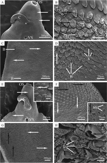
In the apical cone region, moderately severe and widespread swelling of the tegument between the spines was seen in all of the specimens, together with blebbing (Fig. 4A). Some specimens displayed patches of more severe disruption, in which tegumental swelling had occurred (both around and between the spines), and a carpet of blebs covered the surface. Sloughing of the tegument from the tips of the spines occurred in a few specimens (Fig. 4B), but no spine loss was observed in any specimen.
Scanning electron micrographs (SEMs) of Fasciola hepatica (Cullompton isolate) which had been treated in vitro with TCBZ.SO (50 μg mL−1) for 8 h (A–D) or a pre-treatment in verapamil (1×10⁻⁴m) for 2 h followed by a combination of verapamil (1×10⁻⁴m) and TCBZ.SO (50 μg mL−1) until they became inactive (2+6 h) (E–H). (A) Oral cone region. Swelling (arrows) of the tegumental syncytium can be seen behind the oral sucker (OS). C, cirrus; *, gonopore; VS, ventral sucker; (B) A higher-power SEM of the apical cone region, showing some sloughing (black arrows) at the tips and on the backs of the spines and also small blebs (B) on the tegumental surface; (C) Midbody region. Widespread blebbing (B) and swelling (arrows) of the tegument are present, both at the lateral margin and towards the midline of the fluke; (D) A higher-power SEM of the midbody region reveals severe tegumental swelling (arrow), which has engulfed a number of spines (S). Blebs (B) occur on the surface of the tegument between and covering the spines; (E) SEM of the apical cone region showing areas of severe swelling (arrows) of the tegumental syncytium between the oral sucker (OS) and the ventral sucker (VS). The cirrus (C) has everted from the gonopore. Inset shows a higher-power SEM of swollen tegument (arrow); (F) A higher-power SEM of the apical cone region which shows sloughing (black arrows) of the tegumental syncytium between the spines and also from the back of the spines, leaving empty spine sockets. A large area of severe swelling (white arrows) can also be seen. Inset shows tegumental sloughing (black arrow) and empty spine sockets (SS); (G) Midbody region. Sloughing (black arrows) of the tegumental syncytium at the spine tips, resulting in spine loss, can be seen along the lateral margin. Towards the midline of the fluke, severe swelling and blebbing (white arrows) are evident; (H) A higher-power SEM of the midbody region, showing a dense carpet of blebs (B) over the surface and sloughing and splitting (arrows) of the tegumental covering around the spines.

In the midbody region, swelling and blebbing were the only forms of disruption seen and were more severe towards the lateral margins. Severe swelling and blebbing covered approximately one-third of the surface in most specimens, with a greater area (up to two-thirds) showing some level of tegumental swelling (Fig. 4C and D).
In the tail region, swelling and blebbing were again the only forms of disruption observed, but were less widespread than in the midbody, with generally about one-third of the surface affected.
Cullompton isolate treated with a combination of R(+)-VPL (1×10⁻⁴m) and TCBZ.SO (50 μg mL−1) until flukes became inactive (2+6 h)
In the oral cone region, severe swelling and blebbing covered approximately half of the surface in all the specimens examined (Fig. 4E). Sloughing of the tegument covering the spines was also seen in the majority of specimens and was generally more apparent towards the lateral margins. In the most severe cases, this sloughing resulted in spine loss (Fig. 4F).
In the midbody region, in a very small number of specimens, small patches were observed where the tegument had sloughed away to reveal the basal lamina. However, sloughing and separation of the tegument from the spine tips and loss of spines from the tegument were more common and were seen to some degree in most specimens (Fig. 4G and H). Sloughing of the tegument was far more pronounced at the lateral margins. Swelling of the tegument, both between and covering the spines, and blebbing of the surface were evident in all specimens and covered up to half of the surface in most of the specimens examined (Fig. 4G and H).
In the tail region, no spine loss was seen in any of the specimens and sloughing at the spine tips was only seen in isolated patches. Swelling and blebbing were observed in all specimens, but were less severe and widespread than that evident in the midbody region.
Cullompton isolate treated with TCBZ.SO (50 μg mL−1) until flukes became inactive (9 h)
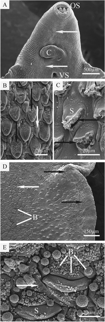
In the oral cone region, the predominant form of disruption observed was swelling of the tegument between the spines. It was widespread in all of the flukes examined and usually was concentrated towards the midline of the flukes (Fig. 5A and B), although it affected the entire region. Large areas of severe swelling were seen in half of the flukes and blebbing of the tegumental surface was generally present, but not severe, in these areas. In many of these flukes, the tegument covering the spines had also swollen, giving them an enlarged and bulbous appearance, and sloughing of the apical plasma membrane was evident on the back and tip of the spines (Fig. 5B). In the other half of the specimens, swelling was not as severe and the spines projected from the surface; however, the tegument covering the spines had split, exposing the spines (Fig. 5C).
Scanning electron micrographs (SEMs) of Fasciola hepatica (Cullumpton isolate) which had been treated in vitro with TCBZ.SO (50 μg mL−1) until they became inactive (9 h). (A) Oral cone. Swelling and blebbing (arrows) of the tegument are evident along the midline of this region, extending from the oral sucker (OS) to the ventral sucker (VS). The cirrus (C) is everted; (B) A higher-power SEM of the oral cone, showing swelling of the tegument between the spines (white arrow) and sloughing of the apical plasma membrane (black arrow) from the back and tip of the spines; (C) SEM of the oral cone region showing splitting (arrows) of the tegument covering the tip of the spines (S); (D) Midbody region. Sloughing of the tegument can be seen as a strip along the lateral margin of the fluke (black arrows). Where the tegument is intact, it is swollen (white arrow) and surface blebbing (B) is evident; (E) A higher-power SEM of the midbody region, showing a carpet of large and small blebs (B) covering the surface. Swelling (arrow) of the tegument between and covering the spines (S) can also be seen.

The midbody region was consistently the most severely affected region. In half of the specimens examined, sloughing of the tegument around the spine tips was observed, leading to the loss of spines. Other flukes showed complete sloughing of the syncytium along the lateral margin (Fig. 5D). Where the tegument remained, it was swollen between and around the spines and was covered by a carpet of large and small blebs (Fig. 5D and E).
In the tail region, an almost identical pattern of disruption to that described for the midbody region was seen, but was not as severe. Sloughing of the syncytium to expose the basal lamina was only seen in one specimen and spine loss and disruption at the spine tips was less common. The severity of swelling and blebbing was very similar to that seen in the midbody region, but was less widespread, with generally less than half of the region affected.
Summary of results
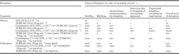
A summary of the SEM results is presented in Table 3.
Summary of scanning electron microscopy results

Key: –, no noticeable disruption; x, mild disruption; xx, widespread mild disruption; xxx, moderately severe disruption; xxxx, severe disruption; xxxxx, very severe disruption; TCBZ.SO, triclabendazole sulphoxide; VPL, R(+)-verapamil.
discussion
From the results of this study, it is evident that co-incubation of TCBZ.SO with R(+)-VPL, a Pgp inhibitor, can potentiate drug activity against the TCBZ-resistant Oberon isolate of F. hepatica. The combined drug treatment also increased the impact of TCBZ.SO on the TCBZ-susceptible Cullompton isolate, but not to such a great extent, as judged by the degree of surface disruption and the time taken to immobilize the flukes. Treatment with R(+)-VPL on its own resulted in only slight swelling of the tegument of both isolates, even after 24 h incubation. The phenomenon of greater disruption of the Oberon isolate was more distinct at the higher TCBZ.SO concentration used.
For the Oberon isolate, with TCBZ.SO at the lower concentration of 15 μg mL−1, there was limited surface disruption (even after 24 h incubation), which took the form of swelling and blebbing. The changes observed were similar to those described by Walker et al. (Reference Walker, McKinstry, Boray, Brennan, Trudgett, Hoey, Fletcher and Fairweather2004) and Devine et al. (Reference Devine, Brennan, Lanusse, Alvarez, Trudgett, Hoey and Fairweather2009). The flukes were actively moving at the end of the incubation period. In the presence of R(+)-VPL, very limited movement was seen in only half the flukes examined and the disruption was greater (see Table 3). At the higher concentration of 50 μg mL−1 for TCBZ.SO, the flukes were motionless on recovery at 24 h. There was more widespread blebbing and spine disruption, along with spine loss and some sloughing of the tegument over the oral cone region in a number of specimens. The changes paralleled those detailed by Walker et al. (Reference Walker, McKinstry, Boray, Brennan, Trudgett, Hoey, Fletcher and Fairweather2004). More extensive sloughing and spine loss was observed when R(+)-VPL was included in the incubation medium and the flukes were inactive.
A similar level of disruption was seen when the combination experiment was stopped at the point when the flukes became inactive. However, when the changes were compared with those observed in the TCBZ.SO time-match specimens, the difference caused by inhibition of the drug efflux pumps was more striking, as the main change induced by TCBZ.SO on its own involved swelling of the tegument (Table 3). Moreover, the TCBZ.SO-treated flukes remained active, albeit at a reduced level. So, for the Oberon isolate, the concentration of TCBZ.SO employed was an important determining factor in the level of disruption attained. Time was far less of a factor, indicating that no further tissue disruption occurred after the cessation of movement.
With the Cullompton isolate, and a TCBZ.SO concentration of 50 μg mL−1, the combination drug treatment markedly reduced the time taken for the flukes to become inactive (cf. Fairweather et al. Reference Fairweather, Holmes and Threadgold1984). Furthermore, the surface impact was significantly greater than that seen in the TCBZ.SO time-matched flukes, especially in relation to spine disruption and loss, but also with regard to tegumental sloughing (Table 3). Although there was a relatively short time difference between the time-matched specimens and the TCBZ.SO only-treated flukes recovered at the point of inactivity, the surface changes were greater, with far more extensive tegumental sloughing and higher levels of blebbing, plus spine disruption and loss in the latter (Table 3).
From previous recordings of fluke motility, it is known that flukes can remain active in even simple saline media for prolonged periods of time (>3 days) and their morphology remains normal for a minimum of 30 h (Fairweather et al. Reference Fairweather, Holmes and Threadgold1983: figs 3, 5, 7). In another experiment, flukes recovered after 48 h incubation in NCTC were active and their morphology normal (Halferty et al. Reference Halferty, O'Neill, Brennan, Keiser and Fairweather2009: fig. 1A). So, a 24-h time period is well within the normal activity span of F. hepatica and the fact that some of the timings (especially in the combination experiments) were much shorter than this indicates that the effects on motility were due to drug action and not to prolonged cultivation in vitro.
The potentiation of TCBZ.SO activity by in vitro co-incubation with R(+)-VPL is similar to that observed with ketoconazole (KTZ) (Devine et al. Reference Devine, Brennan, Lanusse, Alvarez, Trudgett, Hoey and Fairweather2010c). Although KTZ is a recognized CYP inhibitor (Newton et al. Reference Newton, Wang and Lu1995; Pelkonen et al. Reference Pelkonen, Turpeinen, Hakkola, Honkakoski, Hukkanen and Raunio2008), it has dual activity in that it is also regarded as an inhibitor of Pgp-linked drug efflux pumps (Siegsmund et al. Reference Siegsmund, Cardarelli, Akasentjevich, Sugimoto, Pastan and Gottesman1994; Zhang et al. Reference Zhang, Hsieh, Izumi, Lin and Benet1998; Kim et al. Reference Kim, Wandel, Leake, Cvetkovic, Fromm, Dempsye, Roden, Belas, Chaudhary, Roden, Wood and Wilkinson1999; Sandstrom et al. Reference Sandstrom, Knutson, Knutson, Jansson and Lennernäs1999; Wang et al. Reference Wang, Lew, Casciano, Clement and Johnson2002; Ward et al. Reference Ward, Stelman, Morgan, Zeigler, Azzarano, Kehler, McSurdy-Freed, Proksch and Smith2004; Kageyama et al. Reference Kageyama, Namiki, Fukushima, Ito, Shibata and Takada2005). A number of Pgp and CYP inhibitors are known to have overlapping substrate specificities (Lin, Reference Lin2003). Verapamil has been utilized as a Pgp inhibitor in a number of studies on parasites (Alvinerie et al. Reference Alvinerie, Dupuy, Eeckhoutte and Sutra1999; Molento and Prichard, Reference Molento and Prichard1999; Kerboeuf et al. Reference Kerboeuf, Blackhall, Kaminsky and Von Samson-Himmelstjerna2003; Molento et al. Reference Molento, Lifschitz, Sallovitz, Lanusse and Prichard2004; Bartley et al. Reference Bartley, Mc Allister, Bartley, Dupuy, Menez, Alvinerie, Jackson and Lespine2009; James and Davey, Reference James and Davey2009; James et al. Reference James, Hudson and Davey2009; Kasinathan et al. Reference Kasinathan, Goronga, Messerli, Webb and Greenberg2010a, Reference Kasinathan, Morgan and Greenberg2011; Stitt et al. Reference Stitt, Tompkins, Dooley and Ardelli2011; Tompkins et al. Reference Tompkins, Stitt, Morrissette and Ardelli2011; Kasinathan and Greenberg, Reference Kasinathan and Greenberg2012; Ardelli and Prichard, Reference Ardelli and Prichard2013). Ketoconazole has been exploited more as a Pgp inhibitor than as a CYP inhibitor in some studies (Alvinerie et al. Reference Alvinerie, Dupuy, Kiki-Mvouaka, Sutra and Lespine2008; Bartley et al. Reference Bartley, Mc Allister, Bartley, Dupuy, Menez, Alvinerie, Jackson and Lespine2009, Reference Bartley, Morrison, Dupuy, Bartley, Sutra, Menez, Alvineire, Jackson, Devin and Lespine2012; González-Canga et al. Reference González-Canga, Fernández-Martínez, Sahagún-Prieto, Diez-Liébana, Sierra-Vega and García-Vieitez2009). It has also been employed in its capacity as a CYP inhibitor for in vivo experiments on TCBZ pharmacokinetics (Virkel et al. Reference Virkel, Lifschitz, Sallovitz, Ballent, Scarcella and Lanusse2009) and its impact on TCBZ action against TCBZ-resistant F. hepatica (Devine et al. Reference Devine, Brennan, Lanusse, Alvarez, Trudgett, Hoey and Fairweather2011b, Reference Devine, Brennan, Lanusse, Alvarez, Trudgett, Hoey and Fairweather2012).
Over-expression of Pgp has been linked to the development of resistance against a number of anthelmintics (Xu et al. Reference Xu, Molento, Blackhall, Riberio, Beech and Prichard1998; Molento and Prichard, Reference Molento and Prichard1999; Kerboeuf et al. Reference Kerboeuf, Blackhall, Kaminsky and Von Samson-Himmelstjerna2003; Wolstenholme et al. Reference Wolstenholme, Fairweather, Prichard, Von Samson-Himmelstjerna and Sangster2004; Prichard and Roulet, Reference Prichard and Roulet2007; Ardelli and Prichard, Reference Ardelli and Prichard2008, Reference Ardelli and Prichard2013; James and Davey, Reference James and Davey2009; James et al. Reference James, Hudson and Davey2009; Messerli et al. Reference Messerli, Kasinathan, Morgan, Spranger and Greenberg2009; Stitt et al. Reference Stitt, Tompkins, Dooley and Ardelli2011; Tompkins et al. Reference Tompkins, Stitt, Morrissette and Ardelli2011; Yan et al. Reference Yan, Urdaneta-Marquez, Keller, James, Davey and Prichard2012). Results of experiments carried out in vitro have shown that it is possible to reverse the resistance status (i.e. restore the drug sensitivity) of helminths by co-incubation with Pgp inhibitors such as VPL (e.g. Beugnet et al. Reference Beugnet, Gauthey and Kerboeuf1997; James and Davey, Reference James and Davey2009; Kasinathan et al. Reference Kasinathan, Morgan and Greenberg2011; Stitt et al. Reference Stitt, Tompkins, Dooley and Ardelli2011; Tompkins et al. Reference Tompkins, Stitt, Morrissette and Ardelli2011; Ardelli and Prichard, Reference Ardelli and Prichard2013). The combined use of anthelmintics plus Pgp inhibitors such as KTZ and VPL has been advanced as a strategy to deal with drug resistance, by enhancing drug availability and thereby aiming to improve drug efficacy (Dupuy et al. Reference Dupuy, Larrieu, Sutra, Lespine and Alvinerie2003; Alvarez et al. Reference Alvarez, Merino, Molina, Pulido, McKellar and Prieto2006; Hugnet et al. Reference Hugnet, Lespine and Alvinerie2007; Alvinerie et al. Reference Alvinerie, Dupuy, Kiki-Mvouaka, Sutra and Lespine2008; Lespine et al. Reference Lespine, Alvinerie, Vercruysse, Prichard and Geldhof2008, Reference Lespine, Dupuy, Alvinerie, Comera, Nagy, Krajcsi and Orlowski2009, Reference Lespine, Ménez, Bourguinat and Prichard2012; Kasinathan et al. Reference Kasinathan, Goronga, Messerli, Webb and Greenberg2010a, Reference Kasinathan, Morgan and Greenberg2011; Lifschitz et al. Reference Lifschitz, Entrocasso, Alvarez, Lloberas, Ballent, Manazza, Virkel, Borda and Lanusse2010a, Reference Lifschitz, Suarez, Sallovitz, Cristel, Imperiale, Ahoussou, Schiavi and Lanusseb; Kerboeuf and Riou, Reference Kerboeuf and Riou2011; Kasinathan and Greenberg, Reference Kasinathan and Greenberg2012). Enhanced activity and efficacy of such combinations has been demonstrated by Molento and Prichard (Reference Molento and Prichard1999), Bartley et al. (Reference Bartley, Mc Allister, Bartley, Dupuy, Menez, Alvinerie, Jackson and Lespine2009, Reference Bartley, Morrison, Dupuy, Bartley, Sutra, Menez, Alvineire, Jackson, Devin and Lespine2012) and Lifschitz et al. (Reference Lifschitz, Entrocasso, Alvarez, Lloberas, Ballent, Manazza, Virkel, Borda and Lanusse2010a, Reference Lifschitz, Suarez, Sallovitz, Cristel, Imperiale, Ahoussou, Schiavi and Lanusseb), for example.
In comparison with our knowledge of membrane transporters in nematodes, relatively little is known about the role of Pgp and other ABC transporters in trematodes. A putative ABC transporter homologue, named Fhmdr1, has been identified in F. hepatica and is expressed in immature (4–5 weeks old), but not adult, flukes (Reed et al. Reference Reed, Panaccio, Strugnell and Spithill1998). A separate Pgp gene has been identified that is expressed in adult flukes (Wilkinson et al. Reference Wilkinson, Law, Hoey, Fairweather, Brennan and Trudgett2012). Comparison of flukes from field populations characterized as TCBZ-resistant or TCBZ-susceptible showed that a greater haplotype diversity of the gene occurred in the TCBZ-resistant population. Moreover, one allele was particularly frequent in the TCBZ-resistant population and was characterized by an amino acid substitution associated with a functionally important region of the Pgp molecule (Wilkinson et al. Reference Wilkinson, Law, Hoey, Fairweather, Brennan and Trudgett2012). Analysis of other TCBZ-resistant populations will establish whether the substitution plays a role in the development of TCBZ resistance. Application of antibodies against a number of mammalian transporters has shown that orthologues of these molecules are expressed in Fasciola gigantica, a close relative of F. hepatica (Kumkate et al. Reference Kumkate, Chunchob and Janvilisri2008). The Schistosoma mansoni genome contains >20 genes that are predicted to encode ABC transporters (Kasinathan and Greenberg Reference Kasinathan and Greenberg2012). The genes include the Pgp homologue, SMDR2 (Bosch et al. Reference Bosch, Wang, Tao and Shoemaker1994) and SmMRP1, a homologue of mammalian multidrug resistance-associated protein 1 (MRP1) (Kasinathan et al. Reference Kasinathan, Morgan and Greenberg2010b). Expression of the two proteins increases in response to treatment with praziquantel (Messerli et al. Reference Messerli, Kasinathan, Morgan, Spranger and Greenberg2009; Kasinathan et al. Reference Kasinathan, Morgan and Greenberg2010b). Moreover, expression of SMDR2 is greater in schistosome isolates displaying reduced sensitivity to praziquantel and expression of SMDR2 and SmRP1 is higher in juvenile worms compared with adult worms; the juvenile stage is known to be more refractory to praziquantel action (Messerli et al. Reference Messerli, Kasinathan, Morgan, Spranger and Greenberg2009; Kasinathan et al. Reference Kasinathan, Morgan and Greenberg2010b). Knockdown of expression of these two proteins by means of RNAi leads to disruption of egg production, as does incubation of worms with known inhibitors of mammalian multidrug resistance proteins (including the Pgp inhibitor, R(+)-VPL) (Kasinathan et al. Reference Kasinathan, Morgan and Greenberg2011). Treatment of S. mansoni worms with inhibitors also affects the movement of labelled substrates of Pgp and MRP through the excretory system of the worms (Sato et al. Reference Sato, Kusel and Thornhill2002, Reference Sato, Kusel and Thornhill2004). Praziquantel and VPL inhibit the transport activity of SMDR2 expressed in mammalian cells (Kasinathan et al. Reference Kasinathan, Goronga, Messerli, Webb and Greenberg2010a). Apart from the excretory system, the main immunoreactive site for tissue localization of transporters in S. mansoni and F. gigantica is the gut (Kumkate et al. Reference Kumkate, Chunchob and Janvilisri2008; Messerli et al. Reference Messerli, Kasinathan, Morgan, Spranger and Greenberg2009; Kasinathan et al. Reference Kasinathan, Morgan and Greenberg2010b). The gut is also the predominant site for Pgp expression in nematodes (Lincke et al. Reference Lincke, Broecks, Plasterk and Borst1993; Broeks et al. Reference Broeks, Janssen, Calafat and Plasterk1995; Smith and Prichard, Reference Smith and Prichard2002; Kerboeuf et al. Reference Kerboeuf, Blackhall, Kaminsky and Von Samson-Himmelstjerna2003; Zhao et al. Reference Zhao, Sheps, Ling, Fang and Baillie2004; Ardelli and Prichard, Reference Ardelli and Prichard2013). Both praziquantel and TCBZ are regarded as Pgp substrates/inhibitors (Dupuy et al. Reference Dupuy, Alvinerie, Ménez and Lespine2010; Kasinathan et al. Reference Kasinathan, Goronga, Messerli, Webb and Greenberg2010a; Kerboeuf et al. Reference Kerboeuf, Riou, Neveu and Issouf2010).
As indicated above, there is a growing body of evidence that platyhelminth (trematode) parasites possess drug transporters and that the latter are involved in the development of resistance to anthelmintic drugs such as praziquantel and TCBZ. For TCBZ, the mechanism of resistance may be a multifactorial one, involving both drug metabolism and drug uptake/efflux. This appears to be the case both for the Oberon isolate used in the present study and for another TCBZ-R isolate, the Sligo isolate (Alvarez et al. Reference Alvarez, Solana, Mottier, Virkel, Fairweather and Lanusse2005; Mottier et al. Reference Mottier, Alvarez, Fairweather and Lanusse2006b; Savage et al. Reference Savage, Meaney, Brennan, Hoey, Trudgett and Fairweather2013). Reduced drug uptake would lower the effective concentrations of TCBZ metabolites within the fluke and this, combined with more rapid metabolism and inactivation of the compounds, would reduce their ability to reach the target molecule in sufficient concentration for effective drug action. Moreover, alteration of the activity of transporters such as Pgp by means of inhibitors may be one option to follow in the effort to maintain the effectiveness of TCBZ and other drugs. Clearly, more needs to be learnt about the identity, expression, localization and physiological roles of these molecules in F. hepatica. They represent ‘druggable’ targets in their own right, as suggested by Kasinathan et al. (Reference Kasinathan, Morgan and Greenberg2011), Kasinathan and Greenberg (Reference Kasinathan and Greenberg2012) and Kerboeuf and Riou (Reference Kerboeuf and Riou2011).
FINANCIAL SUPPORT
This work was supported by a BBSRC/Defra grant (C00082X/1) and by a grant from the European Union (Deliver grant, no. FOOD-CT-200X-023025).