Branch pulmonary artery stenosis is a common problem encountered in patients with CHD. It occurs as an isolated lesion, in association with other congenital cardiac defects, and following cardiac surgery.Reference Baum, Khoury, Ongley, Swan and Kincaid1–Reference Patel, Kenny, Gonzalez, Amin, Ilbawi and Hijazi4 Stent implantation for the management of branch pulmonary artery stenosis is a frequently performed transcatheter intervention and has been shown to be an effective treatment modality in the current era.Reference Moore, Vincent and Beekman5–Reference Holzer, Gauvreau and Kreutzer8 While stenting may require further intervention in growing children, long-term outcomes are good.Reference Law, Shamszad and Nugent9–Reference Stanfill, Nykanen, Osorio, Whalen, Burke and Zahn11
A significant portion of stents implanted in the branch pulmonary arteries are for ostial stenosis. In a recent report from the IMPACT database, 43% of patients undergoing branch pulmonary artery stent implantation did so for ostial stenosis. While these patients have high rates of success, there are several unique challenges associated with the procedure.Reference Lewis, Kennedy and Ginns6 These include adequate anatomic definition of an often-complex area of stenosis and overlying adjacent structures by angiography; stent stability during implantation; and stent malposition. Malposition may result in inadequate relief of the ostial lesion and jailing of the upper lobe branch or contralateral branch pulmonary artery. Additionally, malposition may necessitate additional stent implantation. While interventionists are familiar with the challenges of pulmonary artery ostial stenoses, surprisingly, there are no publications that specifically assess the frequency of stent malposition and its associated complications in patients who undergo stent implantation for ostial branch pulmonary artery stenosis or discuss the technical considerations when intervening on this particular lesion. We aimed to evaluate the early and mid-term outcomes and complications of stent placement for ostial branch pulmonary artery stenosis and discuss technical considerations to avoid or minimise complications.
Materials and methods
This study was approved by the institutional review board. A retrospective chart review was conducted to identify all patients with 1.5 or 2-ventricle physiology who underwent stent placement for branch pulmonary artery stenosis at our institution from 1 January, 2013 to 15 November, 2017. Angiograms were reviewed, and the distance from the ostium to the stenosis was measured. Patients with a stenosis within 5 mm of the ostium were considered to have ostial branch pulmonary artery stenosis and were included in the study. We collected demographic and clinical data including cardiac diagnoses, prior procedures (surgical or transcatheter), and interventions during the follow-up period.
Catheterisation data collected included the affected branch pulmonary artery, minimum pulmonary artery diameter, distal pulmonary artery diameter, right ventricle:systemic pressure ratio, and gradient across the stenosis pre- and post-stent placement. Procedural data collected included fluoroscopy time, pre-stent angioplasty, post-stent angioplasty, size of balloons used, size and type of stent used, and stent modifications prior to implantation. Additionally, we assessed the number of different angiographic projections needed to profile a stenosis. Angiograms performed in the main pulmonary artery or affected branch pulmonary artery were included. Patients who underwent bilateral pulmonary artery stent placement at the same catheterisation were not included in this portion of the analysis.
Stent malposition was defined in two categories: proximal malposition resulting in jailing of the contralateral branch pulmonary artery or distal malposition resulting in inadequate coverage of the ostial stenosis with or without jailing of the upper lobe branch. Angiograms and catheterisation reports were reviewed to determine whether there was jailing of the upper lobe branch or the contralateral branch pulmonary artery, whether the stent adequately covered the area of stenosis, as well as other complications.
In general, stents were delivered to the site of stenosis using a long delivery sheath over a stiff guidewire. Pre-stent angioplasty or post-implant dilation was at the discretion of the primary operator. The indications for intervention varied, but usually followed the published 2011 American Heart Association guidelines and included elevated right ventricle pressure, increased gradient from the main pulmonary artery to the affected branch pulmonary artery, a significant flow discrepancy, or pulmonary insufficiency.Reference Feltes, Bacha and Beekman12
Statistical analysis was performed using JMP Pro 14 statistical software (SAS institute Inc., Cary, North Carolina, United States of America). Descriptive statistics are presented as median (range). Assessment of differences between pre- and post-intervention variables was performed using a Wilcoxon signed-rank test. Wilcoxon rank-sum and Chi-squared analyses were performed where applicable statistical significance was set at a p-value <0.05.
Results
Patient profile
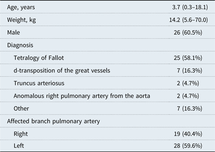
Forty-three patients underwent percutaneous stent placement for ostial branch pulmonary artery stenosis. Three underwent bilateral stent implantation during the same procedure and another underwent bilateral stent implantation during two separate procedures. A total of 47 ostial stenoses were stented. The median age and weight were 3.7 (0.3–18.1) years and 14.2 (5.6–70.0) kg, respectively. The most common diagnosis was tetralogy of Fallot in 58.1% (Table 1).
Baseline characteristics

Values are median (range) or n (%)
Procedural data and technical considerations
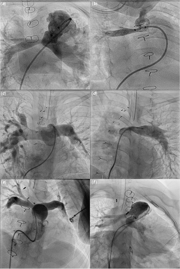
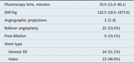
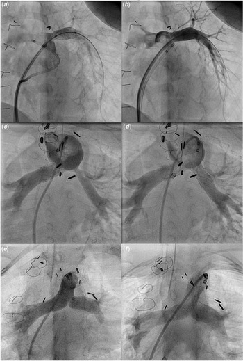
The median dose area product was 132.5 (18.6–1873.6) (µGy × m2)/kg. A median of 3 (2–8) angiographic projections were needed to adequately profile the ostial branch pulmonary artery stenosis for stent positioning due to superimposition of the main pulmonary artery on the area of stenosis or important surrounding structures (Fig 1). In two cases, 3D rotational angiography was utilised. Twenty-five (53.2 %) branch pulmonary arteries underwent angioplasty prior to stent placement. Comparing those who underwent initial pre-stent angioplasty to those who did not, there was no significant differences in the median minimum diameter:distal pulmonary artery diameter ratio [0.40 (0.13–0.57) versus 0.46 (0.19–0.72), p = 0.112]. The balloon diameter used for angioplasty tended to be the same size as the balloon used for stent implantation [8 (4–16) versus 9 (5–16), p = 0.259].
Examples of angiograms in patients with ostial stenosis and angiographic techniques used to assist with adequate anatomic definition. (a and b) Thirteen-month-old with tetralogy of Fallot (TOF) and atrioventricular canal status post-valve sparing TOF repair. (a) The caudal main pulmonary artery (PA) angiogram is suggestive of right PA stenosis, but the origin is not well seen. (b) A right axis oblique and cranial angulation with the catheter in the ostium better identifies the site of stenosis, which involves the ostium. (c and d) Eleven-year-old with TOF status post-repair with a transannular patch. (c) An initial main PA angiogram does not clearly identify the site of stenosis. (d) A left axis oblique (LAO) and cranial angulation with the catheter in the distal PA results in opacification of the ostium prior to the main PA due to pulmonary regurgitation. (e and f) Ten-year-old female with TOF/pulmonary atresia status post-repair with a right ventricle to PA conduit. (e) The ostia of both branch PAs are obscured by the main PA in the LAO and cranial projection. (d) Steep caudle angulation allows for visualisation of both ostia and their relationship to each other and the main PA.

For the initial intervention, 24 (51.1%) Genesis XD stents (Cordis Corp., Warren, New Jersey, United States of America) with varying lengths (19, 25, 29, and 39 mm) were implanted. These were mounted on balloons with a diameter ranging from 5 to 20 mm. Five of these stents were shortened by cutting off 1–3 rows of cells to avoid jailing the ipsilateral upper lobe branch. Twenty-three open-cell Valeo pre-mounted stents (Bard Peripheral Vascular, Tempe, Arizona, United States of America) were implanted (Table 2). Two of these were medium-sized (13 mm maximum diameter) and the rest were large-sized (18 mm maximum diameter) stents. In five cases, the large-sized Valeo pre-mounted stent was remounted onto a smaller balloon (6–8 mm), and in one case it was remounted onto a larger 14 mm balloon. Nine stents were further dilated immediately post-implantation. In six cases, the balloon was larger than the balloon used for stent implantation. In two cases, the balloon was the same size but higher pressure, and in one case the balloon was smaller but higher pressure (due to residual resistant stenosis).
Procedural data

Values are median (range) or n (%)
DAP indicates dose area product in (µGy × m2)
Outcomes
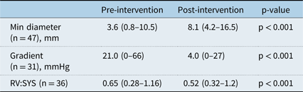
The median minimum ostial diameter improved from 3.6 (0.8–10.5) mm to 8.1 (4.2–16.5) mm (p < 0.001). The gradient across the stenosis was measured in 31 branch pulmonary arteries. The median gradient improved from 21.0 (0–66) to 4.0 (0–27) mmHg (p < 0.001). The right ventricle:systemic pressure ratio, measured in 36 cases, improved from 0.65 (0.28–1.16) to 0.52 (0.32–1.2) (p < 0.001) (Table 3).
Haemodynamic and angiographic data

Values are median (range)
RV:SYS indicates right ventricle to systemic pressure ratio
Stent malposition and complications
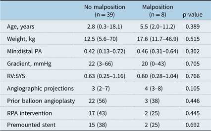
Stent malposition occurred in eight (17%) implantations. Five stents migrated distally and did not cover the ostial stenosis. Three of these migrated during inflation of the balloon, one migrated while re-sheathing the balloon, and one migrated during wire removal. Four of these five migrations resulted in a jailed upper lobe branch. Fortunately, all jailed upper lobe branches continued to have of good flow on post-implantation angiography. A second stent was placed in four of the cases with adequate relief of the ostial stenosis (Fig 2a and b). In the fifth case, the stenosis was relatively mild and likely contributed to the stent embolising so a second stent was not placed. Three stents migrated proximally resulting in partial jailing of the ostium of the contralateral branch pulmonary artery (Fig 2e and f). These three had preserved flow to the contralateral branch pulmonary artery. Table 4 compares those who had stent malposition with those who did not. There were no significant differences between the two groups.
Examples of stent malposition and intentional jailing of the upper lobe branch. (a and b) Five-year-old with tetralogy of Fallot (TOF) status post-repair with a transannular patch. (a) The stent migrated distally, resulting in jailing of the upper lobe branch. (b) A second stent was placed adequately relieving the region of stenosis. (c and d) Twenty-eight-month-old with TOF/pulmonary atresia status post-repair with a right ventricle (RV) to pulmonary artery (PA) conduit. (c) There is ostial stenosis of the left PA present, and the upper lobe branch originates adjacent to the site of stenosis. (d) Stent placement results in intentional jailing of the upper lobe branch, with preserved flow through the “open cells” of the Valeo stent. (e and f) Eight-year-old with TOF/pulmonary atresia status post-repair with an RV to PA conduit. (e) The main PA angiogram demonstrates ostial stenosis of the left PA. (f) Following stent implantation, there is adequate relief of the left PA stenosis. Partial jailing of the right PA is present, but flow is preserved to the right PA.

Comparison of patients without and with stent malposition

Values are median (range) or n (%)
Min:distal PA indicates minimum to distal pulmonary artery diameter ratio
RV:SYS indicates right ventricle to systemic pressure ratio
In four other cases, the distance from the ostial stenosis to the take-off of the upper lobe was shorter than any commercially available large-sized stent and intentional jailing of the ipsilateral upper lobe branch was carried out (Fig 2c and d). An open-cell Valeo stent was selected in three of the four cases. A Genesis XD stent was used in the very first case in the series. While the ostial stenosis was effectively relieved, two cases were noted to have diminished flow to the upper lobe. In the remaining two patients, there was no flow to the upper lobe branch.
Other complications included lung reperfusion edema (1), iliac vein injury (1), transient heart block (2), diminished arterial pulse requiring heparin infusion (1), and conduit tear with haemothorax and placement of a chest a tube (1) during additional Melody valve implant.
Follow-up
During a follow-up period of 2.4 (0.3–4.9) years, 18 ostial stents underwent subsequent catheterisation. The median minimum diameter:distal pulmonary artery diameter ratio was 0.76 (0.41–0.93). The median gradient was 11 (0–35) mmHg. Fifteen ostial stents underwent further dilation due to distal pulmonary artery growth or residual stenosis, and in two of these cases, there was concomitant significant in-stent intimal proliferation (defined as reduction in the stent lumen by at least 50%). One had a second stent placed within the existing stent for significant flow differential to affected lung. The median time to reintervention was 0.9 (0.4–3.3) years. Two of the stents underwent a second follow-up catheterisation with further stent dilation. One patient with tetralogy of Fallot underwent surgical conduit replacement for conduit stenosis.
Among eight jailed upper lobe branches, only three had a subsequent catheterisation during the follow-up period. Angioplasty through the side cell of the Valeo stent was performed in one patient with diminished flow. The other two continued to have good flow to the jailed upper lobe and did not require additional intervention. Of the three partially jailed contralateral pulmonary arteries, two had follow-up catheterisation during which the stent was further dilated. There was not obstruction to flow or difficulty accessing the contralateral pulmonary artery.
Discussion
This is the first report specifically describing outcomes of percutaneous stent implantation for ostial branch pulmonary artery stenosis in children with specific attention towards rates of stent malposition. We demonstrated that stent implantation is an effective treatment for ostial branch pulmonary artery stenosis; however, there are high rates of stent malposition that appear to be in excess of rates reported in larger series of patients undergoing stent implantation for proximal branch pulmonary artery stenosis. The patients in our cohort had significant improvements in branch pulmonary artery size, gradient across the stenosis, and right ventricle pressure due to relief of a proximal obstruction with a large distal vascular bed. Haemodynamically, our outcomes mirror those reported by Lewis et al from the IMPACT registry.Reference Lewis, Kennedy and Ginns6 Stapleton et al reported outcomes of bilateral stents for bifurcation stenoses in pulmonary arteries. Of the 35 patients with proximal branch pulmonary artery stenosis in that series, many likely had ostial stenosis. However, the emphasis was not the challenges and issues that are unique to ostial branch pulmonary artery stenosis, which we have focused on in this study.Reference Stapleton, Hamzeh and Mullins13
The important distinctions in ostial stenosis from other proximal pulmonary artery stenosis include difficulty defining ostial anatomy (Fig 1) and inherent issues with stent stability during implantation due to the short length of stenosis. Given these unique challenges, we suspected that patients with ostial stenosis may experience higher rates of stent malposition compared to patients with other forms of pulmonary artery stenosis, which is supported by our findings. Malposition occurred in eight stents (17%), and four of these required placement of a second stent. Additionally, intentional jailing of the ipsilateral upper lobe branch with adequate coverage of the ostium occurred in an additional four (8.5%) stent implantations. Therefore, a total of 12 (25.5%) stent implantations resulted in suboptimal stent positioning.
In the recent era, rates of stent malposition for proximal branch pulmonary artery stenosis, defined as stenosis proximal to the upper lobe branch, have been reported at 5% in two multicentre studies.Reference Ing, Khan, Kobayashi, Hagler and Forbes7, Reference Holzer, Gauvreau and Kreutzer8 The higher rates of stent malposition in our cohort are most likely associated with some of the difficulties that are unique to ostial branch pulmonary artery stenosis when compared to proximal branch stenosis that does not involve the ostium. Adequate imaging and delineation of the ostial anatomy can be particularly problematic. The ostial region can be obscured by the adjacent dilated main pulmonary artery and right ventricular outflow tract. We found that a steep caudal angulation can often profile both branch pulmonary artery ostia, which can help to determine how much distance the stent has before jailing the contralateral ostium. Furthermore, ostial stenosis may occur secondary to folding of the branch pulmonary artery against a pulsating dilated main pulmonary artery resulting in a very short length of stenosis limited to the ostium. These factors may result in suboptimal positioning of the stent prior to balloon inflation and contributed to the partial jailing of the contralateral branch pulmonary artery in two of the cases. We only used 3D rotational angiography in two cases, but this may be a useful tool to further define the relationship between the branch pulmonary arteries in these lesions and selecting the optimal angles for angiography and intervention.Reference Aldoss, Fonseca and Truong14 Additionally, overlay of 3D imaging (3DRA, CT, or MRI) may allow for more precise stent positioning and implantation and can be performed without adding a significant amount of time to the procedure.Reference Fagan, Kay, Carroll and Neubauer15, Reference Goreczny, Dryzek, Morgan, Lukaszewski, Moll and Moszura16 One technique we utilised to better assess the ostial anatomy in patients with pulmonary insufficiency is a selective branch pulmonary artery angiogram distal to the stenosis, allowing the regurgitant contrast to fill the ostium before the main pulmonary artery, which can otherwise obscure the ostium (Fig 1d).
Jailing of the ipsilateral upper lobe branch also occurred in 17% of stents placed, although in four of the cases this was intentional. Side branch jailing has been reported to occur in 10.5% of cases for proximal pulmonary artery stenting per the initial safety report from the IMPACT registry.Reference Moore, Vincent and Beekman5 In our series, intentional jailing was performed due to the limited commercially available stent lengths when a short distance from the upper lobe branch to the ostial stenosis was present. This, at least in part, was related to the small size of the patient. Fifty-five per cent of our entire cohort weighed less than 15 kg. Of the four patients with intentional jailing of the ipsilateral upper lobe branch, three weighed less than 15 kg. In the fourth patient who weighed 58 kg, the upper lobe branch originated abnormally close to the stenosis, making jailing of this branch unavoidable. One of the techniques we utilised to minimise the risk of jailing the upper lobe branch was modifying the stent to fit the anatomy. In five cases, a Genesis XD stent was shortened to accommodate the short length between the ostium and upper lobe branch. This new technique has been shown to be effective in relieving branch pulmonary artery stenosis with a relatively low rate of associated complications, particularly in small patients. Stents with sigmoidal hinges can be trimmed by removing 1–2 rows of stent cells using sharp scissors and leaving the sigmoidal hinge on the stent. The stent can be mounted on the balloon in the usual fashion. While there is theoretical concern for causing a pinhole in the balloon, the cut sigmoidal hinges are relatively soft and move away as the balloon inflates. The rates of pinhole rupture were relatively low (7.3%) in a previously published series.Reference Sullivan, Liou and Takao17 While malposition did not occur with the modified stents in our cohort, stability of the shortened stent may be compromised and therefore require more precise placement to avoid malposition. Alternatively, we used pre-mounted Valeo stents, which have an “open-cell” design by which side cells can be dilated up to 12 mm, hence preserving flow to the jailed upper lobe if needed (Fig 2d). Additionally, the large-sized Valeo stents can reach a maximum diameter of 18 mm and can be safely remounted on smaller or larger balloons as we did in several of the cases.Reference Travelli, Sullivan, Takao and Ing18 This technique allows implantation of larger stents at the ostium that can be further dilated to the normal adult vessel size over time. An alternative method, which we did not utilise is intentionally crossing the contralateral branch pulmonary artery with an open cell and dilating the side cell to the crossed pulmonary artery.Reference Boudjemline, Legendre and Ladouceur19
Stent malposition can also occur due to instability within the ostium in cases of a severe or highly resistant stenosis resulting in proximal or distal migration of the stent. Balloon angioplasty prior to stent implantation is often performed to assess for stability of the balloon during inflation and the initial waist on the balloon can serve as a reference for precise positioning of the stent. Interestingly, the severity of stenosis did not correlate with use of pre-stent angioplasty. In the three cases with stent malposition during inflation, balloon angioplasty was not performed. The type of stenosis may also affect stent stability. A fold or twist at the ostium may not provide adequate radial force to secure the stent during manipulation of catheters or wires within the stent. In these situations, prior balloon angioplasty may be beneficial to further characterise the compliance properties of the stenosis.
Limitations
There were several limitations to our study. This was a retrospective study and the data are from a single centre with a relatively small number of patients. The population was heterogenous, but all patients did have 1.5 or 2-ventricle physiology. Additionally, there were several operators with varying practice habits, which contributed to the heterogeneity of the data in this series. Finally, we were only able to provide medium-term follow-up data. We hope to have long-term follow-up data regarding further dilation of the stents, stent fractures, and the ability to access jailed vessels at subsequent catheterisations.
Conclusion
Stent implantation is a safe and effective procedure for ostial branch pulmonary artery stenosis. It results in significant improvements in branch pulmonary artery size, gradients across the stenosis, and right ventricle pressure. However, there are high rates of stent malposition and jailing of the upper lobe branch or the contralateral branch. The use of multiple angiographic projections, especially steep caudal projection, 3D rotational angiography, stents with open-cell design, and modification of stents may help mitigate some of these issues. At medium-term follow-up, there are low rates of in-stent restenosis, but further dilation may be necessary to accommodate somatic growth. Long-term follow-up in larger series is important.
Acknowledgements
This work was supported by the Children’s Hospital Los Angeles Heart Ambassadors and the Hearst Foundation.
Conflicts of Interest
The authors declare that they have no conflict of interest.