The somatosensory processes supporting perception and action discussed by Dijkerman & de Haan (D&dH) do not involve major hemispheric asymmetries, as illustrated by their Figure 1. These are briefly mentioned with reference to tactile object recognition and tactile agnosia. The role of spatial processing and of the right hemisphere, and the effects of unilateral spatial neglect after damage to the right posterior parietal cortex (PPC), are considered. D&dH's conclusion is that tactile agnosia may be caused by – and possibly through different underlying pathological mechanisms – both left- and right-sided brain damage.
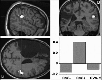
Physiological effect of left caloric vestibular stimulation (CVS) on touch in a left-brain–damaged patient (L6). The area of significant interaction of left CVS with right-sided touches in Patient L6 (left) is displayed on her magnetic resonance imaging (MRI) scan. The bar plot shows the average fMRI blood oxygenation level–dependent signal change (compared with rest in that brain region) before CVS (−), after CVS (+), and at a 30-minute delay (CVS-2). The values of signal change are mean-centered. Images are displayed with the anatomical right (R) on the right side. (Reprinted from Bottini et al. Reference Bottini, Paulesu, Gandola, Loffredo, Scarpa, Sterzi, Santilli, Defanti, Scialfa, Fazio and Vallar2005, figure 2, with permission of Neurology.)

A hemispheric asymmetry exists, however, at the much more basic level of pain sensation and proprioception. In a study of a large, unselected series of left- and right-brain-damaged patients, Sterzi et al. (Reference Sterzi, Bottini, Celani, Righetti, Lamassa, Ricci and Vallar1993) reported that these somatosensory deficits, as assessed by a clinical, standardized neurological exam, are more frequent after right-brain damage than after left-brain damage. This result contrasts with the time-honored view, which still informs clinical neurology (Ropper & Brown Reference Ropper and Brown2005), that there is no hemispheric asymmetry in the more “elementary,” non-cognitive stages of sensation. The asymmetry is not confined to the somatosensory domain and extends to the visual and motor systems: Both visual half-field and motor deficits (hemianopia and hemiplegia), contralateral to the side of a hemispheric lesion, are more frequent after right-hemispheric damage (Sterzi et al. Reference Sterzi, Bottini, Celani, Righetti, Lamassa, Ricci and Vallar1993).
One interpretation relates the higher incidence of left somatosensory deficits to disorders of spatial processing and, more specifically, to unilateral spatial neglect, which also are more frequent and severe after right-brain damage (Bisiach & Vallar Reference Bisiach, Vallar, Boller, Grafman and Rizzolatti2000). In line with this view, in right-brain–damaged patients the detection of left single and simple somatosensory stimuli (touches) is definitely, though temporarily (for about 30 min), improved by the physiological maneuvers, such as caloric vestibular stimulation, that also ameliorate other aspects of the neglect syndrome, in both extra-personal and personal space (for reviews, see Rossetti & Rode Reference Rossetti, Rode, Karnath, Milner and Vallar2002; Vallar et al. Reference Vallar, Guariglia, Rusconi, Thier and Karnath1997). Although the empirical data are definite, the precise nature of the relationships between somatosensory awareness and spatial processing is open to interpretation.
I take the view that in the somatosensory, as well as in the visual domain, perceptual awareness of a sensory event – that necessarily occurs in space – involves not only the more peripheral stages of sensory analysis, but also its localization in the sector of extra-personal (as in the case of visual stimuli), or personal, bodily (as in the case of somatosensory stimuli) space where the event takes place. Accordingly, in patients with right-brain damage, visual and somatosensory (left-sided in retinotopical and somatotopical reference frames) deficits improve, when the stimuli are presented in the right, preserved, side of space, with regard to spatial egocentric coordinate frames (Kooistra & Heilman Reference Kooistra and Heilman1989; Smania & Aglioti Reference Smania and Aglioti1995). A partially preserved spatial processing may give rise, in the tactile modality, to the phenomenon of “blind touch” (analogous to “blind sight”), whereby patients can localize by pointing – possibly by using more automatic and implicit sensorimotor loops – stimuli of which they are not explicitly perceptually aware (i.e., unable to detect and identify) (see also the review by Gallace & Spence Reference Gallace and Spence2007).
The hemispheric asymmetry of the neural underpinnings of these spatial reference frames, that support perceptual awareness in the somatosensory domain, is further elucidated by a recent study: Bottini et al. (Reference Bottini, Paulesu, Gandola, Loffredo, Scarpa, Sterzi, Santilli, Defanti, Scialfa, Fazio and Vallar2005) showed that right hemianesthesia in left-brain–damaged patients is also temporarily improved by caloric vestibular stimulation, provided, however, that the type (cold water) and the side (left ear) of stimulation are those which activate the right cerebral hemisphere, and have proved to be able to ameliorate left hemianesthesia in right-brain–damaged patients (Vallar et al. Reference Vallar, Bottini, Rusconi and Sterzi1993). Interestingly, in two brain-damaged patients the brain regions activated by vestibular stimulation during the temporary recovery of contralesional somatosensory deficits included the insula (right-brain–damaged patient R.F.; Bottini et al. Reference Bottini, Paulesu, Sterzi, Warburton, Wise, Vallar, Frackowiak and Frith1995), the right temporoparietal junction, the secondary somatosensory cortex (SII), and the supramarginal gyrus (left-brain–damaged patient L6; Bottini et al. Reference Bottini, Paulesu, Gandola, Loffredo, Scarpa, Sterzi, Santilli, Defanti, Scialfa, Fazio and Vallar2005) (See Figure 1 here.)
The observation that a caloric vestibular stimulation (cold water in the left ear canal), activating mainly the right hemisphere, temporarily improves both left hemianesthesia in right-brain–damaged patients and right hemianesthesia in left-brain–damaged patients, suggests that a right-hemisphere–based neural network supports a representation of both sides of the body (Bisiach & Vallar Reference Bisiach, Vallar, Boller, Grafman and Rizzolatti2000; Mesulam Reference Mesulam, Karnath, Milner and Vallar2002). Specifically, there is some evidence that in the right hemisphere the somatosensory representation of the ipsilateral right side may be based on non-crossed afferent projections to SII (Bottini et al. Reference Bottini, Paulesu, Gandola, Loffredo, Scarpa, Sterzi, Santilli, Defanti, Scialfa, Fazio and Vallar2005).
The hypothesis of a right-hemisphere–based network supporting a bilateral representation of the body is also consistent with the well-known hemispheric asymmetry of disorders involving delusional beliefs about the ownership of body parts (somatoparaphrenia) (Bisiach & Geminiani Reference Bisiach, Geminiani, Prigatano and Schacter1991), and of anosognosia for hemiplegia (Pia et al. Reference Pia, Neppi-Modona, Ricci and Berti2004; Vallar & Ronchi Reference Vallar and Ronchi2006). As to the relevant brain regions, there is neuropsychological evidence in brain-damaged patients that associates lesions of the right insula with disownership of contralesional body parts (patient F.B. reported by Bottini et al. [Reference Bottini, Bisiach, Sterzi and Vallar2002] and patient 4 reported by Cereda et al. [Reference Cereda, Ghika, Maeder and Bogousslavsky2002]; see also the two patients reported by Moro et al. [Reference Moro, Zampini and Aglioti2004]). These impairments may be listed under the rubric of body image disorders, separate from the mainly proprioceptive-motor, and largely unconscious, body schema (Vallar & Papagno Reference Vallar, Papagno, Code, Wallesch, Joanette and Lecours2003). Within bodily awareness, and the “body image,” a distinction should be drawn, however, between the sense of ownership of body parts (the deficit being somatoparaphrenia) and the monitoring of motor function (the deficit being anosognosia for hemiplegia) (Vallar & Ronchi Reference Vallar and Ronchi2006). In the latter disorder, the main lesion correlate may be the right frontal premotor cortex (Berti et al. Reference Berti, Bottini, Gandola, Pia, Smania, Stracciari, Castiglioni, Vallar and Paulesu2005), rather than the right insula (Karnath et al. Reference Karnath, Baier and Nägele2005). The close association of these higher-order disorders of different aspects of the internal representation of the body (somatoparaphrenia and anosognosia) with unilateral spatial neglect may reflect the damage to common components of a network, specifically related to the integration in spatial reference frames of somatosensory and motor representations. Relevant components may include SII, the PPC, the insula, and, as regards the monitoring of the motor function of the body, the frontal premotor cortex. Damage to single components of this network may gives rise to selective, and spatially constrained, disorders of bodily awareness, ranging from the inability to report single somatosensory stimuli to the higher-order deficits of somatoparaphrenia and anosognosia for hemiplegia.
In sum, D&dH's flow chart (target article, Fig. 1) may be enriched by three additions: (1) What is termed “higher spatial representations,” and is based on the activity of networks comprising the PPC and the insula, is involved not only in “tactile object recognition,” but also in the more basic ability of detecting somatosensory stimuli, and, more generally, in perceptual and motor bodily awareness. (2) Bodily awareness, as far as the monitoring of motor function is concerned, involves the premotor cortex. (3) There is a hemispheric asymmetry, with the right side of the brain supporting a bilateral representation of the body.