INTRODUCTION
The superfamily Ostreoidea Rafinesque, 1815, includes marine and brackish water species adapted to the sessile epifaunal communities attached to hard substrates. The taxon comprises ~80 species with a widespread distribution, which includes oceanic islands (Huber, Reference Huber2010). Most of these species live in hot intertropical waters. Both tolerance and resistance to environmental variations allowed the Ostreoidea to colonize different marine habitats, from coastal regions with clear waters and high salinity levels, to estuarine environments with turbid waters and variable salinity levels (Quayle, Reference Quayle1981).
The genus Crassostrea Sacco, Reference Sacco1897 includes several commercially important species (Rios, Reference Rios1970, Reference Rios1975, Reference Rios1994, Reference Rios2009; Abbott, Reference Abbott1974; Boss, Reference Boss and Parker1982). Species in this genus show a characteristic attachment mark on the left valve resulting from the cementation on mangrove roots (Sowerby II, Reference Sowerby1870–1871) or on offshore or coastlines. They also lack denticles (chomata) near the inner edge of the shell, which are characteristic of other ostreids, such as Ostrea Linnaeus, 1758, Saccostrea Dollfus & Dautzenberg, 1920 and Striostrea Vyalov, 1936.
In Brazil, Crassostrea species are popularly known as mangrove-oysters. They can form banks both in the intertidal and subtidal zones, which are usually attached to rocky substrata and to the rhizomes of Rhizophora mangle Linnaeus, 1753, an estuarine tree (Absher, Reference Absher1989). Crassostrea rhizophorae (Guilding, Reference Guilding1828) is sometimes reported as the only valid species of Crassostrea occurring in Brazil, allegedly from the southern Caribbean to Uruguay (Weisboard, 1964; Rios, Reference Rios1994, Reference Rios2009).
Individuals with a flat right valve and a wider, more convex left valve that is adhered to the substrate are commonly classified under the genus Crassostrea. The resilium is longitudinally striated. The outer surface of the shell is usually coloured light grey to white, brownish and/or purple. The inner surface is white, with an adductor muscle scar near the median region, which can be purple or non-pigmented (Harry, Reference Harry1985). The genus is dioescious, without sexual dimorphism. Females produce from 30 to 50 million eggs per spawning (Boss, Reference Boss and Parker1982).
The inconsistent identity of the species of Crassostrea occurring on the Brazilian coast is noteworthy (Absher, Reference Absher1989). Crassostrea rhizophorae has been considered the single occurring species by some authors (e.g. Rios, Reference Rios2009). Likewise, C. brasiliana has been considered the single occurring species by many others (e.g. Wakamatsu, Reference Wakamatsu1973; Absher, Reference Absher1989; Nascimento, Reference Nascimento and Menzel1991). Both species are also mentioned in different ways to designate estuarine or marine populations, without further explanations (e.g. Nascimento, Reference Nascimento and Menzel1991). On the other hand, the African species C. gasar (Dautzenberg, 1891) has been reported in the literature as occurring in Brazil (Lapégue et al., 2002), and even C. brasiliana has been considered in its synonymy (Varela et al., Reference Varela, Beasley, Schneider, Sampaio, Marques-Silva and Tagliaro2007). A detailed comparative morphological study on these species has never been performed before, and molecular studies are inconclusive (Littlewood, Reference Littlewood1994; Jozefowicz & Ó'Foighil, Reference Jozefowicz and Ó'Foighil1998; Muchmore et al., Reference Muchmore, Moy, Swanson and Vacquier1998; Canapa et al., Reference Canapa, Barucca, Cerioni and Olmo2000; Ó'Foighil & Taylor Reference Ó'Foighil and Taylor2000; Lapégue et al., 2002; Martínez-Lage et al., 2002; Klinbunga et al., Reference Klinbunga, Pripue, Khamnamtong, Puanglarp, Tassanakajon, Jarayabhand, Hirono, Aoki and Menasveta2003; López-Flores et al., Reference Lópes-Flores, Herra, Garrido-Ramos, Boudry, Ruiz-Rejo and Ruiz-Rejo2004, 2010).
In the present paper we perform a detailed morpho-anatomical and taxonomic study of the Crassostrea species from Brazil, aiming at a better understanding of their identity. Specimens of the type species C. virginica and the invasive C. gigas are also investigated to widen the basis for reviewing the taxonomy and systematics of the genus in Brazil. Samples from the type locality of C. rhizophorae (Carriacou Island, Granada) are also studied in this manner for the first time. From this has arisen the necessity of describing the Brazilian species, previously considered to be C. rhizophorae, as a new species. So, herein we formally describe a new species Crassostrea mangle sp. nov., from Brazilian mangrove areas. Additionally, we provide an analysis of the currently available data and make environmental and geographical inferences focused on supporting complementary activities, such as oyster culturing.
MATERIALS AND METHODS
A list of the examined material is given after each species description. A significant part of the material belonged to the mollusc collection of the Museu de Zoologia da Universidade de São Paulo (MZUSP). Additional samplings have been carried out aimed at obtaining fresh specimens allowing for better visualization of the mantle edge and cavity, as well as for in vivo analysis. The samplings were carried out along rocky seashores, mainly on the south-eastern Brazilian coast, near the Cananéia municipality (the largest oyster producing region in São Paulo state)—ESEC Tupiniquins, Peruíbe—Juréia-Itatins Ecologic Station, Ilhabela, Paraty-RJ, in addition to specimens coming from the north-east region. The most important sampling was carried out in the type locality of C. rhizophorae, Carricaou, Grenada, in the south-east Caribbean Sea. Specimens of C. gigas cultivated in south-eastern Brazil were also purchased, reinforcing the comparison with the natural specimens.
The collected material was preserved in 70% or 90% ethanol and deposited in the mollusc collection of MZUSP. The specimens were immersed in ethanol and dissected by standard techniques, under a stereomicroscope (Simone, Reference Simone1997, Reference Simone2009, Reference Simone2011). All systems (cavity of mantle, mantle edge, muscles, digestive, circulatory, excretory and nervous systems) and organs were investigated in detail. The drawings were obtained with the aid of a drawing tube.
In the figures, the following abbreviations are used: ah, accessory heart; ai, adductor muscle impression; am, adductor muscle; an, anus; at, artery; au, auricle; ba, branchial artery (ctenidial vein); bf, branchial filament; cs, crystalline style; db, demibranchs; dh, dorsal hood; e1, e2, e3, depressions of ducts of digestive diverticula; ef, outer mantle fold; fc, caecum selection area; fo, thick fold of stomach; ig1–ig2, portions of intestinal groove; if, inner mantle fold; gi, gill; gs, gastric shield; lb, left pouch; ma, mantle; mb pallial muscles; me, medial portion of mantle border; mg, midgut opening; mf, middle mantle fold; mp, pallial muscles; oe, oesophagus; pe, pericardium; pl, papillae of mantle border folds; qm, Quenstedt muscle; re, resilium; rs, resilifer; rt, rectum; sa2, selection area; ss, stylet sac; ty, higher typhlosole; tm, minor typhlosole; ve, ventricle.
SYSTEMATICS
Genus Crassostrea Sacco, Reference Sacco1897
DIAGNOSIS
Shell usually large, ponderous, pleurothetic. Hinge line narrow; chromate absent. Right valve cup-shaped, with sub-umbonal space in some. Promyal chamber in right side present. Non-incubatory. Type by original designation: Ostrea virginica Gmelin, Reference Gmelin and Linné1791.
Crassostrea mangle, sp. nov.
(Figures 1, 2)
Crassostrea rhizophorae: Rios, Reference Rios1970: 170–171; Reference Rios1975: 204-205; Reference Rios1994: 245 (pl. 85, figure 1202); Reference Rios2009: 500–501 (figure); Oliveira et al., Reference Oliveira, Rezende and Castro1981: 425; Singarajah, Reference Singarajah1980: 833–847; Nascimento, Reference Nascimento and Menzel1991: 125–128; Ignacio et al., Reference Ignacio, Absher, Lazoski and Sole-Cava2000: 987–991; Pereira et al., Reference Pereira, Henriques and Machado2001: 19–28; Làpegue et al., 2002: 232–242; Alvarenga, Reference Alvarenga and Nalesso2006: 163–169; Christo, Reference Christo2006: 1–146; Varela et al., Reference Varela, Beasley, Schneider, Sampaio, Marques-Silva and Tagliaro2007: 1–6; Pie et al., Reference Pie, Ribeiro, Boeger, Ostrensky, Falleiros and Ângelo2006: 1598–1600; Christo & Absher, Reference Christo and Absher2008: 71–77; Carranza et al., Reference Carranza, Defeo and Beck2008: 1–12 (non Guilding, 1828).
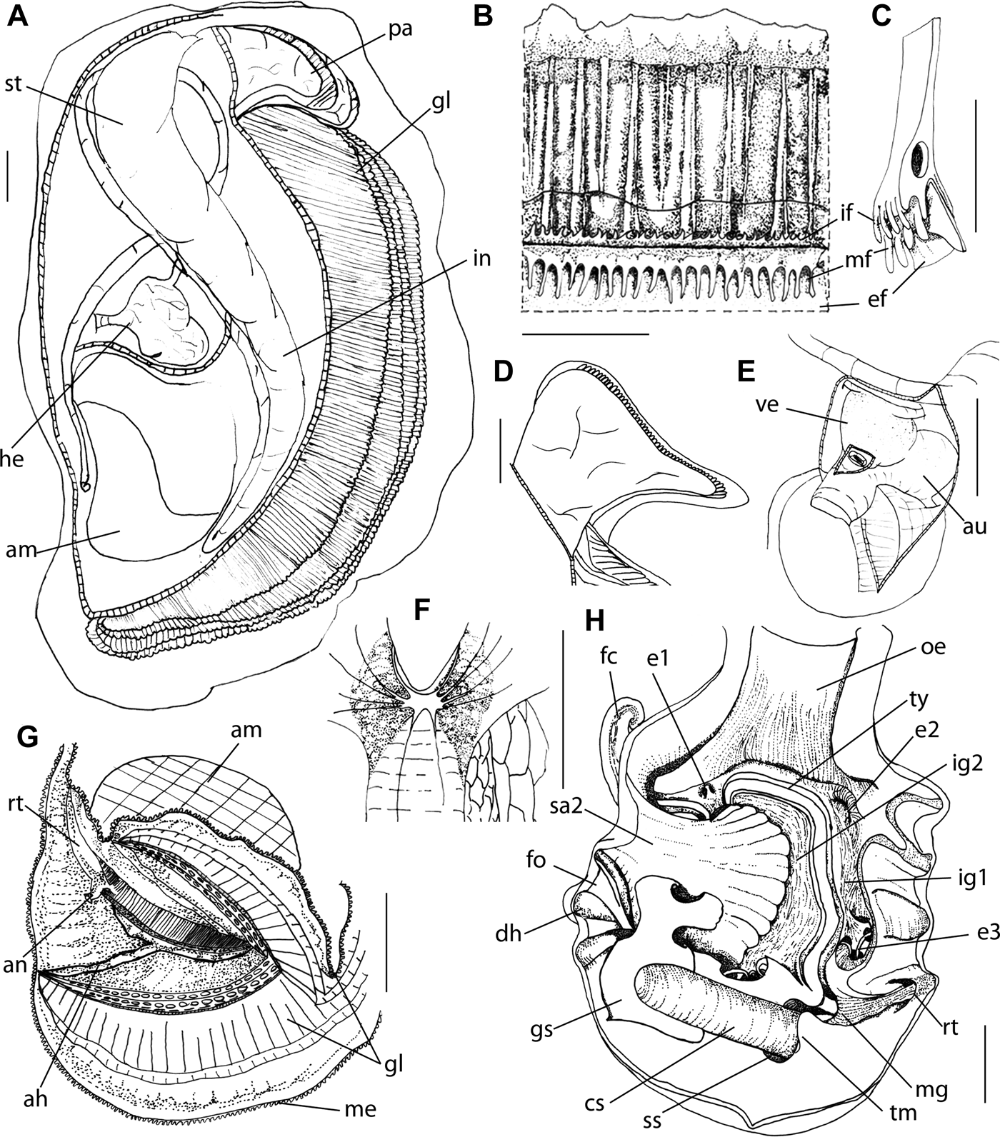
Crassostrea mangle sp. nov. (Holotype) shell and anatomy: (A) external view of shell, right valve (L 56 mm); (B) same, left valve; (C) internal view of shell, left valve, detail of sub-umbonal cavity; (D) same, right valve, detail of ligament; (E) same, detail of adductor muscle impression (ai); (F) right palp, right view, right hemipalp deflected (side 5 mm); (G) general view of right side, Quenstedt muscle in detail (qm) (L 60 mm); (H) gills, ventral edge, ventral view of middle portion, showing alimentary gutter (side 5 mm); (I) inner demibranch, longitudinal section at middle portion, showing internal filaments of folds (side 2 mm); (J) pericardium, right view, pericardial membrane removed (side 5 mm); (K) left mantle lobe, postero-dorsal region, inner view (side 10 mm), detail of accessory heart (ah); (L) right lobe, detail of medial portion of mantle border (me), showing papillae (pl) of median and internal fold (side 3 mm); (M) supra-branchial cavity, region dorsal to adductor muscle, detail of rectum (rt) and anus (an); scale 1 mm; (N) same portion, detail of anus (an) (side 1 mm).

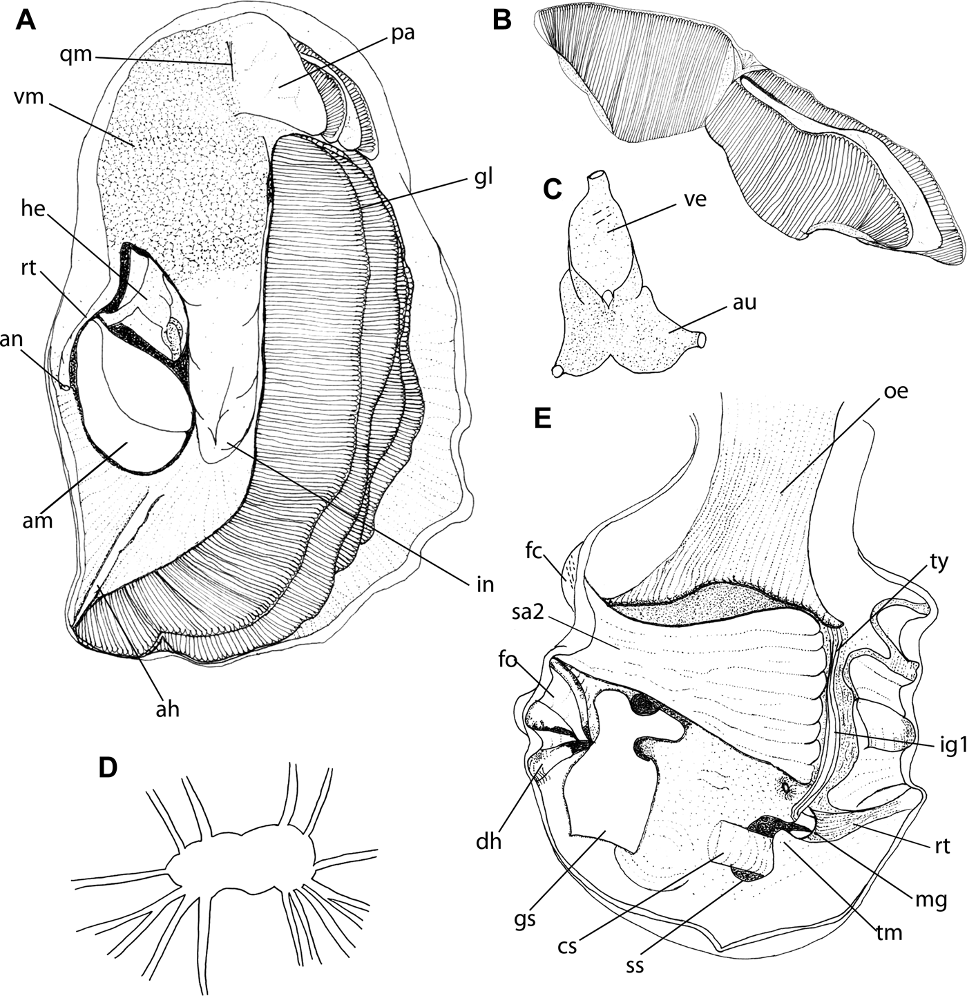
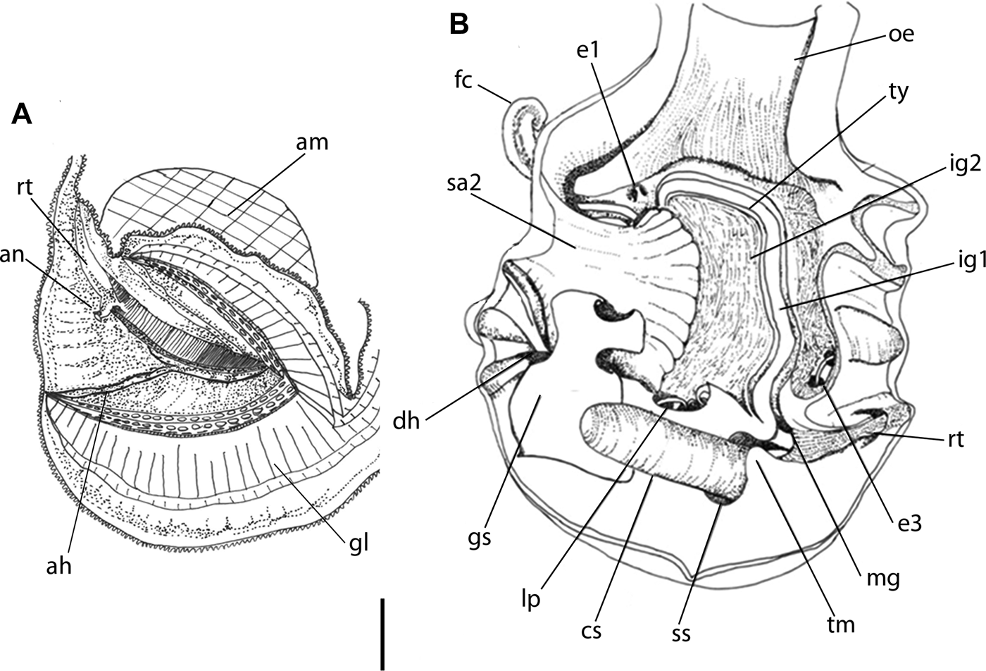
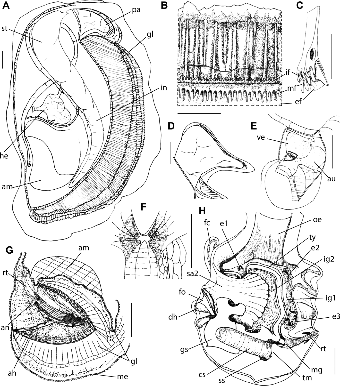
Crassostrea mangle sp. nov. Anatomy: (A) general right view, valve and mantle lobe removed (L 85 mm); (B) mantle border, inner view of median portion (side 5 mm); (C) transverse section at middle region of mantle border (side 5 mm); (D) detail of palps. (E) Detail of heart in pericardium, (ve) ventricle, (au) auricles; (F) detail of visceral ganglia; (G) posterior region of pallial cavity, posterior half of gills sectioned and deflected, showing accessory heart (ah) (side 40 mm) (adapted from Galtsoff, Reference Galtsoff1964); (H) stomach, dorsal view, opened longitudinally, showing inner surface, scale 1 mm.
DIAGNOSIS
Size up to 120 mm; fixed in roots of mangroves, shape cupped; external colour varying from brown to reddish-brown, internal white opalescent; right valve concave, operculum-shaped; left valve larger in edge than right valve. Muscle impression not coloured; adductor muscle more central in postero dorsal region, occupying 1/2 of total size of animal. Accessory heart Y-shaped, composed of three branches of similar length, starting from a common centre. Internal faces of palps plicate, ending before edge, leaving free, narrow and smooth margins.
TYPES
Crassostrea mangle sp. nov.: MZSP 89462 (holotype) and MZSP 100495, MNRJ and USNM (paratypes).
DESCRIPTION
Shell
(Figure 1A–E) Cup shaped, size variable (both height and thickness) up to 120 mm long. Colour varying from white, yellowish, brown to purple. Right (upper) valve flat or concave, operculum-shaped and slightly protuberant near hinge to accommodate visceral portion. Left valve fixed to substrate, cupped and larger than right valve. Umbonal cavity of left valve. External coloration of shell highly variable: white, yellowish, brown, clear and opaque, background, mixed with miscellaneous pigments, forming irregular spots or bands across height of shell. Hinge massive and bulging, protruding from right valve and from respective shallow depression in left valve. Ligament alivincular. Inner surface of valves smooth. Interior of valves predominantly white, with little brightness or opaque. Purple pigmentation close to margin, in most inner regions, or absent. Scar of adductor muscle mostly located in posterocentral region, somewhat central; form crescent to oval, with a concavity towards hinge; occupying about 1/4 shell height. Muscle scar sometimes strongly pigmented with purple, blue, brown bands or a combination (Figure 1E). Scar of Quenstedt muscle oval-shaped, marked by shallow and colourless depression; located in each valve, near to ventral inner edge of ligament area, occupying 1/8 size of adductor muscle.
Muscle system
Adductor muscle
(Figure 1G) Position posterocentral, slightly displaced to dorsal region. Comprised of two different portions, one dorsal, ovate, translucent, occupying 1/2 and 2/3 of total body. Other ventral, crescent shaped and of opaque white-milky aspect; very resistant.
Quenstedt muscle
(Figure 1G) Thin and narrow, fixed to valves close to ventral margin of ligament area or subcentral to it. Fibres crossing mantle in direction to base of labial palps. Portion connected to valve enlarged, almost fan-shaped; becoming thinner through visceral mass.
Mantle
(Figures 1G, K, L, 2B, C) Mantle lobes connected in hinge region by length equivalent to half of valve length; anterior–ventral region forming deeper concavity named sub-umbonal region. Lobes fused to each other only by short portion at posterior end of gill, named pallio-branchial junction. Promyal chamber, or promyal passage, an extension of right epibranchial chamber, heading to posterior margin of body; limited from base of gill to portion of visceral mass, size ~1/3 of visceral mass. Inner surface of two side mantle lobes fixed to base of demibranch and coating adductor muscle, limiting other exhalant passage, cloacal passage. External fold devoid of papillae, exceeding margin of shell when extended. Middle fold with two different types of fingerlike papillae: short with small diameter, and long (more than 1 mm) with enlarged base; arrangement of papillae not following regular pattern, normally presenting up to six short papillae between each two long papillae; both types of papillae of middle fold showing gradual increase in diameter and length from ventral region towards dorsal region. Internal fold with short papillae (Figures 1L, 2B, C). Edge pigmented in variable shades of brown or depigmented. Pallial muscles (Figures 1L, 2B) forming broad bundles around adductor muscle, fixed to visceral mass or to adductor muscle and extending towards free margin of mantle. Each bundle opening like a fan, ending inside three edge folds. Incurrent chamber located in anterior half, extending vertically. Excurrent chamber of ~1/4 size of shell. Anus and rectum located in dorsal–posterior region of body, close to posterior face of adductor muscle.
Gills
(Figures 1H, I) Gills shape elongated, triangular, becoming narrower in region surrounding ventral face of adductor muscle, until fusion to mantle lobes at pallial–branchial junction region. Free anterior–dorsal gill end slightly arched, located between ventral margins of labial palps. External demibranch ending between palp and marginal food groove, reaching base of external palp. Internal demibranch decreasing gradually in height anteriorly; marginal groove extending to base of internal hemipalp. Free margin of external demibranch, i.e. ascending lamella, attached to mantle. Septa of inter-lamellar junction, perpendicular to branchial axis, forming series of aqueous ducts. Gills transversally plicate; each fold ending at free margin of demibranchs, delimiting food groove. Folds comprising average of 13 filaments.
Visceral mass
(Figures 1G, 2A) Occupying ~1/4 total animal's size; located from ventral region of ligament to anterior surface of adductor muscle. Digestive diverticula located in anterior and dorsal region of palps, occupying ~1/4 of visceral mass. Follicular gonads beige, occupying ~1/2 of visceral mass, covering digestive diverticula externally. Renopericardial structures occupying ~1/4 of visceral mass, located in posterior anterior–ventral region of adductor muscle.
Circulatory and excretory systems
(Figures 1J, 2E) Arteries parallel to ventral margin, with several centripetal branches. Left and right circumpallial vessels united to each other in pallial–branchial junction. Pair of accessory hearts (Figures 1K, 2G) with three branches of similar length, starting from a common centre, forming ‘Y’, with basal branch leaning in ventral–posterior direction until its connection to pallial–branchial junction; basal branch bifurcating near adductor muscle, with branch extending towards rectum and anus, and other to epibranchial chamber. Pericardium (Figures 1J, 2E) located between visceral glands and adductor muscle. Two renopericardial channels opening at right and left sides of ventro-posterior wall of pericardium, providing direct communication with kidney. Heart located anteriorly to adductor muscle, with three chambers arranged obliquely in pericardium, connected by aortic root on one side and ctenidial veins on left side. Ventricle pyriform, walls with thin bundles of muscle fibres crossing ventricular lumen, dividing it incompletely into two chambers. Auricles pigmented or depigmented, varying from yellowish-cream to brown. Walls of auricles thinner and lighter than those of ventricle, trabecular. Pair of kidneys triangular, tubular; mottled by light brown marks by translucence of visceral mass. Each kidney with direct communication with pericardium and gonad on medial side and laterally communicating by short chamber. Renal opening located on other side of cerebro-visceral connective.
Digestive system
(Figures 1F, 2D, G)
Palps
(Figures 1F, 2D) Located in anterior–ventral region, under sub-umbonal region of mantle, occupying ~1/8 of animal's body. Hemipalps flattened, elongated, base united throughout its length to visceral mass. Margins slightly arched anteriorly and ventrally free, converging towards ventral region to form apex. Internal faces plicate, folds extending transversely, ending in order to leave free, narrow and smooth margin. External surface smooth, with irregular grooves and ridges. External hemipalps dorsally united over mouth, forming arched dorsal lip. Internal hemipalps ventrally united, forming ventral mouth lip. Lips height similar to median part of hemipalps. Mouth slot-shaped.
Oesophagus
(Figure 2H) Dorso-ventrally flattened, length ~1/8 of that of palps, slightly longer than wide; insertion in stomach wide. Internal surface of distal end striated, with slight folds becoming more evident near stomach.
Stomach
(Figure 2H) Occupying ~1/4 of animal's size, laterally flattened, with irregular external invaginations. Stomach inner surface with small portion of posterior surface of stomach coated by gastric shield (gs), almost same area in transversal section of oesophagus; minor typhlosole (tm) projecting into stomach, ending after intestine opening; major typhlosole (ty) emerging as elbow-shaped structure, adjusting around intestinal opening, extending afterwards along left side of stomach floor; close to oesophageal opening, diverting almost 90° to right, penetrating into purse or caecum (fc). Caecum beginning in floor of anterior right gastric region, under oesophagus and going towards opening of intestine; major typhlosole penetrating deeply in distal end of ventral branch of caecum, returning along its left wall, penetrating and finishing into distal end of dorsal branch; two typhlosoles occupying ~1/3 of stomach length. Caecum (fc) composed of two branches, one longer and flattened, emerging laterally, under stomach floor; other branch smaller in length, projecting to posterior region, parallel and at left side of oesophagus, coiled in a spiral of about 1.5 turns; occupying ~1/8 of stomach width. Dorsal hood (dh) close to roof and on right side of stomach, inside which two thick, broad and slightly smooth folds beginning on right side of gastric wall, ascending to ceiling and invading dorsal hood; opening of ventral margin of dorsal hood protected by fold of gastric shield; right purse (rp) aligned to dorsal hood, consisting of two compartments, one in blind bottom, immediately ventral to dorsal hood opening, other deeper, located on floor of stomach, close to origin of ducts of digestive diverticula; upper edge of blind compartment coated by fold of gastric shield. Intestinal groove (ig1) beginning near main opening of right purse, extending in anterior direction, along left side of major typhlosole, inside dorsal branch of caecum. Intestinal groove returning parallel to opposite face of major typhlosole, comprising ‘ig’ segment, extending inside initial portion of intestine, united to style sac. Style sac occupying ~1/2 of stomach length. Striated tissue in stomach floor beginning inside right purse, extending inside caecum, always right of intestinal groove (ig2), occupying ~1/3 of stomach length. Ducts to digestive diverticula opening inside right purse; additional diverticula originating in small depression (e1) in anterior wall of caecum, very close to its opening, occupying ~1/16 of stomach width. Second depression (e2) on left side of esophagus opening, larger and deeper than ‘e1’, where other ducts of digestive diverticula open. Another shallow depression (e3) wide, appearing on left wall, close to intestinal origin; also receiving ducts of digestive diverticula. Both, e1 and e3, occupying ~1/8 of stomach width; ‘e1’, ‘e2’ and ‘e3’ walls showing narrow folds advancing inside opening of ducts of digestive diverticula. Depression (h) located in stomach ceiling, posterior to dorsal hood, extending ventrally along right wall, forming deep trench (rt), ending at intestine opening. Cylindrical portion of stomach emerging opposite to oesophagus insertion, represented by style sac and intestine. Intestine and style sac fused with each other, running up to ventral end of adductor muscle, surrounded by reproductive and excretory apparatus, forming pyloric process. Style sac walls transversally striated with ~1/8 of stomach width.
Intestine
(Figure 2A, H) Distinguished from distal end of style sac by 180° loop; afterwards passing through pericardium, following ovoid portion of visceral mass in ‘U’-shaped loop, passing posteriorly to stomach, close to connection with esophagus, extending to right face of ovoid portion of visceral mass. It changes again course to ventral and posterior direction.
Rectum
(Figures 1M, N, 2G) Crossing pericardium, running in contact with posterior face of adductor muscle; internal surface with well-developed typhlosole, with broad base and grooved free end up to anal opening.
Anus
(Figures 1M, N, 2G) Sessile, surrounded by flat edge-shaped fold, bell-shaped. occupying 1/3 of height of adductor muscle.
Reproductive system
(Figure 1G) Gonad formed by two lobes of follicular aspect, occupying ~1/4 of animal size. Dorsally and ventrally spread in left lobe and right lobe; shape indistinct from each other. Two pairs of separate systems of genital channels, as described above (excretory system).
Central nervous system
(Figure 2F) Cerebral ganglia located at base of labial palps, about 5% oesophagus. Cerebral commissure U-shaped, dorsally surrounding oesophagus 15 times longer than ganglia. Pair of visceral ganglia slightly ovoid, with projections; antero-ventral to adductor muscle, in depression formed by junction of translucent and opaque parts of adductor muscle, about 1/12 of size of adductor muscle. Circumpallial nerve travelling along mantle. Pedal ganglion absent.
DISTRIBUTION AND HABITAT
Brazil, Pará to Santa Catarina, always associated with the mangroves.
EXAMINED MATERIAL
Brazil
Ceará: Fortaleza; Mangue da Foz do Rio Ceará, 3°42′9″S 38°35′31″W, MZSP 101785, 50 specimens (V.S. Amaral col., 9 November 2011). Rio Grande do Norte: Ceará Mirim, Estuário do Rio Ceará Mirim, 05°40′658″S 35°13′578″W, MZSP 107753, 50 specimens (M.D. Tavares col., 18/xi/2009). Paraíba; João Pessoa; Cabo da Ponta do Seixas, 07°08′42.3″S 34°48′12.2″W to 07°08′46.5″S, 34°47′52.7″W, MZSP 41344, 12 specimens (Simone col. 27 April 2004), 07°08′43.3′S 34°48′11.3″W, MZSP 45287, 4 specimens, (Simone col. 14 January 2005). Pernambuco: Itamaracá, manguezal, MZSP 104522, 20 specimens (V.S. Amaral col., 27 February 2012). Alagoas: Maceió, lagoa de Mundaú, MZSP 46141, 3 specimens (José Geraldo Marques col., 23 September 1974); Barra de Camaragibe, estuary of Rio Camaragibe (M.D. Tavares et al., col.), MZSP 89462, 30 specimens, MZSP 89518, 20 specimens (21 October 2008) (holotype), MZSP 100491, 15 specimens (8 January 2011), MZSP 100495, 40 specimens (8 January 2011) (paratype); MZSP 100497, 30 specimens (8 January 2011); MZSP 100498, 50 specimens (8 January 2011); MZSP 100499, 30 specimens (8 January 2011); MZSP 107743, 50 specimens (June 2011); MZSP 107746, 20 specimens (June 2011); MZSP 107758, 2 specimens (June 2011); MZSP 109156, 4 specimens (27 March 2010). Bahia: Salinas da Margarida, MZSP 46068, 9 specimens (Iracema Nascimento col.); Boipeba, 13°38.528″S 038°54.315′ W MZSP 101849, 15 specimens (M.D. Tavares col., August 2011); Ilhéus, Olivença, MZSP 101553, 10 specimens, (M.D. Tavares col., 27 May 1994). Rio de Janeiro: São João da Barra, Praia de Manguinhos, MZSP 15286, 1 specimen, 1 valve (‘Exp. Dep. Zool.’; August 1963). São Paulo: São Sebastião, Araçá, 23°48′54.89″S 45°24′31.73″W MZSP 101922, 20 specimens (V.S. Amaral col., 18 August 2011), MZSP 106009, 8 specimens, (Simone col., 21 January 2012); Bertioga, MZSP 101984, 15 specimens (Francisca do Val col., May 2011); Praia Grande, Manguezal do Portinho, 23°58′59″S 46°24′53″W MZSP 101980, 15 specimens (Luiza Saad col., 19 July 2011); Cananéia, Cooperostra, MZSP 89670, 60 specimens (Fausto Campos Pires leg., March 2008); IPESCA, MZSP 89673, 3 specimens (Patrícia de Oliveira leg., June 2008); Portinho, MZSP 89676, 50 specimens (V.S. Amaral col., 8 January 2009). Santa Catarina: Porto Belo, Ponta Norte da Praia de Porto Belo, MZSP 36319, 4 specimens (Cenemar col., 12 October 2002).
(Figures 3, 4)
Ostrea rhizophorae Guilding, 1828: 542; Reeve, 1889: 9, 17a–b; Dall, Reference Dall1889: 687; Lamy, Reference Lamy1929: 95–99, 100, 108, 135, 137, 139, 271; McLean, Reference McLean1941: 5–7; Mattox, Reference Mattox1949: 339–356; Olsson & McGinty, 1958: 20.
Crassostrea rhizophorae shell and anatomy: (A) external view of right valve (L 65 mm); (B) right valve, internal view, detail of adductor muscle impression (ai); (C) view, detail of sub-umbonal region (sb) and adductor muscle (am); (D) left valve, lateral view; (E) general view of right side; (F) anterior view, detail of Quenstedt muscle; (G) right lobe, detail of accessory heart (ah); (H) right lobe: mantle edge (external, middle and internal folds with papillae) and pallial muscles; (I) right palp (pa), right view; (J) general view of digestory system; (K) gills, detail of alimentary chamber.
Crassostrea rhizophorae anatomy: (A) general right view, valve and mantle lobe removed; (B) detail of palps, internal view; (C) detail of heart, ventricle and auricles; (D) visceral ganglia in detail; (E) stomach, dorsal view, opened longitudinally showing inner surface.
Ostrea parasitica Mörch, 1878: 16; Arango & Molina, 1878: 273; Ihering, Reference Ihering1907: 359, 374, 426, 450; Richards, 1935: 236; Jaume & Farfante 1942: 38. (auct. non Gmelin, Reference Gmelin and Linné1791)
Ostrea (Crassostrea) rhizophorae McLean, 1951: 25 (pl. 4, figure 6).
Crassostrea rhizophorae Gunter, Reference Gunter1950: 44–45; Abbott, Reference Abbott1974: 456 (figure 5273); Abbott, 1954: 374–375; Nowell & Usticke, 1959: 9; Warmke & Abbott, 1961: 173 (figure 35b); Buroker et al., 1979a: 171–184; Buroker, Reference Buroker1984: 550–557; Castillo-Rodriguez & García-Cubas, Reference Castillo-Rodríguez and García-Cubas1984: 1–57; Hedgecock & Okazaki, Reference Hedgecock and Okazaki1984: 535–545; Angell, Reference Angell1986: 1–37; Arakawa, Reference Arakawa1990: 1–13; Márquez, 1992: 51–55; Littlewood, Reference Littlewood1994: 221–229; Abbott & Dance Reference Abbott and Dance2000: 318 (figure 5); Wang et al., Reference Wang, Xu and Guo2004: 46–54; Vaisman et al., Reference Vaisman, Marins and Lacerda2005: 582–288; Erse & Bernardes, Reference Erse and Bernardes2008: 57–93; Guimarães et al., Reference Guimarães, Antonio, Peixoto and Oliveira2008: 118-122; Mikkelsen & Bieler, Reference Mikkelsen and Bieler2008: 112–113 (figure 1).
TYPES
Ostrea rhizophorae: NHMUK (possibly lost) (Carriacou, near Grenada).
DIAGNOSIS
Size up to 80 mm; fixed in roots of mangrove, shape cupped; colour varying from white, yellowish to brown; right valve operculum-shaped; left valve; larger and sling than right valve. Adductor muscle postero-dorsal located, occupying 1/15 of total size of animal; mantle with several bindle of pallial muscles; colour of mantle edge brown or yellowish. Accessory heart with one only branch. Palps with folds extending transversally up to superior margin.
DISTINCTIVE REDESCRIPTION
Shell
(Figure 3A–D). Shape cupped, size up to 80 mm long. Colour varying from white, yellowish, to brown. Right (upper) valve flat or slightly concave, operculum-shaped, no protuberant near hinge to accommodate visceral portion. Left (lower) valve fixed to substrate, cupped and almost same width than right valve. Umbonal cavity on left valve small. Interior of valves predominantly white, with little brightness. Scar of adductor muscle unpigmented, mostly located in posterodorsal region, more lateralized; form oval, with a slightly concavity towards hinge; occupying about 1/15 of valve surface. Scar of Quenstedt occupying 1/20 of size of adductor muscle.
Muscle system
Adductor muscle
(Figure 3C, E) Posterodorsal elongated, close to dorsal region, occupying 1/15 of total size body.
Quenstedt muscle
(Figure 3F) Thin and narrow, fixed to valves close to ventral margin of ligament or subcentral to it.
Mantle
(Figure 3E–H) Mantle lobes connected in hinge region by equivalent to 2/3 of valve width–sub-umbonal region. Promyal passage, size ~1/4 visceral mass. External fold devoid of papillae, exceeding margin of shell when extended. Middle fold with two different types of fingerlike papillae: short with small diameter, and medium; arrangement of papillae not following clear regular pattern, normally presenting up to two short papillae for one medium papillae; both types of papillae of middle fold showing gradual increase in diameter and length from ventral region towards dorsal region. Internal fold with only elongate middle papillae. Edge pigmented in variable shades of brown. Anus and rectum located in dorsal–posterior region of body, close to middle face of adductor muscle.
Gills
(Figure 3K) Gills shape elongated, triangular, becoming narrower in region surrounding ventral face of adductor muscle, until fusion to mantle lobes at pallial–branchial junction region. Gills transversally plicate; each fold ending at free margin of demibranchs, delimiting food groove. Folds comprising average of 13 filaments.
Visceral mass
(Figure 3E, I) Occupying ~1/4 of total animal size. Digestive diverticula located in anterior and dorsal region of palps, occupying ~1/4 of visceral mass. Follicular gonads beige, occupying ~1/2 of visceral mass, covering digestive diverticula externally. Renopericardial structures occupying ca 1/6 of visceral mass, located in posterior anterior–ventral region of adductor muscle.
Circulatory and excretory systems
Arteries parallel to ventral margin, with several centripetal branches. Left and right circumpallial vessels united to each other in pallial–branchial junction. Pair of accessory hearts (Figures 3G, 4A) with only one branch, connecting pallial–branchial junction to near adductor muscle, not surrounding adductor muscle. Heart with ventricle elongate, auricle pyriform, walls formed by thin bundles of muscle fibres, dividing it incompletely into two chambers. Colour lacking or cream-yellowish (Figure 4C).
Digestive system
Palps
(Figures 3J, 4A, B) Located in anterior–ventral region, occupying ~1/12 of animal's body. Hemipalps flattened, elongated, base united throughout its anteroventral region to visceral mass; margins slightly arched anteriorly and ventrally free, converging towards ventral region to form apex; internal faces plicate, folds extending transversally up to superior margin; external surface smooth, which irregular grooves and ridges. External hemipalps dorsally united over mouth, forming arched dorsal lip. Internal hemipalps ventrally united, forming ventral mouth lip.
Oesophagus
(Figures 3J, 4E) Dorso-ventrally flattened, same length as palps, slightly longer than wide; insertion in stomach wide; internal surface of distal end striated, with slight folds becoming more evident near stomach.
Stomach
(Figures 3J, 4E) Occupying ~1/6 of animal's size. Two typhlosoles occupying ~1/4 of stomach length. Dorsal hood (dh) close to roof and on left side of stomach, inside which two thick, broad and slightly smooth folds beginning on right side of gastric wall, ascending to ceiling and invading dorsal hood; opening of ventral margin of dorsal hood protected by fold of gastric shield; upper edge of blind compartment coated by fold of gastric shield. Style sac walls transversally striated occupying ~1/8 of stomach length.
Intestine
(Figures 3J, 4A, E) distinguished from distal end of style sac by 180° loop; afterwards passing through pericardium, following ovoid portion of visceral mass in ‘S’-shaped loop, passing posteriorly to stomach, close to connection with esophagus, extending to right face of ovoid portion of visceral mass. Intestine and style sac fused with each other, running up to ventral end of adductor muscle, surrounded by reproductive and excretory apparatus, forming pyloric process.
Anus
(Figure 4A) Sessile, surrounded by flat edge-shaped fold, occupying 1/4 of height of adductor muscle.
Reproductive system
(Figures 3E, 4A) Gonad formed by two lobes of follicular aspect, occupying ~1/4 of animal's size. Dorsally and ventrally spread in left lobe and right lobe; shape indistinct from each other.
Central nervous system
Cerebral ganglia, almost 10% of area of oesophagus. Cerebral commissure U-shaped, dorsally surrounding esophagus, 30 times more long of cerebral ganglia. Pair of visceral ganglia (Fig. 4D) slightly ovoid, with two anterior projections, one lateral and three posterior projections; antero-ventral to adductor muscle, in depression formed by junction of translucent and opaque parts of adductor muscle, occupying about 1/30 of size of adductor muscle.
DISTRIBUTION AND HABITAT
Caribbean, always associated with mangroves.
EXAMINED MATERIAL
Grenada. Carriacou: Tyrrel Bay, Oyster Bay, 12°27′51.51″N 61°45′18.67″W, MZSP108492, 50 specimens (Amaral & Simone col., 9 October 2012) (type locality).
Crassostrea brasiliana (Lamarck, Reference Lamarck1819)
(Figures 5, 6)
Ostrea brasiliana Lamarck, Reference Lamarck1819: 205; Maury, Reference Maury1936:283; Ranson, Reference Ranson1967: 189–191; Bianchi, Reference Bianchi1969: 3–40; Figueiras & Sicardi, Reference Figueiras and Sicardi1979: 107–161.
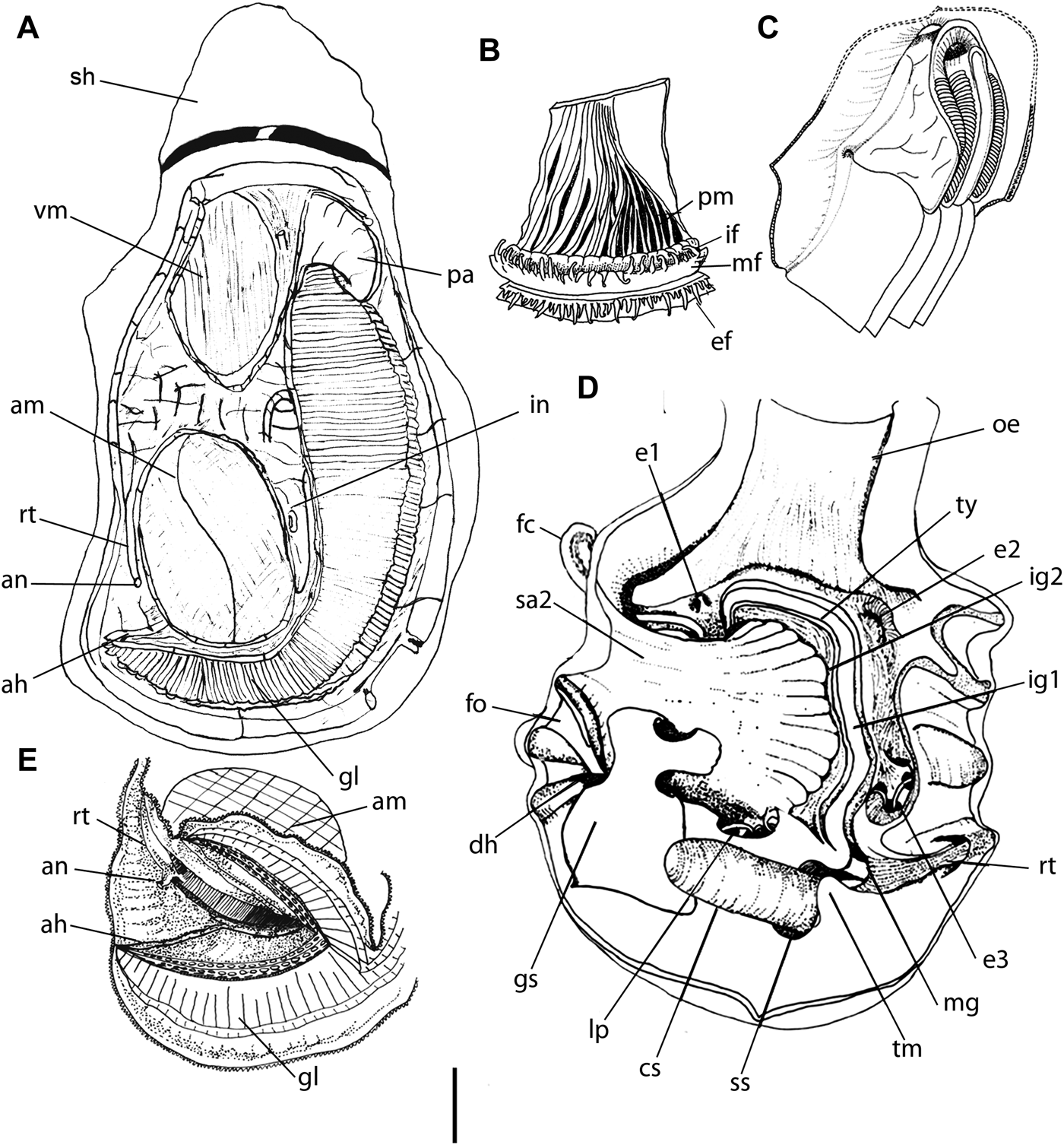
Crassostrea brasiliana shell and anatomy: (A) external view of shell, right valve; (B) same, left valve; (C) internal view, right valve, detail of muscle impression; (D) same, left valve (L 65 mm); (E) general view of right side, valve and mantle lobe removed; (F) right lobe, middle portion of mantle border (side 20 mm), detail of papillae of internal and middle folds; (G) right palp, right view, outer hemipalp deflected; (H) pericardium, right view, pericardial membrane removed; (I) gill, detail of ventral edges of demibranchs, ventral view, showing alimentary chamber; (J) same, longitudinal section of median portion of internal demibranch, showing internal filaments and folds; (K) supra-branchial cavity, region dorsal to adductor muscle, detail for a rectum and anus; (L) left mantle lobe, postero-dorsal region, internal view, showing accessory heart (ah). Scales: 1 mm.
Crassostrea brasiliana anatomy: (A) general right view, valve and mantle lobe removed (L 62 mm); (B) mantle border, internal view of median portion (L 5 mm); (C) palps, right-slightly ventral view (L 30 mm); (D) posterior region of pallial cavity, with posterior half of gill longitudinally cut and deflected showing accessory heart (ah) (am, adductor muscle; rt, rectum; an, anus, me, mantle edge; gi, gill), scale 1 mm (adapted from Galtsoff, Reference Galtsoff1964); (E) stomach opened longitudinally, dorsal view, inner surface (L 20 mm).
Crassostrea parahibanensis Singarajah, Reference Singarajah1980: 5 (figures 2–3): (new synonym). Harry, Reference Harry1981:44.
Crassostrea brasiliana: Singarajah, Reference Singarajah1980: 833–847; Muniz et al., Reference Muniz, Jacob and Helm1986: 77–138; Nascimento, Reference Nascimento and Menzel1991: 125–134; Galvão et al., Reference Galvão, Pereira, Machado and Henrique2000: 147–462; Ignacio et al., Reference Ignacio, Absher, Lazoski and Sole-Cava2000: 987–991; Pereira et al., Reference Pereira, Machado, Henriques, Galvão and Bastos2000: 49–62; Pereira et al., Reference Pereira, Machado, Henriques, Galvão and Bastos2001: 85–95; Làpegue et al., 2002: 232–242; Machado et al., Reference Machado, Maio, Kira and Carvalho2002: 13–18; Lazoski, Reference Lazoski2004: 197–212; Christo, Reference Christo2006: 1–146; Pie et al., Reference Pie, Ribeiro, Boeger, Ostrensky, Falleiros and Ângelo2006: 1598–1600; Christo & Absher, Reference Christo and Absher2008: 71–77; Carranza et al., Reference Carranza, Defeo and Beck2008: 1–12; Guimarães et al., Reference Guimarães, Antonio, Peixoto and Oliveira2008: 118–122; Christo et al., 2010: 645–650; Lazoski et al. Reference Lazoski, Gusmão, Boudry and Solé-Cava2011: 197–212.
Crassostrea parahibensis: Rios, Reference Rios1994: 245 (err).
Crassostrea gasar Làpegue et al., 2002: 232–242; Varela et al., Reference Varela, Beasley, Schneider, Sampaio, Marques-Silva and Tagliaro2007: 1–6; Melo et al., Reference Melo, De Paula, Beasley and Tagliaro2007: 1; Carranza et al., Reference Carranza, Defeo and Beck2008: 1–14. (non Dautzenberg, 1891).
TYPES
Ostrea brasiliana: Syntype: MNHN no number (figure 82). Crassostrea parahibanensis: Holotype: NHMUK 1978114; Paratypes: NHMUK 1978115, MNRJ 3815-3816 [Paraíba River estuary, Brazil] (figures 80, 81) (all examined).
DIAGNOSIS
Size up to 130 mm; fixed in roots of mangrove submerse and in rock; shape cupped or oval; colour varying from white, brown, purple to greenwish; right valve slightly operculum-shaped; left valve, fixed in substrate, more larger than right valve. Muscle impression is purple and adductor muscle, oval central in posterior region. Adductor muscle postero-dorsal located, occupying 1/5 of total size of animal; hood present and fully filled by palps and gonads; colour of mantle edge usually brown. Accessory heart of three branches of similar length, starting from common centre, forming ‘Y’. Palps with margin superior free.
DISTINCTIVE DESCRIPTION
Shell
(Figure 5A–D) Ovoid, wide and thin, height 50–130 mm. Colour greenish to brownish with white bands. Right (upper) valve opercular and wide. Left (lower) valve shallow cupped with slight depression under umbo (um). Ornamentation present or absent on external face of valves (since directly influenced by tidal action). Internal portion white, smooth, with little brightness, sometimes showing orange pigmentation on edge. Adductor muscle impression (ai) located in posterior–dorsal region, always rounded in shape, proportionally larger, occupying ~1/6 of shell size. Purple pigmentation sometimes present. Light Quenstedt muscle impression located just below ligament area, ~1/10 of size of adductor muscle impression.
Muscle system
Adductor muscle
(Figures 5E, 6A) Section rounded, dorsal portion ovoid, translucent, vertical portion crescent shaped with opaque white-milky aspect. Occupying ~1/5 of animal size.
Quenstedt muscle
(Figure 6A) Widened origin in portion linked to shell and anteriorly directed towards inside of visceral mass, inserted in base of palps. Size ~1/10 of adductor muscle.
Mantle
(Figures 5F, L, 6B, E) Blood vessels wide, apparent in pallial muscles (me), near palp. Sub-umbonal region (sb) filled by palps and gonads. Mantle edge (me) usually brown pigmented. External fold devoid of papillae, height similar (~2% of pallial lobe height). Middle fold with long, medium and short papillae (pl), two last finger-like from 2 to 4 medium or short papillae to one long papilla. Papillae uniform in size and distribution, 2–3 times more numerous in middle fold than in internal fold. Internal fold with long papillae, more concentrated in pallial–branchial junction of mantle lobes, sometimes pigmented (Figures 5F, 6B). Pallial muscles evident, with more abundant bundles near adductor muscle, forming fan. Radial muscles fainter throughout length of mantle border; occupying ~30% of mantle lobe surface. Circumpallial veins well-developed. Accessory heart (ah) (Figures 5L, 6E) of left lobe with thicker tissue, smaller in diameter than C. mangle, bifurcating around adductor muscle towards gills. Portion around muscle reduced or absent. Accessory heart of right lobe devoid of bifurcations comprised a single vessel ending in branchial artery.
Gills
(Figure 5I, J) Similar to anterior species in size and number of lamellae, having 12–13 bundles. Marginal alimentary groove wide, with ~1/2 width mantle border plicae.
Visceral mass
(Figures 5E, 6A) Occupying ~1/5 of animal total volume. Located in ventral region of ligament to anterior surface of adductor muscle. Digestive diverticula, located at dorsal region of palps, occupying about half of significant part of visceral mass volume. Gonads beige-yellowish, covering whole visceral mass, including digestive diverticula, externally. Renopericardial structures occupying ~1/6 of visceral mass, located in antero-ventral posterior region of adductor muscle.
Circulatory and excretory systems
(Figure 5H) Pericardium wide, anteriorly and ~1/2 size of adductor muscle. Ventricle (ve) thin, reticulate, usually translucent. Auriculas (au) thin, less reticulate and non-pigmented than ventricle. Excretory organ (renal opening and kidneys) occupying same position as C. mangle, over each side of visceral mass. Pigmented mark absent.
Digestive system
(Figures 5G, 6C, D) Same as C. mangle, except for mouth accompanying animal's size, oesophageal plicae following to straight and wide esophagus.
Palps
(Figures 5G, 6C) Located under sub-umbonal region of mantle filling free space, occupying ~1/6 of animal size. Slightly flattened, sickle-shaped, joined throughout length of their base to visceral mass. Free margins, anterior and ventral, slightly ventrally arched forming apex. Internal faces intensively plicate; upper internal face of palps showing free region. External faces smooth or with slight striations and irregular grooves. Anterior and posterior portions wide and devoid of plicae. Dorsally united external palps over mouth covering inner palps.
Oesophagus
(Figure 6D) Slightly long, longer than wide, reaching stomach; inner surface striated, with striations becoming fainter near stomach.
Stomach
(Figure 6D) Same as C. mangle, except for longer selection area (sa2) ending near base of major typhlosole. Shorter selection caecum (fc), not completing one and a half turn.
Intestine
Narrow in portion surrounding adductor muscle ventrally; internal structures similar to C. mangle.
Reproductive system
(Figures 5E, 6A) Gonad composed of abundant follicular formations, beige coloured, covering mantle and between digestive diverticula, comprising visceral mass and extending ventrally to end of pyloric process, where they open to mantle cavity. Genital opening located at terminal level of style sac, ventral of adductor muscle.
DISTRIBUTION AND HABITAT
On rocky coasts, but may appear in mangroves (including cultures, e.g. Cooperostra, 2009- Cananeia, SP). It occurs on rocky coasts and in mangroves (subtidal).
EXAMINED MATERIAL
Brazil. Paraiba: Conde, Mouth of Rio Gramame (beach), 07°14′07.0″S 34°48′14.7″W, MZSP 45372, 2 specimens (Simone col.; 12 January 2005), (mangrove), 07°14′13.2″S 34°48′23.2″W, MZSP 45395, 1 specimen (Simone col., 11 January 2005). Rio Grande do Norte: Nísia Floresta, Praia de Búzios, MZSP 32656, 7 valves, (Simone & Souza col., 27 July 1999); Natal, Balsa, 05°47′46.44″S 35°13′45.02″W, MZSP 79689, 10 specimens (Simone col., August 2006); Praia da Ponta Negra, 05°53′04.68″W 35°09′52.73″W, MZSP 79690, 3 specimens, 1 valve, MZSP 79692, 4 specimens; Barra da Praia de Canhaú, MZUSP 89672, 4 specimens (C.M. Cunha leg., purchased in Búzios beach, 20 October 2007). Alagoas: Maceió, Lagoa Mundaú, Canal do Calunga, close to sea, MZSP 46129, 6 specimens (J.B. Pereira-Barros col., 12 June 1970). Espírito Santo: MZSP 87473, 2 specimens, (G.H.P. Filho & F. Corteleti col.; 25 March 2008). Rio de Janeiro: Búzios, Praia da Ferradura, MZSP 48451, 30 specimens, (Exp. IMP, 19 March 2005); Arraial do Cabo, Praia do Forno, 3 m deep, MZSP 50375, 1 specimen, 5 valves, (Exp. IMP, 17 March 2005); São João da Barra, Praia de Manguinhos, MZSP 15286, 1 specimen, 1 valve (Exp. Dep. Zool.; August 1963); Niterói, MZSP 26349, 6 specimens; Angra dos Reis, Praia Grande do Frade, MZSP 46126, 10 specimens (Exp. Dep. Zool.; 24 July 1966); Ilha dos Coqueiros, MZSP 46231, 1 specimen (Exp. Dep. Zool.; 5 July 1966); Baía de Ilha Grande, MZSP 46090, 25 valves, (Exp. Dep. Zool.; 27 April 1966); Praia dos Meros, MZSP 86222, 1 specimen, [seed collector (cultivation of Nodipecten nodosus, 3° PET)]; Paraty, Trindade, Caixa d'aço, natural pool, 23°21′35.05″S 44°43′55.87″W, MZSP 55644, 2 specimens (‘Exp. IMP’, 8 December 2005); Praia da Lula, 23°11′38.5″S 44°38′06.5″W, MZSP 55827, 2 specimens, MZSP 55937, 1 specimen (10 December 2005), 55937, 2 specimens (10 December 2005); Praia do Catimbau, 23°11′26.4″S 44°37′25.5″W, MZSP 55946, 2 specimens (Exp. IMP, 10 December 2005; in Cymatium pathernopeum and Spondylus americanus). São Paulo: Ubatuba, Rio Puruba, MZSP 91799, 20 specimens (Beto Chagas col, March 2009); Ilhabela, Praia da Vila, 0–3 m, MZSP 41743, 3 specimens (Simone et al., col., 5 May 2004); Praia de Barra Velha, MZSP 81167, 2 specimens (Simone, 27 October 1996); Praia do Sino, MZSP 89675, 40 specimens (V.S. Amaral col., 1 December 2008); São Sebastião, Praia de Boissucanga, 13 m deep, MZSP 46133, 12 valves (‘Emilia’, 20 April 1959); Bertioga, MZSP 89674, 3 specimens, (E.P. Gonçalves col., May 2008); Praia de Indaiá, MZSP 46111, 8 specimens, (Isabel Cristina N. dos Santos col., March 1999); Santos, Ponta da Praia, MZSP 90847, 20 specimens (C.M. Cunha col., April 2009); São Vicente, Ilha de Urubuqueçaba, intertidal, MZSP 85718, 1 specimen (C.H. Guimarães col., 24 February 2007); Praia Grande, Praia do Forte, MZSP 82576, 1 specimen, (Simone col., 1990); Peruíbe, Estação Ecológica Juréia-Itatins, lade of Rio Perequê, Marina, 24°21′39.2″S 46°58′49.5″W, MZSP 86030, 15 specimens (V.S. Amaral col., 4 September 2007); Ponta da Juréia, adjacent to Praia do Rio Verde, MZSP 26992, 30 specimens (A.E. Migotto, 5 October 1982); Cananéia, Cooperostra, MZSP 91800, 20 specimens (V.S. Amaral leg., January 2009), Ilha do Castilho, 0–9 m deep, ESEC Tupiniquins, 25°16′17.3″S 47°57′31.6″W, MZSP 83306, 13 specimens, MZSP 83309, 6 specimens, MZSP 83335, 2 specimens (C.H. Guimarães et al., 26 January 2007); Ilha Comprida, 23°10′16.45″S 44°40′41.62″W, MZSP 86972, 10 specimens (C.M. Cunha et al., col.; 18 January 2008). Paraná: Paranaguá, 40–50 m, MZSP 37135, 1 specimen (Barco de Camaroneiro col., C.M. Cunha leg., May 2000); Iate Club de Paranaguá, MZSP 87739, 10 specimens (M. Galvão leg., 2008). Santa Catarina: Arvoredo, MZSP 46088, 1 specimen (Manoelito Ribas col., ‘Dalila’, 17 June 1960).
Crassostrea virginica (Gmelin, Reference Gmelin and Linné1791)
(Figures 7, 8)
Synonymy in: Rodriguez & García-Cubas (1984: pp. 28–29)
Complement:
Ostrea elongata Lightfoot, Reference Lightfoot1786: 151 (non Born, 1778).
Crassostrea virginica shell and anatomy: (A) external view, right valve; (B) left valve, sub-umbonal cavity, internal view (L 36 mm); (C) same, detail of adductor muscle impression; (D) same, external view, detail of ornamentation of the shell edge; (E) gill, internal surface of middle portion; (F) general view of right side, shell and mantle lobe removed (L 48 mm); (G) mantle border, middle portion (side 5 mm); (H) pallial cavity, postero-dorsal region, right view, showing accessory heart (ah) (L 30 mm); (I) pericardium and heart, right view, pericardial membrane removed. Scales: 1 mm.
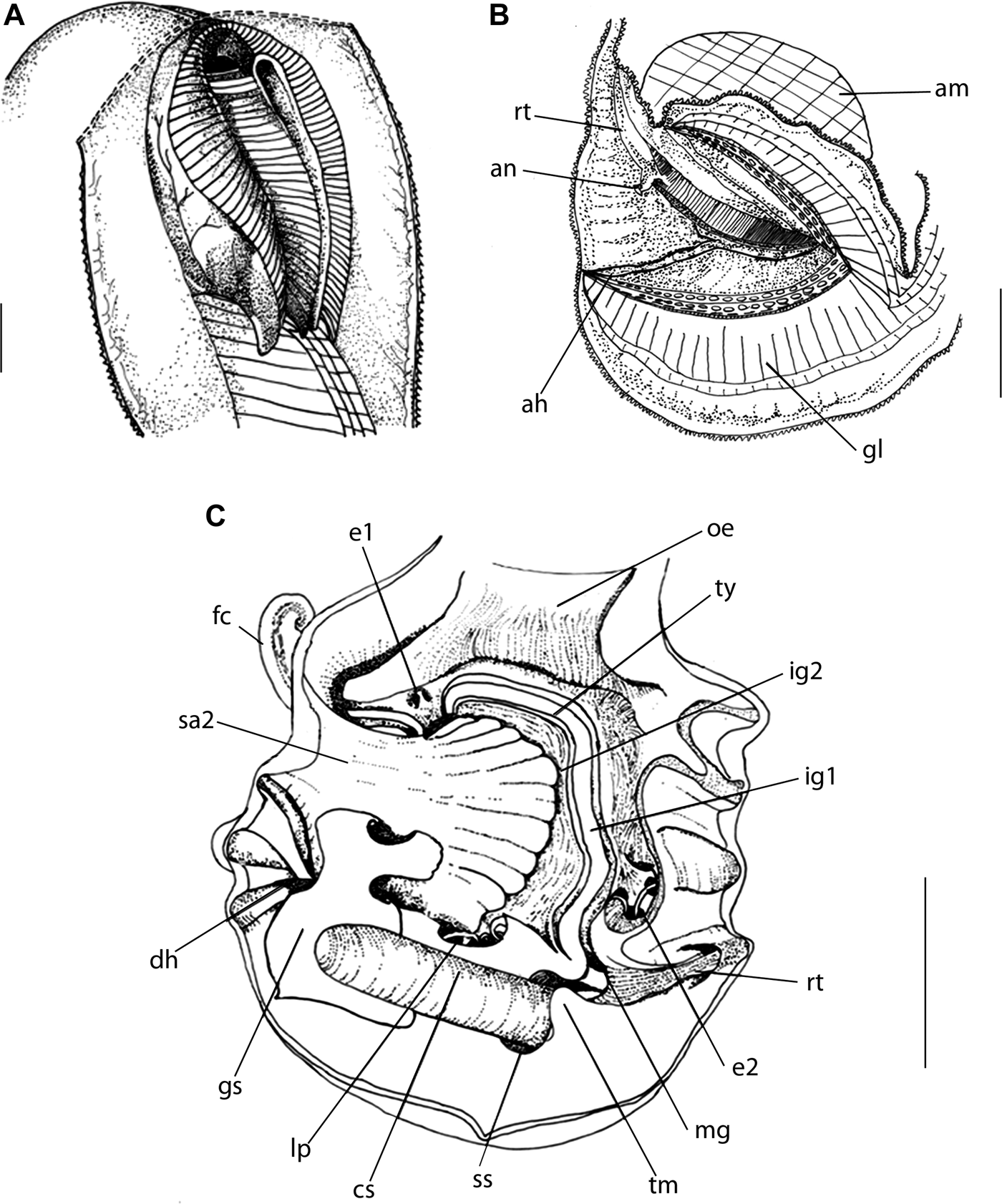
Crassostrea virginica anatomy: (A) palps and adjacent structures, right-slightly ventral view, adapted from Galtsoff (1954); (B) pallial cavity, posterior region, with posterior half of gill cut and deflected, detail for a accessory heart (ah) (adapted from Galtsoff, Reference Galtsoff1964); (C) stomach opened longitudinally, dorsal view showing internal structures. Scale: 1 mm.
Ostrea virginica Gmelin, Reference Gmelin and Linné1791: 3336; Galtsoff, 1937: 356, Reference Galtsoff1938a: 461–486, Reference Galtsoff1938b: 75; Reference Galtsoff1940: 117–135; Walzl, Reference Walzl1937: 125–140; Nelson, Reference Nelson1938: 1–61, Reference Nelson1960: 163–203; Lunz, 1938: 367; Medcof, Reference Medcof1939: 287–301, Reference Medcof1944: 209–216; Loosanoff, Reference Loosanoff1939: 177–178; 1949: 122; Taylor, 1941: 278–281; Medcof & Needler, Reference Medcof and Needler1941: 253–257; Menzel, Reference Menzel1955: 69–153; Numachi, Reference Numachi1962: 211–217.
Ostrea (Crassostrea) virginica: Sacco, Reference Sacco1897: 23 (pl. 15).
Crassostrea virginica: McLean, Reference McLean1941: 8 (pl. 1, figures 1–4); Warmke & Abbott, Reference Warmke and Abbott1962: 173; Galtsoff, Reference Galtsoff1964: 1–458; Stenzel, Reference Stenzel and Moore1971: 967–1172; Abbott, Reference Abbott1974: 456 (figure 5274); Brusca & Ardill, 1974: 111–131; Menzel, Reference Menzel1974: 453–456; Ahmed, Reference Ahmed1975: 357–397 (figure 6); Andrew, 1979: 293–335; Haley & Newkirk, Reference Newkirk and Haley1982: 399–401; Buroker, Reference Buroker1983: 99–112, Reference Buroker1984: 550–557; Castillo-Rodriguez & García-Cubas, 1984: 1–57; Hedgecock & Okazaki, Reference Hedgecock and Okazaki1984: 535–545; Harry, Reference Harry1985: 152; Quayle, Reference Quayle1981: 1–241; Arakawa, Reference Arakawa1990: 1–13; Brock, Reference Brock1990: 59–63; Reeb & Avise, Reference Reeb and Avise1990: 397–406; Márquez, 1992: 51–55; Hu et al., Reference Hu, Fuller, Castagna, Vrisenhoek and Lutz1993: 471–496; Lawrence, Reference Lawrence1994: 185–202; Littlewood, Reference Littlewood1994: 221–229; Kennedy et al., Reference Kennedy, Newell and Eble1996: 1–734; Beninger & Veniot, Reference Beninger and Veniot1999: 179–188; Leitão et al., Reference Leitão, Boudry, Labat and Thririot-Quiévreux1999: 175–186; Coan et al., Reference Coan, Valentich-Scott and Bernard2000: 218 (pl. 38); Wang et al., Reference Wang, Xu and Guo2004: 46–54; Lópes-Flores et al., Reference Lópes-Flores, Herra, Garrido-Ramos, Boudry, Ruiz-Rejo and Ruiz-Rejo2004: 181–188; Wang et al., Reference Wang, Guofan, Liu and Guo2008: 489–493.
DIAGNOSIS
Size up to 130 mm; fixed in rocks; shape cupped elongate; colour varying between white and purple; right valve slightly operculum-shaped; left valve, fixed in substrate, more larger than right valve with ornamentation in edge shell. Muscle impression is elongated and pigmented purple bands. Adductor muscle postero-dorsal located, occupying 1/6 of total size of animal; sub-umbonal region present and free; colour of mantle edge normally brown or orange. Accessory heart in left lobe is bifurcate around adductor muscle reduced, but thick. Right lobe presenting only one branch of accessory heart. Palps superior margin with plicae.
DISTINCTIVE DESCRIPTION
Shell
(Figures 7A–D) Large, ~130 mm, thick, with well-defined cupped shape. Surface ornamented with waves and layered lamellae. Right valve opercular and protruding near hinge. Left valve slightly larger than right valve, with high umbo. External coloration variable, from white to purple, with orange or brownish spots on borders. Inner portion of valves predominantly white, sometimes opalescent brightness like in other species. Adductor muscle impression oval, not elongated, located in postero-dorsal region, concavity towards hinge, occupying ~1/6 of valve area. Muscular impression highly pigmented, forming purple bands. Quenstedt muscle impression oval, marked by slightly deep and colourless depression, located in middle of anterior quadrant, occupying ~1/20 of size of adductor muscle impression.
Muscle system
Adductor muscle
(Figure 7F) Rounded in section.
Quenstedt muscle
Inserting in most basal portion of palps, near gill.
Mantle
(Figures 7F–H, 8B) Thin, with well-defined and abundant pallial muscles. Sub-umbonal region free, not filled by palps. Pallial muscles mainly located around adductor muscle. Accessory heart of left lobe with thin walls, with wide diameter. Bifurcation around adductor muscle reduced, but thick. Other branch leaves pallial branchial junction and follows towards gills, like in C. mangle. Right lobe presenting only one branch of accessory heart. Mantle borders slightly pigmented, brownish or orange. Middle fold with uniformly distributed medium to long papillae. Internal fold with well distributed long papillae.
Gills
(Figure 7E) Similar to other species in size or number of lamellae. Margin of alimentary groove thinner, with ~1/2 size of alimentary channel of C. brasiliana.
Visceral mass
(Figure 7F) Occupying ~1/4 of animal's size. Located from ventral region of ligament to anterior surface of pericardium. Digestive diverticula, located in dorsal region of palps, occupying ~1/4 of visceral mass.
Circulatory and excretory systems
(Figure 7I) Ventricle large, reticulated, sometimes pigmented. Auricles slightly thinner and less reticulated, devoid of pigmentation. A pair of kidneys occupying relatively same position of other species.
Digestive system
Palps
(Figure 8A) Somewhat small, ~1/5 size of animal. Palps located under mantle in sub-umbonal region, not filling whole free space of sub-umbonal region. Thin, sickle-shaped, joined throughout length of its base towards visceral mass. Upper margin with plicae over border of palps. Anterior and posterior portions reduced, but devoid of plicae. Internal faces intensively plicate, plicae thinner than in other species. External face smooth, but striations may be present.
Oesophagus
(Figure 8C) Short, ~1/6 size of palps; internal surface intensively striated.
Stomach
(Figure 8C) Same as other species, except for smaller globose region with ~1/8 of animal's size, laterally flattened, with numerous irregular external and internal invaginations. Style sac and intestine separated by thinner pyloric process, slightly longer than in other species. Digestive diverticula well distributed and not very abundant.
Intestine
Thin in portion between adductor muscle and gills; internally similar to other species. Minor and major typhlosoles thin. Rectum shorter, not surrounding curved portion of muscle.
Reproductive system
(Figure 7F) Gonads abundant, occupying ~1/5 of animal's height. Genital opening not observed.
DISTRIBUTION AND HABITAT
From western Canada to Florida and Mexican Gulf (Abbott, Reference Abbott1974; Mikkelsen & Bieler, Reference Mikkelsen and Bieler2008). Only on rocky coasts.
EXAMINED MATERIAL
USA. Florida: Wabasso, Sand and flat on rest area, on causeway, state route 512, 27°49.3′N 80°25.257′W, MZSP 46930, 1 specimen (Simone col.; 5 August 2004); Fort Pierce, sand bar on north side of Fort Pierce Inlet, and of Little Jim Island, 27°28.147′N 80°18.503′W, MZSP 46961, 3 specimens (Simone col., 6 August 2004); Monroe, Field Museum of Natural History FMNH 314566, 1 specimen (D. Gonsalves-Jackson, 4 August 1999); Off Florida, Gulf of Mexico, USMN 833964, 1 specimen (CSA for MMS, 9 December 1984).
Crassostrea gigas (Thünberg, 1793)
(Figures 9, 10)
Ostrea gigas Thünberg, 1793: 140–142 (pl. 6, figures 1–3); Lischke 1896: 174.
Crassostrea gigas shell and anatomy: (A) external view, right valve; (B) left valve, inner partial view (L 97 mm); (C) same, detail of adductor muscle impression; (D) same, detail of sub-umbonal region at anterior portion; (E) same, detail of postero-ventral mantle edges showing pallial muscles; (F) general right view, valve removed (L 85 mm); (G) palps, anterior—slightly ventral view (side 20 mm); (H) edge of right lobe, middle-ventral portion, showing papillae of middle and internal folds (side 6 mm); (I) left mantle lobe, detail of postero-dorsal region, internal view, detail for a accessory heart (ah) (side 20 mm); (J) gill, internal surface of middle portion; (K) detail for a rectum (rt) and anus (an). Scale: 1 mm.
Crassostrea gigas anatomy: (A) pallial cavity, posterior region, posterior half of the gills sectioned from each other and deflected (H 55 mm), detail of accessory heart (ah) (adapted from Galtsoff, Reference Galtsoff1964); (B) stomach opened longitudinally, dorsal view, inner surface exposed, scale 1 mm.
Ostrea (Crassostrea) gigas: Hirase, Reference Hirase1930: 49–55.
Crassostrea gigas Ahmed & Sparks, Reference Ahmed and Sparks1967: 2155–2158; Abbott, Reference Abbott1974: 456 (figure 5276); Menzel, Reference Menzel1974: 453–456; Ahmed, Reference Ahmed1975: 375 (figure 10); Andrew, 1979: 293–335; Buroker et al., Reference Buroker, Hershberger and Chew1979a: 157–169; Buroker et al., Reference Buroker, Hershberger and Chew1979b: 171–184; Torigoe, Reference Torigoe1981: 304–305, 325 (pl. 2, figure 1); Castillo-Rodriguez & García-Cubas, 1984: 1–57; Harry, Reference Harry1985: 152; Arakawa, Reference Arakawa1990: 1–13; Brock, Reference Brock1990: 59–63; Littlewood, Reference Littlewood1994: 221–229; Li, Reference Li1989: 239–255; Banks et al., Reference Banks, McGoldrick, Morgeson and Hedgecock1994; Boudry et al., Reference Boudry, Heurtebise, Collet, Cornette and Gérard1998: 279–291; Ó'Foighil, et al., Reference Ó'Foighil, Gaffney, Wilbur and Hilbish1998: 497–503; Abbott & Dance, Reference Abbott and Dance2000: 318 (figure 7); Coan et al., Reference Coan, Valentich-Scott and Bernard2000: 217 (pl. 37); Hayami, Reference Hayami and Okutani2000: 925; Yu et al., Reference Yu, Kong, Zhang, Guo and Xiang2003: 31–37; Christo, Reference Christo2006: 101–110; Pie et al., Reference Pie, Ribeiro, Boeger, Ostrensky, Falleiros and Ângelo2006: 1598–1600; Camara et al., Reference Camara, Davis, Sekino, Hedgecock, Li, Langdon and Evans2008: 313–322; Christo & Absher, Reference Christo and Absher2008: 71–77; Xia et al. Reference Xia, Yu and Kong2009: 1–8.
DIAGNOSIS
Size up to 140 mm; fixed in rocks; shape cupped elongate, with crenulations and ornamentation in edge shells; colour varying from white, brown, purple to yellowish spots; right valve and left valve almost of same size; left valve cupped-shaped. Adductor impression purple and is oval. Adductor muscle postero-dorsal located, occupying 1/4 of total size of animal; sub-umbonal region present and usually free; mantle edge unpigmented. Accessory heart of left lobe thick, bifurcated portion around adductor muscle very reduced; in right lobe with single branch. Palps with margin superior with plicae.
DISTINCTIVE DESCRIPTION
(More information and figures in Galtsoff (Reference Galtsoff1964).)
Shell
(Figure 9A–C) Large, ~140 mm, thick and with well-defined, elongated cupped shape. Surface ornamented by waves in borders and many layered lamellae in upper shell surface. Right valve almost same size as left valve, presenting a protruding area near hinge. Left valve cupped-shaped, a little larger than right valve, with internal depression that advances under a deep umbo. External coloration variable, sometimes with white, brown and yellowish spots. Inner portion of valves predominantly white, with opalescent brightness sometimes present like other species. Adductor muscle impression (ai) oval, slightly elongated, located in postero-dorsal region, concavity towards hinge; highly pigmented with purple bands. Quenstedt muscle impression located in middle of anterior portion of valves.
Muscle system
Adductor muscle
(Figure 9F) Outline ovoid. Distinction of quick and slow portions clear, of similar size.
Mantle
(Figures 9D–F, H, J, 10A) Thick, with well-defined pallial muscles, stronger around adductor muscle. Accessory heart (Figures 9H, 10A) of left lobe thick, diameter varying from narrow to wide. Bifurcated portion around adductor muscle very reduced. Other branches leaving pallio–branchial junction towards gills, like preceding species. Accessory heart of right lobe with single branch. Mantle border unpigmented. External fold almost two times larger than middle and internal folds. Middle fold with small and middle finger-like, uniformly distributed papillae, more numerous than those of internal fold. Internal fold with small and uniform papillae (Figure 9J). Sub-umbonal region not filled by palp.
Gills
(Figure 9I) Same as other Crassostrea in size. Number of filaments of lamellae ~15. Marginal alimentary groove thin.
Visceral mass
(Figure 9F) Occupying ~1/6 of animal's size. Located from ventral region of ligament to anterior surface of pericardium. Digestive tubes located in anterior region, posterior to palps, occupying ~1/4 of visceral mass, with diverticula. Gonads yellowish, covering digestive diverticula externally.
Circulatory and excretory systems
Ventricle triangular-shaped, very reticulated, usually unpigmented. Auricles thinner and less reticulated, unpigmented.
Digestive system
(Figures 9G, 10B) Alimentary channel fully involved by gonads, diverticula of digestive gland and kidneys. All structures as in preceding species.
Palps
(Figure 9G) Large, located under mantle sub-umbonal region, of free space of sub-umbonal region. Thick, wide sickle-shaped, joined throughout length of their base to visceral mass. Anterior and posterior portions wide and devoid of plicae. Anterior and ventral free margins slightly arched in ventrally to form apex. Internal faces intensively plicate, including upper region of palps. External faces smooth, in which there may be striations and irregular grooves.
Oesophagus
(Figure 10B) Oesophagus dorso-ventrally flattened, relatively longer than short. Internal surface intensively striated.
Stomach
(Figure 10B) Same as other species. Selection wall portion shorter than C. mangle and C. brasiliana. Digestive diverticula fewer than in other species, with fewer connections to stomach. Selection area (sa2) smaller than other species.
Reproductive system
(Figure 9F) Gonads abundant, occupying ~1/4 of animal's height.
DISTRIBUTION AND HABITAT
Crassostrea gigas is the most cultivated species around the world, but it appears naturally in estuaries and rocky coasts of Japan and south-eastern Asia. It is usually found from the intertidal to subtidal zones (NIMPIS 2002). In Brazil, it is largely cultivated in the Southern region of the country. It appears only on rocky coasts.
EXAMINED MATERIAL
Brazil. Santa Catarina: Florianópolis, cultivation, MZSP 89671, 12 specimens (Simone leg., August 2008).
Crassostrea praia (Ihering, Reference Ihering1907) (revalidated)
(Figure 11A, B)
Ostrea (Crassostrea) parasitica praia Ihering, Reference Ihering1907: 426 [not figured].
Types. Crassostrea praia shell type MZSP 10916: (A) left valve, detail of ornamentation; (B) same, external view, inner view (H 80 mm). Crassostrea parahibanensis shell paratype. MNRJ 3815; (C) left valve, external view, detail of ornamentation; (D) same, inner view (H 220.5 mm). Crassostrea brasiliana syntype; (E) right valves, inner view.
Crassostrea rhizophora praia: Rios, Reference Rios1970: 171; Reference Rios1975: 205 (in syn.); 1994: 245 (in syn.); 1985: 229 (in syn.).
Crassostrea rhizophorae praia: Rios, Reference Rios2009: 501 (in syn.)
DIAGNOSIS
Size up to 100 mm; left valve shape cupped elongate and thick, with crenulations and ornamentation on entire shells; colour varying from white to purple. Muscle impression purple. Adductor muscle oval–elongate, occupying 2/3 of height of shell. Sub-ligamentary area long; sling umbonal cavity present.
TYPES
Syntypes MZSP 10916 (figures 81, 82).
DIFFERENTIAL DESCRIPTION
Shell
(Figure 11A, B) Shape cupped, elongate, up to 100 mm long. Colour varying from white to purple, with longitudinal bands of these. Right valve not seen. Umbonal cavity of left valve sling. Interior of valves predominantly white, with little brightness or opaque. Purple pigmentation close to margin, in most inner regions. Scar of adductor muscle mostly located in anterocentral region, somewhat middle; form long crescent, occupying about 2/3 of shell height. Muscle scar sometimes strongly pigmented with purple, brown bands or a combination.
DISCUSSION
The new species described herein, Crassotrea mangle, has been referred to in Brazil as C. rhizophorae. A comparison with specimens of C. rhizophorae from the type locality (Carriacou) revealed significant differences, which endorse the specific separation. The introduction of a new species was necessary, as no previous name was available. Ostrea arborea Dillwyn Reference Dillwyn1817, a possible name for the Brazilian species, is considered herein a nomen dubium, as neither the too brief description nor the locality (Atlantic and Indian Oceans, Moluccas Island and Curaçao) were precise enough to allow for its recognition (Dillwyn, Reference Dillwyn1817; Rios, Reference Rios1970). The type specimen has not been found. Therefore, O. arborea is here understood as a senior synonym of C. rhizophorae. Furthermore, Parodiz (Reference Parodiz1948) described the morphological differences between O. arborea, O. spreta d'Orbigny, 1845 and O. puelchana d'Orbigny, 1842 (Parodiz, Reference Parodiz1948; Castellanos, Reference Ageitos de Castellanos1968), and highlighted the confusion involving these taxa.
Another previously described name and possible synonym, Ostrea parasitica praia Ihering, Reference Ihering1907, occurs in Rio Grande do Sul State (Parodiz, Reference Parodiz1948). The shell characteristics differ from those of C. mangle and C. rhizophorae (more information below). Crassotrea praia is available according to ICZN Art. 12 (Bouchet, 2013, personal communication)
Shell
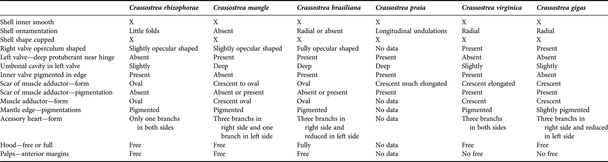
The shells of the Caribbean C. rhizophorae are generally the smallest, up to 60 mm long, cup-shaped and thicker; the colour varies from brown, yellowish to white, with intermediary shades (Figure 2E, F). Crassotrea mangle shells are also cup-shaped, with an average of 70 mm long, and a more elongated outline (Figure 1A–C). Crassotrea brasiliana is large, with an average length of 100 mm and a more rounded outline when occurring in open sea, or cup-shaped in mangroves (Figure 4B, C). Crassotrea praia are elongated but not as large as C. gigas, average 100 mm (Figure 9E, F). Crassotrea virginica has an ornamented shell, with foliaceous layers and undulations, which externally distinguishes it from the other species (Galtsoff, Reference Galtsoff1964; Abbott, Reference Abbott1974) (Figures 5L, 6A, B). Crassotrea gigas has a larger (with average of 120 mm) and a thicker shell, ornamented by foliaceous layers coupled with undulating margins (Figure 7F, G). The exterior of the valves is ornamented by projections of the prismatic layer forming foliaceous blades with variable number of folds or scales, low and short, arranged radially in C. brasiliana, C. virginica and C. gigas (Galtsoff, Reference Galtsoff1964). In C. brasiliana and C. mangle, such ornamentation can be inconspicuous or absent, depending on the environment (Nascimento, Reference Nascimento and Menzel1991). In contrast, C. rhizophorae presents small folds on the right valve. In C. praia the ornamentation consists of longitudinal undulations.
The outer colour is highly variable in all species; it may vary in C. rhizophorae, C. mangle and C. brasiliana from brown, yellow, white to purple, including a combination of these colours. Crassotrea virginica and C. praia have shades of purple and brown, with white spots. Crassotrea gigas varies from brown to orange with white spots. Natural erosive agents may cause fading, especially in the older specimens (Harry, Reference Harry1985).
The right valve of C. mangle and C. rhizophorae is flattened or slightly arched (convex), opercular, and in C. mangle it is slightly protuberant towards the hinge (it accommodates the visceral portion of the oyster's body). In C. brasiliana, C. virginica and C. gigas, the right valve is arched-convex, opercular and very protuberant towards the hinge. The right valve of C. praia was not observed in the present study.
The left valve of C. rhizophorae, C. mangle, C. brasiliana, C. virginica, C. gigas and C. praia, as is the case in any ostreoidean, is fixated to the substrate. It is cup-shaped, and particularly much larger than the right valve (Figures 1D, 2E, 4C, 5L, 7F, 9E). All of them have a deeper umbonal cavity, which may be shallower (Gunter, Reference Gunter1950; Thompson, Reference Thompson1954; Galtsoff, Reference Galtsoff1964; Yonge, Reference Yonge1966) in some specimens.
The internal surface of the six species is usually glossy, but some specimens have it opaque. The pallial line is absent in ostreids (Morton, Reference Morton and Wilburn1983), possibly due to the fibres of radial muscles being fixed directly to the region surrounding the adductor muscle and to the visceral mass, and not to the region close to the shell border, as is the case in most bivalves (Galtsoff, Reference Galtsoff1964). The Quenstedt muscles scar is very small, marked by a shallow and colourless depression, close to the internal ventral margin of the ligamentary area; it is similar in position and size in all species. The adductor muscle impressions of C. mangle (Figure 1D) and C. brasiliana (Figure 4B, C) may be pigmented or unpigmented; it is unpigmented in C. rhizophorae (Figure 2G). The different pigmentation has been mentioned in the literature as a distinguishing feature between species (Nascimento, Reference Nascimento and Menzel1991), however, this proves to be inaccurate according to our findings, as a high degree of variation is present. A pigmented adductor muscle scar is also commonly found in C. praia (Figure 11B), C. virginica (Figure 7C) and C. gigas (Figure 9C). The free margin of the shell and the cavity that comprises the visceral mass in C. rhizophorae, C. mangle, C. brasiliana and C. praia, can be coloured light purple (Weisboard, 1964; this study), which has been regarded as a characteristic of tropical populations (Harry, Reference Harry1985).
Musculature
ADDUCTOR MUSCLE
The adductor muscle of C. rhizophorae (Figure 3C, D), C. mangle (Figure 1E), C. brasiliana (Figure 5C), C. virginica (Figure 7C) and C. gigas (Figure 9C) show variable outlines among specimens, including crescent, oval, oval–elongated in cross-section. This variation has been pointed out in the literature (Nascimento, Reference Nascimento and Menzel1991 for C. rhizophorae; Galtsoff, Reference Galtsoff1964 and Kennedy et al., Reference Kennedy, Newell and Eble1996 for C. virginica), and possibly is associated with the high level of shell plasticity.
The loss of the post-larval anterior adductor muscle and the enlargement of the posterior adductor muscle are characteristic of the ostreids. These are obviously related features. In Gryphaea Lamarck, 1801 the fibres of the opaque portion of the adductor muscle occupy about 30% of the adductor cross-section area (Thompson, Reference Thompson1954); this contrasts with more than 50% in Ostrea and is significantly reduced in Pycnodonte Waldheim, 1835. This proportion has been considered in the diagnosis of the three genera (Ostrea, Pycnodonte and Gryphaea) (Stenzel, Reference Stenzel and Moore1971). However, the diagnostic value of such character is not applicable in Crassostrea, since C. rhizophorae, C. brasiliana and C. virginica show individual variations in the proportions of the opaque and translucent parts. The opaque part may be found in proportions higher than 30%, but is always smaller than the translucent portion (Galtsoff, Reference Galtsoff1964 and Kennedy et al., Reference Kennedy, Newell and Eble1996 for C. virginica; this study).
QUENSTEDT MUSCLES
The pair of Quenstedt muscles has no differentiation regarding the origin on the shell and the insertion in the base of the palps in the five species; they are thicker in the portion connected to the shell in C. mangle (Figure 1G), C. rhizophorae (Figure 3F) and C. brasiliana (Figure 5E) if compared to C. virginica and C. gigas. The Quenstedt muscles in the five species are not difficult to find, differently from what has been postulated in the literature (Galtsoff, Reference Galtsoff1964; Stenzel, Reference Stenzel and Moore1971; Mesquita, Reference Mesquita1993). The direct observation of the muscle is possible mainly in large (>40 mm) specimens. From their origin on the shell, the muscles run through the anterior visceral mass up to the insertion in the base of the palps. This peculiar morphological arrangement shows improbable a homology with the anterior adductor muscle or any foot muscles (suggested by Galtsoff, Reference Galtsoff1964; Stenzel, Reference Stenzel and Moore1971). They may retract the gills and the palps, helping in transferring particles to the mouth.
Pallial structures
MANTLE
The mantle lobes in C. mangle (Figure 1L), C. rhizophorae (Figure 3H), C. brasiliana (Figure 5F) and C. gigas (Figure 9F) are very similar. They are robust, with evident blood vessels mainly near the palps. The pallial musculature is strong in all species, clustering near the adductor muscle. C. virginica (Figure 7F) differs by having thin mantle lobes (Galtsoff, Reference Galtsoff1964).
The sub-umbonal region in Crassostrea is formed by the anterior junction of the mantle lobes; it is actually an inner mould of the depth of the apex of the left valve. In Ostrea, this structure is not present, since the shells are not cup-shaped. The sub-umbonal region may be completely filled by the anterior region of the palps, as is the case in C. brasiliana (Figure E), or free, not filled by palps, as in C. mangle (Figure 1G), C. rhizophorae (Figure 3E), C. virginica (–Figure 7F) and C. gigas (Figure 9D).
The mantle edges of C. rhizophorae are thick and pigmented; the external fold is devoid of papillae and may exceed the shell margins in length when extended (Figure 3H). The middle fold has two different kinds of fingerlike papillae: short and thin, and long (more than 1 mm) with an enlarged base. The papillae are arranged normally presenting up to six short papillae alternated by two longer ones; both types of papillae show a gradual increase in diameter and length from the ventral region towards dorsal region. The edge is pigmented in various shades of brown or depigmented. In C. mangle, the mantle edge is thick, and can be pigmented or unpigmented; the three folds have similar sizes; the middle fold has sparsely distributed, short and long papillae; the successive arrangement may be two long papillae and up to six short papillae; the internal fold has evenly distributed papillae (Mesquita, Reference Mesquita1993; this study) (Figure 1L). The mantle edges of C. brasiliana are always brown coloured; the middle fold has long, medium and short papillae, having from 2 to 4 medium or short papillae for each long one (Figure 5F). In C. virginica the mantle edge can be brown or orange coloured; the middle fold shows intercalated medium and long papillae; the internal fold has evenly distributed, long papillae, intercalated with the middle fold papillae (Figure 7G). The mantle edge of C. gigas is pigmented; the middle fold shows medium and small papillae; the internal fold has small digitiform folds. In general, the internal fold of the mantle margin is extensible and highly vascularized (Figure 9J).
PROMYAL PASSAGE
One of the outstanding characters of Crassostrea is the promyal passage on the right side of the visceral sac. This additional opening significantly improves the cleaning mechanisms on the animal's dorsal side, providing a wide area closer to the hinge from which the water can be expelled. Additionally, as it is closer to the support between the valves, the water leaving by the promyal chamber can be ejected more vigorously, by using the same intensity of the adductor contraction (Nelson, Reference Nelson1938). The promyal passage was first described in C. virginica by Kellogg (Reference Kellogg1892), and has been pointed out as associated with the higher capacity of growth and survival of C. gigas when compared to the sympatric and promyal-wanting Ostrea lurida Carpenter, 1864 by Elsey (Reference Elsey1935).
Otherwise, and beyond the promyal passage, the oysters with a cup-shaped left valve, such as Crassostrea, have a more efficient cleaning mechanism of the pallial cavity, which allows for the establishment of populations close to river mouths. In such environment, the turbidity is higher and the suspended sediment is significant enough to preclude the survival of oysters not provided with a promyal passage. Galtsoff (Reference Galtsoff1964) did not agree with the opinion of Nelson (Reference Nelson1938), stating that ‘nothing is known about the adaptive meaning of such [promyal] passage, in addition to the fact that it provides an additional way for the water flowing through the gills’. The positioning and function of the promyal passage undoubtedly characterize a more effective water flowing mechanism, besides preventing the accumulation of particles in the pallial cavity.
GILLS
The gills of all species are similar to each other, with the number of lamellae increasing with the animal's size. The general shape of the gills of all species studied herein is similar to that described for Ostrea edulis Linné, 1758, C. virginica and C. angulata in the literature (Ridewood, Reference Ridewood1903; Yonge, Reference Yonge1926, Reference Yonge1966; Atkins, Reference Atkins1937a, Reference Atkinsb, Reference Atkins1938; Nelson, Reference Nelson1938; Galtsoff, Reference Galtsoff1964). The gills of the studied species differ only by the thickness of the marginal food channel. Crassostrea mangle (Figure 1H), C. rhizophorae (Figure 3K) and C. brasiliana (Figure 5I) have a broader food channel, while C. virginica (Figure 7E) and C. gigas have a narrower one. In all examined species, the gills reach ~80% of the specimen's length. The presence of a ventral particle groove is another particularity of an adult pseudolamellibranch gill (the Pectinidae do not possess such a groove) (Cannuel & Beninger, Reference Cannuel and Beninger2006).
The number of gill folds and filaments is a variable character, apparently increasing proportionally to the animal's size. However, the numbers can be discrepant between individuals of similar size. Ridewood (Reference Ridewood1903) mentioned 9–12 filaments per fold in O. edulis, while Atkins (Reference Atkins1937a) mentioned 8–17. Atkins (Reference Atkins1937a) mentioned 8–17 in C. virginica; Nelson (Reference Nelson1938) mentioned 8–15. Galtsoff (Reference Galtsoff1964) mentioned 10–16 filaments per fold in C. angulata, while Atkins (Reference Atkins1937a) referred to 15–16 and Nelson (Reference Nelson1938) 8–14 filaments per fold. In the present paper we verified that the number of filaments per fold in C. rhizophorae, C. mangle, C. brasiliana and C. virginica varies from 12 to 13, reaching 15 in C. gigas. Based on the proximity of the numbers, this parameter seems to be of little use in distinguishing the examined species.
The mucous secretion of the gill plays an important role in the selection mechanisms. It has been verified that, in O. edulis Linnaeus, 1758, the common filaments have twice more mucous cells than the main ones, concluding that the material reaches the front face of the common filaments and has twice the chance of causing sufficient mucous secretion for its rejection when reaching the palps (Nelson, Reference Nelson1960). Those characteristics have been observed in C. mangle, C. brasiliana, and C. gigas in the analyses of functional anatomy performed here.
Circulatory system
HEART
The hearts of the analysed species differ in colour, arrangement and thickness of the ventricular and auricular fibres. The ventricle of C. rhizophorae is elongate and thicker than the auricles; the auricle walls are formed by thin bundles of muscle fibres crossing the ventricular cavity, dividing it incompletely in two chambers. The colour varies from cream-yellowish to depigmented (Figure 4C). Crassostrea mangle has a more developed, thick-walled ventricle, with internal fibres crossing in several directions; the auricles can be pigmented or unpigmented; it has a thinner external tissue, with also thinner fibres, forming a trabecular structure (Figure 1J). Crassostrea brasiliana has a thin, well-reticulated, usually translucent ventricle; the two auricles are less reticulated and unpigmented (Figure 5H). Crassostrea virginica, as reported by Galtsoff (Reference Galtsoff1964) (Figure 7I) and C. gigas have a broad, reticulated ventricle, either pigmented or unpigmented; the auricle, in contrast, has fewer fibres and is colourless.
Accessory heart
The accessory heart, present symmetrically in both mantle lobes, is characteristic of the genus, and shows significant differences between the four species. The primary function of the accessory hearts is to promote the blood circulation inside the mantle and facilitate the gaseous exchanges (Galtsoff, Reference Galtsoff1964). In C. rhizophorae, the accessory heart has a single branch, and lies near the adductor muscle on both sides (Figure 3G). In C. virginica, it has three long and well-defined branches (Y-shaped) on both lobes, as described by Galtsoff (Reference Galtsoff1964) and Stenzel (Reference Stenzel and Moore1971) for C. virginica (Figure 7H). In C. mangle (Figure 1K), C. brasiliana (Figure 5L) and C. gigas (Figure 9H), the branch surrounding the adductor muscle towards the anus on the left lobe is reduced or absent; while the left lobe has a single vessel. Additionally, in all species, the accessory heart also varies concerning the gauge of vessels and tissue thickness, as stressed in the descriptions.
Digestive system
PALPS
The labial palps are evident organs, located fully or partially inside the mantle's sub-umbonal region. The palps of C. mangle are flattened and elongated, slightly arched anteriorly, with ventral free margins; the internal surfaces have folds, which extend themselves transversally. The joining of the palps constitutes a ventral lip (Figures 1F, 2D). In C. rhizophorae the palps are flattened, elongated, the base is united throughout its length to the visceral mass; the margins are slightly arched anteriorly and ventrally free, converging towards the ventral region to form the apex; the internal faces are plicate, the inner folds extend transversally, ending in the free, narrow and smooth inner margin (Figures 3I, 4B). The palps of C. brasiliana are slightly flattened and sickle-shaped; the superior margin of the folds are free and smooth (Figures 5G, 6C). In C. virginica (Figure 8A), the palps are thinner and smaller than in the prior species, and have no free upper margin such as in C. brasiliana. In C. gigas (Figure 9G), the palps are large and thick and lack a free upper margin, such as those of C. virginica and C. rhizophorae.
The arrangement of the palps of C. mangle is similar to the palps of Ostrea and Crassostrea described above, but there are significant differences concerning their functionality. The currents in the smooth (external) surface of the four hemipalps are similar to each other and work in order to lead all particles towards the ventral (aboral) margin of the palps.
OESOPHAGUS
The oesophagus is dorso-ventrally flattened in all species. It varies only concerning the length and width: in C. mangle (Figure 2A, H), C. brasiliana (–Figure 6D), and in C. rhizophorae (Figures 3J, 4E) it is slightly longer than wide; in C. virginica, the oesophagus is clearly the shortest (Figure 8C); and in C. gigas it is slightly longer than short. In all species the oesophagus have similar internal striae (Figure 10B).
STOMACH
The stomach is slightly flattened dorsal–ventrally in all species. It is located in the globular region of the visceral mass, and shows a higher number of ducts to digestive diverticula in some species (C. rhizophorae and C. brasiliana) or a smaller number in others (C. mangle, C. gigas and C. virginica). Besides the general quantity, these structures are abundant in the globular region. The stomach ducts to the digestive diverticula are much reduced, ~50% in C. gigas when compared to the other species.
The internal structures of the stomach are similar among the species, only differing by some details. The selection area of C. brasiliana (Figure 6D) is longer than that of C. mangle (Figure 2H) and C. rhizophorae (Figure 4E), ending closer to the base of the typhlosoles on the stomach floor. The caecum of the selection area of C. brasiliana is shorter than that of C. rhizophorae, not completing 1.5 turns (Mesquita, Reference Mesquita1993; present study). In C. virginica, the style sac and the intestine are surrounded by a thinner pyloric process and are slightly longer than that of the other species. In C. gigas, the selection area is shorter than that of C. rhizophorae and C. brasiliana.
The stomach of C. virginica has been additionally studied based on latex or resin moulds (Shaw & Battle, Reference Shaw and Battle1957; Galtsoff, Reference Galtsoff1964). Such an organ has been described as bag-shaped, internally divided into a smaller anterior chamber and other larger posterior chamber, by means of a thick fold; such a fold is projected to the organ's lumen and seems to serve as a means to direct particles to the posterior region through a narrow channel. Shaw & Battle (Reference Shaw and Battle1957) saw the caecum organization in C. virginica and named the ventral branch as ‘posterior appendix of the caecum’ and the dorsal branch as ‘anterior appendix’. Purchon (Reference Purchon1957) named such set of bags with a blind bottom in C. parasitica as ‘food sorting caecum – fc’. In C. rhizophorae, C. mangle and C. brasiliana, the base of such fold on the floor can be strong, with grooves that extend to the distal region, or it can be almost smooth. This grooved area corresponds to the sorting area (sa2).
All such stomach characters are similar to those of O. edulis (Yonge, Reference Yonge1926; Graham, Reference Graham1949; Purchon, 1957; Reid. Reference Reid1965), C. parasitica (Shaw & Battle, Reference Shaw and Battle1957) and O. chilensis (Purchon, 1957). Other details of the morphology and physiology of the ostreid digestive tract, such as ciliary mechanisms and stomach structure and operation, can be found in the literature (Shaw & Battle, Reference Shaw and Battle1957; Nelson, Reference Nelson1960; Reid, Reference Reid1965; Yonge, Reference Yonge1966). In this scenario, the internal anatomy of the stomach of C. rhizophorae, C. mangle, C. brasiliana, C. virginica and C. gigas has the same essential characteristics described by Purchon (1957) for C. parasitica, but the author did not mention the anatomy and the currents of the cylindrical portion (style bag and early intestine) of the stomach of that species. Purchon (1957) did not disclose in his schemes the existence of folds on the walls of the recesses of C. parasitica, but in C. rhizophorae, C. mangle, C. brasiliana and C. gigas the structures were very conspicuous. Regardless, the locality of the specimens studied by Purchon (1957) was not mentioned.
When comparing the globular region of the stomach of C. mangle and O. edulis (Yonge, Reference Yonge1926; Graham, Reference Graham1949), a general morphological similarity was detected, with a few exceptions. The caecum of selection in Ostrea edulis has only a ventral branch (= posterior appendix of Shaw & Battle, Reference Shaw and Battle1957), where the largest typhlosole penetrates and ends; two evaginations are missing in the anterior wall of the stomach, as well as the isolated opening of the digestive duct on the right wall, appearing in C. mangle and C. brasiliana. Graham (Reference Graham1949) mentioned the existence of a series of folds and grooves with an irregular arrangement on the right wall and close to the intestine opening of O. edulis; these were considered similar to the folds that constitute the well-developed area of the posterior sorting in Glycymeris glycymeris (Linné, 1758). In this same position, irregular plicae and grooves can be observed in the stomach of C. rhizophorae and C. brasiliana, which lies inside the dorsal hood.
Yonge (Reference Yonge1926) considered the sorting mechanisms inside the selection caecum of O. edulis less complex than that of Modiolus (Lamarck, 1799) and Mya (Linné, 1758), and attributed such simplicity to the higher development of the selection mechanisms of the gills and palps.
INTESTINE
The intestine is individualized from the style sac distal end; it returns in the opposite direction, extending behind the pericardium, superficially immersed in the gonads in the visceral mass. In C. brasiliana, the portion surrounding the adductor muscle is narrower than in C. mangle. In C. virginica it is thinner, like in C. brasiliana and C. rhizophorae, and its typhlosoles are also thinner. The rectum of C. virginica is shorter than that of the other species, keeping the adductor muscle free. C. gigas has a similar to that of C. mangle and C. brasiliana.
Genital system
The gonads of all species are massive, apparently regardless of the gonadal maturation period, and surround the whole visceral mass, in some cases surrounding the region near the pyloric process. They are usually coloured beige or yellowish. The genital opening has been observed in C. mangle and C. brasiliana in the kidney, and distinguishes itself from the opening by a brownish spot.
Nervous system
The relatively simple nervous system of the four species is similar, displaying variations in the size of the commissures in relation to the animal size. The cerebral ganglia are interestingly inconspicuous, because of their small size and deep location in the visceral mass, showing no remarkable differences.
Habitat and distribution
We confirm C. rhizophorae occurs in salty mangroves in the south-east Caribbean Sea; C. mangle has been reported only in mangroves and C. brasiliana in both environments—open sea, rocky shores and mangroves. Crassostrea gigas has been observed forming banks in the Indo-Pacific Ocean and has been introduced in Southern Brazil (Orensanz et al., Reference Orensanz, Schwindt, Pastorino, Bortolus, Casas, Darrigran, Elıas, Lopez-Gappa, Obenat and Pascual2002; Ruesink et al., Reference Ruesink, Lenihan, Trimble, Heiman, Micheli, Byers and Kay2005).
Larval development (survival) and incrustation of Crassostrea are associated with high salinity levels (27–37‰) (Sandison Reference Sandison1966; Nascimento, Reference Nascimento and Menzel1991; Lemos et al., Reference Lemos, Nascimento, Araújo, Pereira, Bahia and Smith1994; Marques-Silva et al., Reference Marques-Silva, Beasley, Gomes, Gardunho, Tagliaro, Schories and Mehlig2006). Crassostrea brasiliana and C. mangle have a quick development and growth in low salinities, but C. brasiliana tends to be more tolerant to salinity fluctuations (8–34‰) than C. mangle (7–28‰) (Nascimento, Reference Nascimento and Menzel1991); C. rhizophorae tolerates salinities of 37–44‰ (Mattox, Reference Mattox1949; Ladrón de Guevara et al., Reference Ladrón de Guevara, Winkler, Rodríguez-Romero and Palma-Rojas1996); and C. virginica of 19–25‰ (Castillo-Rodriguez & García-Cubas, 1984). This explains the presence of C. brasiliana in both estuarine and open sea regions, and C. mangle's restriction to mangroves.
The struggle for space in the environment causes distortions in the shell shape, as described by several authors (e.g. Galtsoff, Reference Galtsoff1964), precluding the immediate visual identification. According to Stenzel (Reference Stenzel and Moore1971), the ‘spatuliform’ or ‘semitubular’ shells (Mesquita, Reference Mesquita1993) are characteristic of overpopulated substrates. This is intuitive and confirmed in the presently studied samples.
Importation and culture of exotic species have gradually obscured nature's long-term patterns of adaptation and evolution (Andrews, Reference Andrews, Giese and Pearse1979).
Despite some authors having considered Crassostrea mangle, wrongly named as C. rhizophorae, as the single species of this genus occurring in Brazil (e.g. Rios, Reference Rios2009); some studies have restricted its occurrence to mangroves, while the name C. brasiliana has been applied to forms restricted to intertidal environments (Absher, Reference Absher1989; Galvão et al., Reference Galvão, Pereira, Machado and Henrique2000). This landscape is only partially confirmed here. As C. brasiliana also occurs in estuarine environments, this concept must change. Further distinction between both species (once more, considering the misidentification of C. mangle as C. rhizophorae) has also been based on biological and ecological differences (Mattox, Reference Mattox1949; Galtsoff, Reference Galtsoff1964; Timothy et al., Reference Timothy, Littlewood and Donovan1988), and growth and larval morphology (Absher, Reference Absher1989; Christo & Absher, Reference Christo and Absher2008, including C. gigas).
Even though Crassostrea praia is endemic to Lagoa dos Patos, Rio Grande do Sul, no records (old or recent) were found in the MORG (Museu Oceanográfico do Rio Grande—‘Prof. Eliéser de Carvalho Rios’). Lagoa dos Patos is under severe influence of the nearby port, as well as the construction of a dam; it may be that the species is already extinct due to environmental changes. A comparison of the syntypes (left valves, strongly elongated with characteristic ornamentation) with other examples found on the Brazilian coast, suggests the specific separation between C. praia and C. rhizophorae = (C.mangle).
Some recent papers (Ignácio, 2002; Lèpegue et al., 2002; Varela et al., Reference Varela, Beasley, Schneider, Sampaio, Marques-Silva and Tagliaro2007) have considered the occurrence of Crassostrea gasar, an African species identified as a synonym of C. brasiliana, in north-eastern and south-eastern Brazil. Based on the anatomical studies made with the voucher material of Varela et al. (Reference Varela, Beasley, Schneider, Sampaio, Marques-Silva and Tagliaro2007), identified as C. gasar, we verified that specimens are, in fact, C. brasiliana. In a study on the molecular phylogeny of the Brazilian mangrove oysters, Varela et al. (Reference Varela, Beasley, Schneider, Sampaio, Marques-Silva and Tagliaro2007) assume that Brazilian oysters identified as C. gasar are C. brasiliana, based on identical sequences of 16S rRNA. Until further studies with reliable African samples of C. gasar are carried out, we assume that the species does not occur in Brazil, being restricted to Africa, and all citations of C. gasar in Brazil must be interpreted as C. brasiliana. A comparative molecular work of this species (and others species) will be included in a future revision of Crassostreinae.
CONCLUSIONS
The comparative and detailed anatomy of the species Crassostrea rhizophorae, C. mangle, C. brasiliana, C. gigas and C. virginica, as well as the conchology of C. praia, allow for the following conclusions.
1. As stated in the descriptions and discussion, important anatomical differences have been found in almost all structures, allowing for the discernment of the species and their identification. This includes the type species of Crassostrea.
2. The distinction between the Crassostrea species based only on shell characters is complicated but not impossible (see Figure 12 and Table 1), since their structure shows phenotypical plasticity and may be subject to environmental influence.
3. Based on conchological, anatomical and geographical differences, C. rhizophorae, has been separated from the supposed conspecific population in Brazil. A new species, C. mangle, is introduced in order to settle the Brazilian entity.
4. In reference to the environment, two of the studied species native to Brazil, C. mangle occurs in mangroves and C. brasiliana occurs in open sea and mangroves. The Indo-Pacific species, C. gigas, has been introduced in Southern Brazilian seashores, mainly in open sea. Crassostrea praia occurs, apparently, only in Southern Brazil.
5. The occurrence of the African species, C. gasar, in Brazil has not been confirmed. The examination of testimonial material allowed us to assert that it is misidentified, and is, in fact, C. brasiliana.
6. The geographical distribution of the studied species is the following: C. rhizophorae occurs in mangrove areas in the south-eastern Caribbean Sea; C. mangle occurs from Pará to Santa Catarina, only in mangroves; C. brasiliana from Maranhão to Santa Catarina, in mangroves and open sea rocky shores; C. virginica occurs from the Atlantic coast of Canada to the Caribbean; C. gigas is originated from the Indo-Pacific; and C. praia is restricted to Rio Grande do Sul. The latter is here understood as a probably extinct endangered species.
Identification key.
Characteristics of species, morphological and anatomical.
ACKNOWLEDGEMENTS
We thank the Instituto de Biociências da Universidade de São Paulo and Museu de Zoologia da Universidade de São Paulo for the infrastructural support in carrying out this work. Special thanks for Dr Philippe Bouchet of MNHN, Dr Paula Spotorno of MORG, José Carlos Tarasconi and Dr Franklin Noel of UFES, for help in taxonomic comments and collection of specimens.
FINANCIAL SUPPORT
This study was developed under governmental support by CNPq (Conselho Nacional de Desenvolvimento Científico e Tecnológico) and FAPESP (Fundação de Amparo à Pesquisa do Estado de São Paulo).