Diaphragmatic flutter, also called Leeuwenhoek’s disease was originally described by Antony Van Leeuwenhoek, the founder of the microscope in the year 1723, when he was afflicted with the disorder.Reference Leeuwenhoek1 This entity remains rare with very few reported cases.Reference Rigatto and De Medeiros2–Reference Jinnai, Takahashi, Shundo, Komine, Gotoh and Fujita5 It is characterized by rapid rhythmic, involuntary contractions of the diaphragm. A rarer pattern of the disease also involving the respiratory muscles is termed respiratory myoclonus.Reference Philips and Eldridge6 Previous surgical interventions may play a role in the pathogenesis.Reference Jinnai, Takahashi, Shundo, Komine, Gotoh and Fujita5
Case report
A 15-year-old girl well known to the Congenital cardiac disease service gave a four month history of episodic “funny turns” felt in the chest and upper abdomen. These episodes were unrelated to exertion or stress. They usually occurred at night and lasted from ten minutes to two hours. Her mother described jerking movements of the abdomen and chest wall with no loss of consciousness. Her heart rate recorded during these episodes seemed normal.
Her past medical history included severe congenital aortic stenosis presenting with cardiac failure as a neonate. She had several aortic valvotomies, followed by a mechanical valve replacement at the age of eight. A permanent pacemaker was implanted postoperatively for complete heart block. Left ventricular dysfunction and pulmonary hypertension persisted. Vasodilator drug therapy previously caused symptomatic postural hypotension and presyncope. Atrial tachyarrhythmias had been documented and treated. Pacemaker function had been satisfactory with no evidence of diaphragmatic pacing. Symptoms secondary to anxiety and hyperventilation also occurred previously.
Investigations included a chest radiograph showing cardiomegaly, the pacemaker and the right hemidiaphragm higher than the left. High resolution computed tomography of the chest showed increased interstitial markings in keeping with cardiac failure and pulmonary artery dilatation secondary to pulmonary hypertension. Echocardiography demonstrated a well seated 21 millimetre mechanical aortic valve replacement with normal haemodynamics but severely impaired left ventricular function and pulmonary hypertension. Baseline lung function testing revealed a mixed restrictive-obstructive pattern (forced expiratory volume in one second of 1.1 litres, vital capacity of 1.54 litres) in keeping with the cardiac history and coincidental asthma. The muscle pressures were in the low normal range with a mean expiratory pressure of 65% and a mean inspiratory pressure of 64% of predicted values.
The initial differential diagnoses for these episodes included diaphragmatic pacing, a tachyarrhythmia, transient obstruction of the mechanical valve, pulmonary hypertensive episodes, drug induced hypotension and hyperventilation related to anxiety. The above were excluded by prolonged monitoring, telemetry, holter tape recording, echocardiography of valve function, and extensive pacemaker interrogation without reproduction of symptoms. The output voltages of the atrial and ventricular pacing leads were progressively increased to the maximum without provoking diaphragmatic pacing.
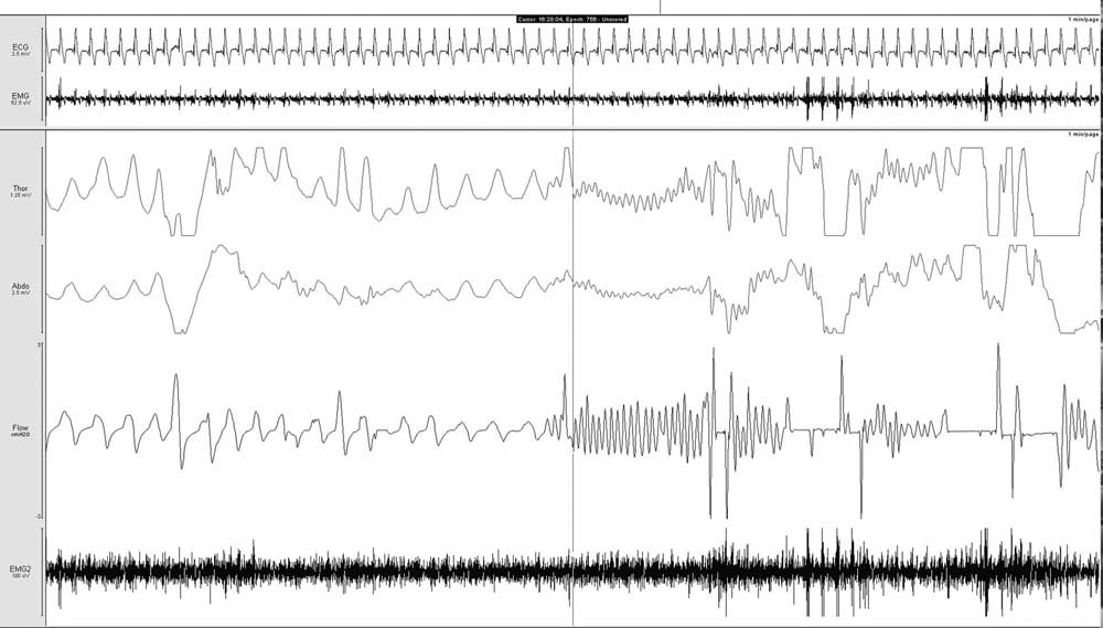
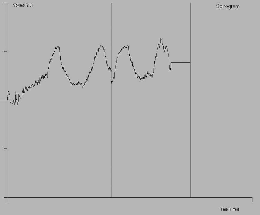
However, an episode was observed, and recorded on the family’s phone camera. Direct observation showed involuntary rapid oscillation of the upper abdomen together with voluntary tidal breathing. Respiratory band monitoring of an episode demonstrated high frequency oscillations associated with high frequency thermistor-detected air-flow at the nose (Fig 1). A volume-time spirogram showed a high frequency wave pattern superimposed on the low frequency rhythm of breathing (Fig 2). Fluoroscopy confirmed bilateral high frequency, rhythmic contractions of the diaphragm unrelated to respiratory effort or cardiac pacing (Video 1, see http://www.journals.cambridge.org/CTY).
Respiratory band monitoring of thoracic and abdominal movements showing the start of high frequency oscillations coincident with patient initiated event-marker (vertical line) correlating with high frequency airflow. From top, ECG – Electrocardiogram monitoring; EMG – Electromyogram surface electrode over diaphragm; Thor – thoracic cage respiratory band; Abdo – abdominal respiratory band; Flow – nasal airflow, EMG2 – Electromyogram surface electrode caudal to first EMG probe.

Volume-time spirogram revealed a high frequency wave pattern superimposed on a low frequency rhythm. The x axis – time (1 minute), y axis – volume (2 litres). Video: Fluoroscopy showing bilateral high frequency rhythmic oscillations of the diaphragm unrelated to respiratory effort.

The symptoms decreased in frequency once the diagnosis was made and she was treated with reassurance. She remained free of the symptoms of diaphragmatic flutter twelve months later.
Discussion
The pathophysiological basis of diaphragmatic flutter is thought to be secondary to abnormal excitation of the phrenic nerve, either by disturbance of the central nervous system or by irritation of the phrenic nerve or the diaphragm itself.Reference Rigatto and De Medeiros2 Suggested etiologies included lung disease or surgery, cardiomegaly in the setting of rheumatic heart disease, irritation of the phrenic nerve or the diaphragmatic muscle from pleurisy, peritonitis, fractured xiphoid process, cervical disc herniation, lymphadenitis, a cervical rib and psychogenic factors.Reference Rigatto and De Medeiros2, Reference Jinnai, Takahashi, Shundo, Komine, Gotoh and Fujita5–Reference Soderstrom9 The etiology in our patient could be central in origin given the bilateral involvement, but multiple previous surgeries may have played a role in local irritation of the phrenic nerve.
Fluoroscopic and physiological examination effectively confirmed the diagnosis in our patient. An electromyogram of the diaphragm demonstrating myoclonic activity superimposed on normal respiration with predominance of large amplitude motor unit action potentialsReference Chen, Remtulla and Bolton3 and a spirogram during an attack showing a dual respiratory rhythm pattern with small high frequency flutter waves superimposed on a background of large slow wavesReference Kondo, Tamaya, Ohta and Yamabayashi10 can be used to confirm the diagnosis.
Treatment of the disorder includes elimination of any identified etiological factor. Several drugs including diphenylhydantoin,Reference Rigatto and De Medeiros2, Reference Kondo, Tamaya, Ohta and Yamabayashi10 carbamazepine,Reference Chen, Remtulla and Bolton3 haloperidolReference Kondo, Tamaya, Ohta and Yamabayashi10 and clonazepamReference Chen, Remtulla and Bolton3 have been used successfully. Phrenic nerve crush surgery has also been used to treat symptoms resistant to pharmacological therapy.Reference Rigatto and De Medeiros2, Reference Cvietusa, Nimmagadda, Wood and Liu4 Psychological reassurance may afford relief of symptoms as occurred in our patient. In some instances this may be temporary and therefore periodic surveillance is required.
The main challenge with our patient lay in making a clinical diagnosis of the disorder in a young patient with a very complicated medical history, coexistence of several conditions with similar clinical manifestations and a known element of anxiety. This case illustrates the importance of thorough investigations before the diagnosis of a non-organic disorder is made. The similarity of the symptoms to anxiety and hyperventilation may lead to a delay in the diagnosis and awareness of this disease entity amongst physicians is essential to ensure early diagnosis and appropriate treatment.
Acknowledgements
The authors thank Dr Richard Slaughter, Director of Medical Imaging; Ms Janet Shaw, Respiratory Investigations Unit; Sleep scientists, Sleep Investigations Unit at The Prince Charles Hospital for their expertise.