A boy aged 4 years and 7 months was admitted to our hospital to evaluate persistent tachycardia of more than 4 years’ duration. His height was 40.6 inches and his weight was 37.0 lbs. The patient did well until 1 month after birth when he presented with vomiting and malaise. Physical examination at the time showed the heart rate as 200 beats per minute (bpm). Electrocardiogram and Holter monitoring showed alternations of atrial flutter, atrial fibrillation, and atrial tachycardia. A right atrial appendage aneurysm (22×36×20 mm) was revealed by echocardiography in another hospital 2 years ago. Digoxin, propafenone, metoprolol, amiodarone, propranolol, and sotalol had been used individually or as combinations to treat his tachycardia during the past 4 years, and his heart rhythm keeps presenting as chaotic atrial tachycardia. Holter monitoring showed total heart beats of 182,880/day, maximum heart rate of 240 bpm, and average heart rate of 110~150 bpm.
His past medical history was nothing special. Personal history was as follows: G1P2, small of twins, and full-term born by Caesarean owing to amniotic fluid insufficiency. Birth weight was 6.06 lbs. There was no history of intrauterine infection or asphyxia hypoxia. The patient’s twin brother is healthy. There was no family history of arrhythmia or CHD.
Physical examination showed that the heart was not enlarged, heart rate was 110 bpm, heart rhythm was regular, and there was no cardiac murmur.
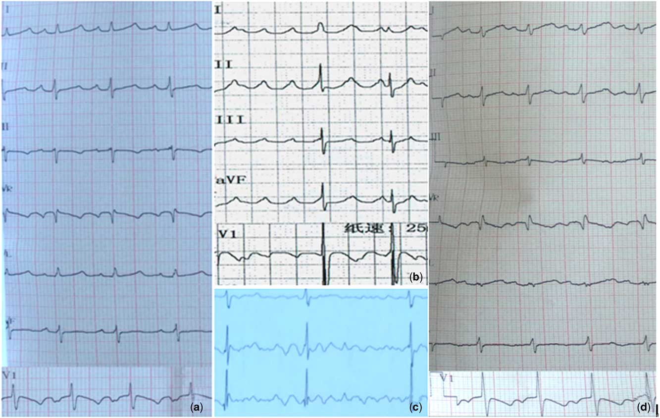
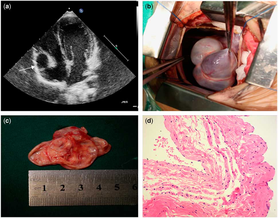
With regard to ancillary examination, electrocardiogram showed atrial tachycardia and atrial flutter (Fig 1a and b). Holter monitoring showed total heart beats of 177,013/day (21.5 hours), maximum heart rate of 238 bpm, minimum heart rate of 72 bpm, and average heart rate of 123 bpm. Persistent alternations of rapid atrial flutter, atrial fibrillation, and atrial tachycardia are shown (Fig 1c). Echocardiography showed a left ventricular diastolic diameter of 35 mm (normal range 34–37), left ventricular ejection fraction of 65%, and significant enlargement of right atrial appendage with a diameter of 15×20 mm (Fig 2a).
(a) A 12-lead atrial tachycardia shown by electrocardiogram. P-wave morphology in V1 is “w” type. P-wave polarity is positive in leads I, II, III, and AVF indicating that the atrial tachycardia originates from the right atrial appendage. (b) Electrocardiogram showed atrial flutter with rate of 300bpm and 2:1–4:1 atrioventricular conduction. (c) Holter monitoring showed impure atrial flutter and atrial fibrillation. (d) Postsurgical 12-lead electrocardiogram showing atrial tachycardia with P-wave polarity negative in leads V1 and III, and positive in leads I, II, AVL, AVF indicating that this atrial tachycardia does not originate from the right atrial appendage.

(a) Echocardiography showed significantly enlarged right atrial appendage with a diameter of ~15×20mm – indicated by arrow. (b) View during surgery of the right atrial appendage excision, showed aneurysmal dilatation of right atrial appendage. (c) Right atrial appendage with extremely thin wall with size ~5×5cm. (d) Focal fibrosis and increased thickness of the endometrium; general fibrosis of myocardium; and partial attenuation of myocardium thickness (90–1000μm). Within parts with minimum thickness, myocardium only contributes to 1/4 and fibrosis contributes to the remaining 3/4. Enlargement of appendage cavity with focal complete fibrosis of atrial wall was detected. Myocardial cell hypertrophy, vacuolar degeneration, increased vascularity, vascular dilation, and tortuous arterioles can be detected. Increased adipose tissue in the pericardium with occasional lymphocyte infiltration was detected. The thickness of the endometrium was slightly increased with proliferation of elastic and collagenous fibres.
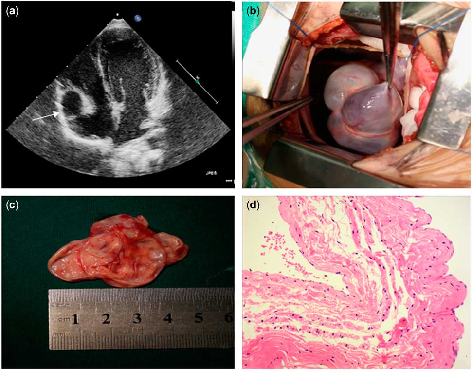
He was admitted to our hospital because of intermittent palpitation and chaotic atrial tachycardia, including atrial flutter, atrial fibrillation, and atrial tachycardia. His height is below age-specific average value, less than the tenth quantile. Rapid atrial tachycardia was the main type of arrhythmia in this patient. P-wave morphology is “w” type in lead V1 and P-wave polarity is positive in leads I, II, III, and AVF, indicating that atrial tachycardia originates from the right atrial appendage (Fig 1a). Owing to resistance to anti-arrhythmic medication therapy, the patient’s right atrial appendage was excised by surgery. His right atrial appendage measured approximately 5×5 cm, and extremely thin right atrial wall was detected during surgery (Fig 2b and c). Postsurgical electrocardiogram showed atrial tachycardia. Owing to changed P-wave morphology and polarity, we speculated that the new atrial tachycardia developed after surgery does not originate from the right atrial appendage (Fig 1d). The maximum heart rate was 200 bpm and the average heart rate was 140–160 bpm. He appeared pale with cold sweating. His blood pressure was hard to maintain. After 3 days’ oral sotalol and propafenone treatment, the patient’s heart rhythm recovered to normal sinus rhythm with an average heart rate of 70–130 bpm. Sotalol 50 mg Q12h (5.36 mg/kg.day) combined with propafenone 50 mg Q8h (8.93 mg/kg.day) was continued for 3 months. After medications were withdrawn, the patient was kept on normal sinus rhythm at 10 months’ follow-up and Holter monitoring showed total heart beats of 121,363/day, maximum heart rate of 133 bpm, and average heart rate of 84 bpm.
Pathology report showed focal fibrosis and increased thickness of the endometrium; general fibrosis of the myocardium; and partial attenuation of myocardium thickness (90–1000 μm). Within parts with minimum thickness, myocardium only contributes to 1/4 and fibrosis contributes to the remaining 3/4. Enlargement of appendage cavity with focal complete fibrosis of atrial wall was observed. Myocardial cell hypertrophy, vacuolar degeneration, increased vascularity, vascular dilation, and tortuous arterioles can be detected. Increased adipose tissue in the pericardium with occasional lymphocyte infiltration was detected. The thickness of the endometrium was slightly increased with proliferation of elastic and collagenous fibres (Fig 2d). Immunohistochemistry:H131806-D: Actin(+); Desmin(+); Myoglobin(+). Conclusion: the child was diagnosed with cardiomyopathy with severe loss of myocardial contractile protein. No significant pathogenic mutations were detected on all-exon high-channel gene sequencing (Beijing Deyi Oriental Transformational Medicine Center).
Discussion
Congenital aneurysm of atrial appendage is a rare anomaly presenting as aneurysmal dilatation of atrial appendage without pericardium focal defect. Solitary left or right atrial appendage aneurysms have been reported. Congenital left and right atrial appendage aneurysms have been reported. Aneurysmal dilatation of the left atrial appendage was first reported in 1922.Reference Ingram and Macfie 1 Compared with left atrial appendage aneurysm, right atrial appendage aneurysm is a rare condition that was first reported in 2001.Reference Mizui, Mori and Kuroda 2 The aetiology of aneurysmal dilation of atrial appendage could be either acquired or congenital. Acquired aneurysmal dilatation of the atrial appendage is often associated with high atrial pressure resulting from stenosis or regurgitation of mitral or tricuspid valve, injury by surgeries, and so on. Congenital aneurysm of atrial appendage is associated with dysplasia of atrial pectinate muscles, bloodstream lashing, and increased atrial pressures and volume load.Reference Sevimli, Gundogdu and Aksakal 3 Left atrial appendage is usually 2–3 cm long. Left atrial appendage aneurysm can be diagnosed if its longevity is more than 3 cm. Left atrial appendage aneurysms will be regarded as “giant” when its longevity is more than 5 cm.Reference Ulucam, Muderrisoglu and Sezgin 4 Congenital aneurysm of atrial appendage can be diagnosed antenatallyReference Mizui, Mori and Kuroda 2 , Reference Ishii, Inamura and Kayatani 5 , and no family-inherited inclination has been identified.Reference Duran, Ertürk and Gündüz 6 Patients usually have no symptoms except that when complications appear such as development of atrial arrhythmias and systemic embolisation caused by embolus formed in the dilated left atrial appendage. Only one-third of patients developed symptoms before 10 years of age. For most patients, this condition has been discovered in the second to the fourth decades of life. Congenital aneurysm of the right atrial appendage is rare in children.Reference Mizui, Mori and Kuroda 2 , Reference Ulucam, Muderrisoglu and Sezgin 4
Atrial arrhythmias including atrial flutter, atrial fibrillation, and atrial tachycardia have been reported in patients with congenital aneurysm of atrial appendage, which were thought to be due to aneurysmal dilatation of atrial appendage. The exact mechanism of tachycardia was not completely understood, and two assumptions exit. One is that there might be ectopic foci in the aneurysm of the right atrial appendage with enhanced automaticity. The other is that reentrant circuit might be created by the recombination of atrial muscle fibres and fibrous tissues.Reference Mizui, Mori and Kuroda 2 Owing to abnormal anatomy, these arrhythmias are usually refractory to medication therapy.Reference Scalia, Stafford and Burstow 7 Previous cases cured by successful resection of abnormal left atrial appendage have been reported.Reference Yildirim and Gölbaşi 8 , Reference Corti, Sciuchetti and Ferro 9 Clinical experience of treatment for right atrial appendage aneurysm is limited. The natural history and optimal management strategy are still unclear.Reference Gulati, Gheta and Chan 10 Mizui et alReference Mizui, Mori and Kuroda 2 reported an infant with ectopic atrial tachycardia owing to an aneurysm of the right atrial appendage who developed congestive heart failure. Although successfully ablated initially, tachycardia recurred 2 days later. The right atrial appendage aneurysm was excised successfully by surgery and the patient was reversed to normal sinus rhythm thereafter. They stipulated that the reason for high recurrence rate after ablation is that the range of tissue responsible for tachycardia is too large to be completely ablated. If tachycardia is resistant to medication treatment or catheter ablation, resection of the abnormal atrial appendage might successfully eradiate the recurrent tachycardia.Reference Mizui, Mori and Kuroda 2
Characteristics of this patient were as follows: first, aneurysm of the right atrial appendage is associated with chaotic atrial tachycardia and the rapid atrial tachycardia originates from the right atrial appendage; second, arial tachyarrhythmia is refractory to medication therapy; third, radiofrequency catheter ablation is limited because of extremely thin atrial appendage wall prone to perforation. As tachycardia originates from the right atrial appendage, resection was chosen. The diagnosis of right atrial appendage aneurysm was confirmed by aneurysmal dilation of right atrial appendage and an extremely thin atrial wall observed during surgery. Pathological exploration showed generalised fibrosis, attenuation, increased adipose tissue, and severe loss of myocardial contractile protein in the right atrial appendage aneurysm that are concordant with previous literature.Reference Gulati, Gheta and Chan 10 Fourth, after resection of the right atrial appendage aneurysm, the chaotic atrial tachycardia was terminated but a new atrial tachycardia developed, which did not originate from the right atrial appendage. After 3 days of oral sotalol and propafenone oral treatment, normal sinus rhythm was regained. The patient maintained sinus rhythm within 10 months of follow up. These suggest that resection of the right atrial appendage aneurysm was effective. Finally no significant pathogenic mutations were found on all-exon high-channel gene sequencing.
Right atrial appendage aneurysm is an extremely rare congenital malformation. Potential complications include arrhythmia, thrombosis, and rupture.Reference Gulati, Gheta and Chan 10 It is refractory to the medication therapy. Resection of aneurysm is the recommended treatment with satisfactory efficacy.
Acknowledgements
The authors thank Dr Zhang Yan for revising English writing.
Financial Support
Dr. Wu Shunde Foundation for research in medical science (202-400-00811).
Conflicts of Interest
None.
Ethical Standards
All procedures performed in this study involving human participants were in accordance with the ethical standards of the institutional and with the 1964 Helsinki Declaration and its later amendments or comparable ethical standards.