Congenital fistulas between the aorta and the right or left atriums are rare, but haemodynamically important, congenital anomalies. Accurate determination of their exact origin and insertion is crucial for successful treatment, and may require cardiac catheterisation and angiography. In our case, catheterisation confirmed the echocardiographic suspicion of a variant that is, to the best our knowledge, previously unreported, and has no obvious embryological explanation
Case report
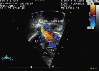
A 7 day old baby, weighing 2.5 kilograms, was referred with signs of congestive cardiac failure for further assessment. He was in moderate distress, with a respiratory rate of 60 breaths per minute, and sub-costal recession. The praecordium was hyperdynamic, with an increased left ventricular impulse. A continuous murmur, graded at 4 from 6, was audible over the entire praecordium, and also radiated to the midscapular region posteriorly, to the right of the spine at the level of fifth and sixth thoracic vertebrae. The electrocardiogram showed sinus rhythm, with a rate of 140 beats per minute, and right axis deviation. The chest X-ray showed cardiomegaly, with a cardiothoracic ratio of 68%. Echocardiography revealed usual atrial arrangement with concordant atrioventricular and ventriculo-arterial connections. The atrial septum was intact, with normal systemic venous drainage, but the left heart was markedly dilated. An abnormal and large vessel was noted behind the heart, with continuous flow through it to the left atrium (Fig. 1). The right upper pulmonary vein was also significantly dilated, with a Doppler gradient of 36 millimetres of mercury at the point of entry of the abnormal vessel to the right upper pulmonary vein. The arterial duct was closed, and there was no evidence of coarctation.
A fistula is seen originating from the descending (desc) aorta and joining the right upper pulmonary vein (RUPV) at its entry into the left atrium (LA). LV=left ventricle. RV=right ventricle.

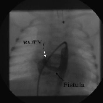
In view of these unusual findings, cardiac catheterisation was performed, both to confirm the anatomy and possibly to attempt closure. Aortography confirmed that a large tortuous blood vessel of 5 millimetres diameter originated from the descending aorta and drained into the right upper pulmonary vein (Fig. 2). Right ventricular pressure was measured at 30 over 4, descending aortic pressure at 36 over 18, and pulmonary arterial pressure at 28 over 8. These pressures were generally low, probably related to the combination of general anaesthesia and the significant run-off through the large fistulous connection, but the procedure was well tolerated. We deemed the proximity of the narrowest point of the fistula to the pulmonary vein to preclude closure with a device, so the patient was referred for surgery. Through a left thoracotomy, the descending aorta was mobilised, and a large rightward-directed vessel was found below the hilum of the right lung. This was ligated uneventfully, and the baby was discharged home on the 5th post-operative day. After 18 months of follow-up, there has been no clinical or echocardiographic evidence of adverse sequels, or residual or recurrent shunting.
Descending aortic angiography, in the antero-posterior view, shows the fistula originating from the descending aorta (black arrow) and joining the right upper pulmonary vein (RUPV) at its entry into the left atrium (white arrow).

Discussion
Fistulous communications within the heart are relatively rare, but have been reported to involve the coronary arteries, the atrial appendages, and the aorta.Reference Khan, Sivasubramonian and Simpson1, Reference Nihoyannopoulos, Sapaford and Oakley2 Occasionally these fistulas communicate between the aorta and the left or right atrium,Reference Coto, Caffarena, Such and Marques3–Reference Elwatidy, Galal, Rhydderch and Ashmeg7 and may arise from either the ascendingReference Coto, Caffarena, Such and Marques3–Reference Rosenberg, Williams and Trusler5 or descendingReference Soler, Mehta, Garcia, Kaiser and Tamer6, Reference Elwatidy, Galal, Rhydderch and Ashmeg7 aorta. A fistulous communication between the descending aorta and a right pulmonary vein, to the best of our knowledge, has not previously been described. Differentiation from a direct aorto-atrial fistula was important, as pulmonary venous return might well have been compromised by inappropriate placement of a device. The embryological origin remains obscure.
Acknowledgment
We thank Dr Frances Bu’Lock for her help in preparing the manuscript.