




No CrossRef data available.
Article contents
Comparison of segmental and global systemic ventricular function at rest and during dobutamine stress between patients with transposition and congenitally corrected transposition
Published online by Cambridge University Press: 22 April 2005
Abstract
The aim of the study was to evaluate segmental and global right ventricular function at rest and during stress in patients having a systemic morphologically right ventricle. We studied 17 patients after atrial correction for transposition, 13 with congenitally corrected transposition, and 11 age-matched controls using cardiovascular magnetic resonance at rest and during stress with dobutamine given at 15 micrograms per kilogram per minute. Blood was drawn to obtain levels of brain natriuretic peptide. Right ventricular ejection fraction was calculated, and wall-motion and wall-thickening were assessed, in 4 segments of a midventricular slice. The findings for the systemic right ventricle were compared to the left ventricle in controls. Patients with transposition showed a decreased ejection fraction at rest (57 percent versus 69 percent, p equal to 0.005), decreased wall motion of the anterior, lateral and septal wall (p less than 0.01, p less than 0.01, and p less than 0.01) and decreased thickening of the lateral wall (p less than 0.01). Patients with congenitally corrected transposition showed normal ejection fraction, wall thickening, and wall motion at rest. During dobutamine stress, we found no significant differences in wall motion and thickening between the two groups. Ejection fraction, also increased to comparable values during stress, at 67 percent versus 66 percent, p not being significant. In both groups, we observed similarly increased levels of brain natriuretic peptide (p equal to 0.02 and 0.03, respectively). We conclude that only patients with transposition showed segmental wall motion and wall thickening abnormalities at rest. After dobutamine stress, however, segmental and global right ventricular dysfunction was similar in both groups.
Information
- Type
- Original Article
- Information
- Copyright
- © 2005 Cambridge University Press
The morphologically right ventricle is subjected to excessive high pressures in patients who have undergone an intra-atrial repair, such as the Mustard or Senning procedure, for transposition, as it is for those with congenitally corrected transposition. These patients with a systemic morphologically right ventricle are at risk of heart failure, accompanied by high mortality.1–3 To our knowledge, it is not known whether both groups carry a similar risk for developing right ventricular dysfunction. Cardiac anatomy and clinical history differ greatly between the groups. Dobutamine stress cardiovascular magnetic resonance appears to be a promising tool for early detection of right ventricular dysfunction.4 Furthermore, analysis of segmental ventricular function may give insight into pathophysiology.5, 6 Finally, measurement of the levels of brain natriuretic peptide, a cardiac neurohormone, may be useful as an early marker for heart failure.7 The purpose of our study, therefore, was to compare these markers for right ventricular dysfunction in patients having systemic morphologically right ventricles in the settings of transposition and congenitally corrected transposition.
Patients and methods
Study subjects
We studied 30 consecutive patients, from 3 tertiary referral centres, with chronic right ventricular pressure overload due to congenital cardiac disease, and having a mean age of 28 years, with a range from 17 to 65 years, comparing them to 11 healthy control subjects with a mean age of 31 years, and a range from 21 to 59 years. The patients consisted of 13 with congenitally corrected transposition, that is discordant atrioventricular and ventriculo-arterial connections, and 17 patients after atrial correction for transposition defined on the basis of discordant ventriculo-arterial but concordant atrioventricular connections. The patients with isolated transposition had undergone an atrial inflow repair using the Mustard or Senning techniques, while 6 of those with congenitally corrected transposition had undergone multiple surgical procedures on the path to physiological repair (Table 1). Data on the type and number of surgical procedures, and follow-up, were obtained from the records. Patients with a pacemaker were excluded from the study because of the contraindication for cardiovascular magnetic resonance. Informed consent was obtained from all subjects and the ethical reviews boards of the institutions approved the study.
Table 1. Clinical characteristics of the study population.

Dobutamine stress cardiovascular magnetic resonance
Subjects were studied using a 1.5 Tesla scanner with high power gradients (Vision, Siemens, Erlangen, Germany) between March 1999 and May 2001. We created a stack of 12 to 14 contiguous images, at a thickness of 10 millimetre with no gap between slices, in the short axis of the ventricle from the base to the apex using an electrocardiographically triggered, ultra fast turbo field echo imaging sequence. The repetition time was based on the R-R interval, the echo time was 4.8 milliseconds, flip angle was 20 degrees, and the spatial resolution better than 1.6 by 1.6 by 10 millimetres.8 Dobutamine was administered through an intravenous line by a digital infusion pump, using an initial dose of 5 micrograms per kilogram per minute, and increasing the infusion after 3 minutes by 5 micrograms per kilogram per minute to a maximum of 15 micrograms per kilogram per minute. Three minutes after reaching the maximal dose, the protocol was repeated with an acquisition time of approximately 10 minutes. The electrocardiogram, heart rate, and blood pressure were monitored continuously throughout the protocol. Patients with an irregular rhythm did not undergo dobutamine stress because of the danger for developing tachyarrhythmias.
Image analysis
MASS (Medis, Leiden, The Netherlands) image analysis software was used to display multislice, multiphase images individually and in a movie loop mode. Ejection fraction and ventricular mass were determined by manual outlining of the endocardial and epicardial contours of end-diastolic and end-systolic frames. Ejection fraction was defined as 100 times stroke volume, this being the end-diastolic minus the end-systolic volume, divided by the end-diastolic volume. When calculating the ventricular mass, we considered the ventricular septum to be part of the systemic ventricle.
Segmental systemic ventricular function was analyzed with wall thickening and wall motion at rest and during stress using the endocardial and epicardial contours of a midventricular slice. Beginning from the posterior attachment of the right ventricle, 100 equally divided chords were created using the modified centreline method (Fig. 1).9 Wall motion was calculated as the motion of each chord relative to the centre of the systemic ventricle. Wall thickening was defined as the percentage change of end-diastolic and end-systolic wall thickness. Wall motion and thickening were analyzed for 4 regions by averaging the values of chords present in that region, choosing the septal, anterior, lateral, and inferior regions.

Figure 1. The figure shows how we determined wall motion and wall thickening. Endocardial and epicardial contours were drawn in a midventricular slice of the systemic ventricles (white lines). Using the centreline method, 100 equally divided chords (black line) were created clockwise beginning at the posterior junction of both ventricles (arrow). These chords were obtained during end-diastole and end-systole yielding end-diastolic wall thickness (EDWT) and end-systolic wall thickness, respectively. The chords were divided into four segments, septal, anterior, lateral, and inferior. The septal segment was included in the estimation of systemic ventricular mass.
Brain natriuretic peptide
At the time of enrolment, blood specimens were collected in citrated tubes and centrifuged for 15 minutes. The plasma was stored at minus 80 degrees Celsius and at the end of the study. Concentrations of the peptide were determined with immunoradiometric assay kits (Shionoria, Osaka, Japan) without knowledge of the clinical outcome. Levels of less than 10 picomoles per litre were considered normal.
Statistical analysis
Data are described as means with standard deviation. Differences between baseline characteristics were sought with independent samples t-testing, and chi-squared testing, when appropriate. Differences in global and regional ventricular function were sought with the independent samples t-test. Reaction to dobutamine stress was sought with a paired t-test. A p value of less than 0.05 was considered statistically significant.
Results
Resting segmental and global function
At rest, end-diastolic wall thickness was lower in the setting of transposition in comparison to controls and congenitally corrected transposition, with p values of less than 0.001, and less than 0.001, respectively, for all segments (Fig. 2). Ejection fraction was significantly decreased, with a p value of 0.005, in patients with transposition, at 57 percent with standard deviation of 11 percent, in comparison to controls where values were 69 percent with standard deviation of 10 percent. They tended to be lower than the values of 64 percent with standard deviation of 13 percent for those with congenitally corrected transposition, this not being statistically significant. Similarly, there was decreased motion of the lateral, anterior and septal wall in those with transposition when compared to controls (p less than 0.01, p equal to 0.01, and p less than 0.01, respectively) and compared to those with congenitally corrected transposition (p less than 0.01, p less than 0.01, and p less than 0.01, respectively; Fig. 3a). Thickening of the lateral wall was decreased compared to controls (p less than 0.01) and those with congenitally corrected transposition (p less than 0.05; Fig. 3b). Increased levels of brain natriuretic peptide were observed in both groups of patients (Fig. 4).

Figure 2. End-diastolic wall thickness of the systemic ventricle for control subjects, those with transposition, and those with congenitally corrected transposition. Note the decreased wall thickness of those with transposition in all segments of the systemic ventricle.

Figure 3. Differences in wall motion (a) and wall thickness (b) are depicted between those with transposition, congenitally corrected transposition and healthy controls at rest and during dobutamine stress.

Figure 4. Brain natriuretic peptide levels are depicted for control subjects, and those with transposition and congenitally corrected transposition. Note the increased levels of brain natriuretic peptide in those with transposition (5.42, with standard deviations of 3.44, picomoles/liter), and those with congenitally corrected transposition patients (7.73, with standard deviation of 6.95, picomoles/liter), in comparison to the controls, at 2.55 with standard deviation of 1.81. The p values are 0.03 and 0.02, respectively.
Reaction to dobutamine stress
Those with congenitally corrected transposition failed to increase their ejection fraction when stressed with dobutamine, levels changing from 64 percent with standard deviation of 13 percent to 66 percent with standard deviation of 7 percent, this change not being statistically significant. The ejection fraction of those with transposition, at 67 percent with a standard deviation of 8 percent, increased to similar levels as those with congenitally corrected transposition, with values of 66 percent and standard deviation of 7 percent. This difference was not statistically significant, albeit that both values were lower when compared to the controls, at 67 percent with standard deviation of 8 percent versus 83 percent with standard deviation of 5 percent, this giving a p value of less than 0.001. Wall thickening and motion in both groups of patients was similarly decreased compared to controls, except for the motion of the lateral wall, which was lower in those with transposition compared to the patients with congenitally corrected transposition (p equal to 0.01; Figs 3a and 3b). We could find no significant differences in wall motion, wall thickening, and ejection fraction between the patients with congenitally corrected transposition who had or had not undergone previous cardiac surgery.
Discussion
To our knowledge, ours is the first study to compare segmental wall motion in patients with systemic morphologically right ventricles. At rest, the patients with isolated transposition, in other words discordant ventriculo-arterial but concordant atrioventricular connections, showed decreased segmental and global systemic ventricular function, in contrast to the patients with congenitally corrected transposition defined as discordant connections at atrioventricular and ventriculo-arterial junctions. During stress, nonetheless, those with congenitally corrected transposition failed to increase segmental and global ventricular function, having comparable values during stress as observed in the patients with transposition. Elevated levels of brain natriuretic peptide levels were also observed in both groups of patients.
Making the assumption that the perfect result after anatomic repair entails normalization of systemic ventricular function and reserve, we used the left ventricle of our healthy controls to compare with the systemic morphologically right ventricle. Abnormal thickening and motion of the wall of the systemic morphologically right ventricle have previously been reported.6, 10, 11 The presence of pre- and peri-operative hypoxia in the clinical history of patients with transposition may underscore some of the differences we observed when comparing them to patients with congenitally corrected transposition at rest. In the setting of congenitally corrected transposition, however, Hornung et al.,6 using echocardiography, showed reduced resting wall motion and thickening in 4 children with congenitally corrected transposition. We could not confirm these findings in our 13 patients using cardiovascular magnetic resonance. Even in the 6 patients with congenitally corrected transposition who had undergone previous surgical procedures, we found no significant differences from our controls.
When stressed with dobutamine, nonetheless, the patients with congenitally corrected transposition showed remarkably reduced wall motion and wall thickening. The patients with isolated transposition, in contrast, were able to increase their segmental and global right ventricular function to comparable values to those observed in the setting of congenitally corrected transposition. During stress, demand ischemia may play an important role in patients with a systemic ventricle. Demand ischemia, as a result of excessive right ventricular hypertrophy, may lead to right ventricular dysfunction.12, 13 Such demand ischemia may play a greater role in the setting of congenitally corrected transposition, as end-diastolic wall thickness is more outspoken in these patients when compared to those with isolated transposition.
During stress, right ventricular function was decreased to comparable levels in both groups of patients. This is consistent with the equally increased levels of brain natriuretic peptide. This suggests that a comparable state of chronic heart failure may be present in both categories.14, 15 A limitation of our study is the relatively small number of patients in each group, which entails loss of statistical power. Our current study, therefore, requires validation using larger groups of patients.
Acknowledgement
This manuscript is supported by a grant from the Netherlands Heart Foundation (NHF-99207).
References
Graham TP Jr, Bernard YD, Mellen BG, et al. Long-term outcome in congenitally corrected transposition of the great arteries: a multi-institutional study. J Am Coll Cardiol 2000; 36: 255–261.Google Scholar
Roos–Hesselink JW, Meijboom FJ, Spitaels SE, et al. Decline in ventricular function and clinical condition after Mustard repair for transposition of the great arteries (a prospective study of 22–29 years). Eur Heart J 2004; 25: 1264–1270.Google Scholar
Piran S, Veldtman G, Siu S, Webb GD, Liu PP. Heart failure and ventricular dysfunction in patients with single or systemic right ventricles. Circulation 2002; 105: 1189–1194.Google Scholar
Tulevski II, van der Wall EE, Groenink M, et al. Usefulness of magnetic resonance imaging dobutamine stress in asymptomatic and minimally symptomatic patients with decreased cardiac reserve from congenital heart disease (complete and corrected transposition of the great arteries and subpulmonic obstruction). Am J Cardiol 2002; 89: 1077–1081.Google Scholar
Holman ER, Buller VG, de Roos A, et al. Detection and quantification of dysfunctional myocardium by magnetic resonance imaging. A new three-dimensional method for quantitative wall-thickening analysis. Circulation 1997; 95: 924–931.Google Scholar
Hornung TS, Bernard EJ, Jaeggi ET, Howman-Giles RB, Celermajer DS, Hawker RE. Myocardial perfusion defects and associated systemic ventricular dysfunction in congenitally corrected transposition of the great arteries. Heart 1998; 80: 322–326.Google Scholar
Tulevski II, Groenink M, Van der Wall EE, et al. Increased brain and atrial natriuretic peptides in patients with chronic right ventricular pressure overload: correlation between plasma neurohormones and right ventricular dysfunction. Heart 2001; 86: 27–30.Google Scholar
Neffke JG, Tulevski II, van der Wall EE, et al. ECG determinants in adult patients with chronic right ventricular pressure overload caused by congenital heart disease: relation with plasma neurohormones and MRI parameters. Heart 2002; 88: 266–270.Google Scholar
van Rugge FP, van der Wall EE, Spanjersberg SJ, et al. Magnetic resonance imaging during dobutamine stress for detection and localization of coronary artery disease. Quantitative wall motion analysis using a modification of the centerline method. Circulation 1994; 90: 127–138.Google Scholar
Millane T, Bernard EJ, Jaeggi E, et al. Role of ischemia and infarction in late right ventricular dysfunction after atrial repair of transposition of the great arteries. J Am Coll Cardiol 2000; 35: 1661–1668.Google Scholar
Vogel M, Derrick G, White PA, et al. Systemic ventricular function in patients with transposition of the great arteries after atrial repair: a tissue Doppler and conductance catheter study. J Am Coll Cardiol 2004; 43: 100–106.Google Scholar
Redington AN, Rigby ML, Oldershaw P, Gibson DG, Shinebourne EA. Right ventricular function 10 years after the Mustard operation for transposition of the great arteries: analysis of size, shape, and wall motion. Br Heart J 1989; 62: 455–461.Google Scholar
Hornung TS, Bernard EJ, Celermajer DS, et al. Right ventricular dysfunction in congenitally corrected transposition of the great arteries. Am J Cardiol 1999; 84: 1116–1119, A10.Google Scholar
Tulevski II, Hirsch A, Dodge-Khatami A, Stoker J, van der Wall EE, Mulder BJ. Effect of pulmonary valve regurgitation on right ventricular function in patients with chronic right ventricular pressure overload. Am J Cardiol 2003; 92: 113–116.Google Scholar
Bolger AP, Sharma R, Li W, et al. Neurohormonal activation and the chronic heart failure syndrome in adults with congenital heart disease. Circulation 2002; 106: 92–99.Google Scholar
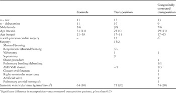
Table 1.

The figure shows how we determined wall motion and wall thickening. Endocardial and epicardial contours were drawn in a midventricular slice of the systemic ventricles (white lines). Using the centreline method, 100 equally divided chords (black line) were created clockwise beginning at the posterior junction of both ventricles (arrow). These chords were obtained during end-diastole and end-systole yielding end-diastolic wall thickness (EDWT) and end-systolic wall thickness, respectively. The chords were divided into four segments, septal, anterior, lateral, and inferior. The septal segment was included in the estimation of systemic ventricular mass.

End-diastolic wall thickness of the systemic ventricle for control subjects, those with transposition, and those with congenitally corrected transposition. Note the decreased wall thickness of those with transposition in all segments of the systemic ventricle.

Differences in wall motion (a) and wall thickness (b) are depicted between those with transposition, congenitally corrected transposition and healthy controls at rest and during dobutamine stress.

Brain natriuretic peptide levels are depicted for control subjects, and those with transposition and congenitally corrected transposition. Note the increased levels of brain natriuretic peptide in those with transposition (5.42, with standard deviations of 3.44, picomoles/liter), and those with congenitally corrected transposition patients (7.73, with standard deviation of 6.95, picomoles/liter), in comparison to the controls, at 2.55 with standard deviation of 1.81. The p values are 0.03 and 0.02, respectively.
 You have
Access
- 21
- Cited by