

Introduction
Constraint is a ubiquitous feature of phenotypic evolution (Futuyma Reference Futuyma2010), whether arising from genetic interactions, phylogenetic history and contingency, biomechanical limitations, or restricted developmental pathways (e.g., Arnold Reference Arnold1992; Brakefield Reference Brakefield2006; Brakefield and Roskam Reference Brakefield and Roskam2006; de Vos et al. Reference de Vos, Dawid, Sunderlikova and Tans2015). These factors can limit morphological disparity along a lineage, resulting in stasis or lower levels of evolutionary novelty in certain features relative to those of less constrained clades. A classic example of this restriction on phenotypic novelty can be found in the vertebral column of mammals and other synapsids (e.g., Cuvier Reference Cuvier1835; Flower and Lydekker Reference Flower and Lydekker1891; Leboucq Reference Leboucq1898; Schultz Reference Schultz, Hofer, Schultz and Stark1961; Galis Reference Galis1999). Whereas sauropsid (≈ reptilian) clades possess great plasticity in the number of presacral vertebrae (Müller et al. Reference Müller, Scheyer, Head, Barrett, Werneburg, Ericson, Pol and Sánchez-Villagra2010), the synapsid line shows little variation (Müller et al. Reference Müller, Scheyer, Head, Barrett, Werneburg, Ericson, Pol and Sánchez-Villagra2010; Head and Polly, Reference Head and Polly2015; Jones et al. Reference Jones, Angielczyk, Polly, Head, Fernandez, Lungmus, Tulga and Pierce2018), with mammals and their close relatives possessing a highly conserved axial column (Buchholtz Reference Buchholtz, Asher and Müller2012), particularly in the cervical region (Jenkins Reference Jenkins1971; Crompton and Jenkins Reference Crompton and Jenkins1973). This mammalian constraint appears to stem from a combination of developmental (Narita and Kuratani Reference Narita and Kuratani2005; Buchholtz Reference Buchholtz2014) and biomechanical (Galis et al. Reference Galis, Carrier, van Alphen, van der Mije, Van Dooren, Metz and ten Broek2014) factors, although there are exceptions to this conservative morphology. Among mammals, sloths (Buchholtz and Stepien Reference Buchholtz and Stepien2009) and sirenians (Buchholtz et al. Reference Buchholtz, Wayrynen and Lin2014) are the only taxa to have evolved variation in the number of cervical vertebrae, and afrotherians have a high amount of interspecific variation in thoracolumbar count relative to other mammalian groups (Narita and Kuratani Reference Narita and Kuratani2005; Sánchez-Villagra et al. Reference Sánchez-Villagra, Narita and Kuratani2007). Therefore, evolutionary conservation of vertebral count—and the rare instances of morphological novelty—are key features in the evolution of the synapsid Bauplan.
The sacrum, composed of the vertebrae that articulate with the ilium via sacral ribs (Mivart and Clarke Reference Mivart and Clarke1879), plays a central role in nearly all tetrapod locomotion as the sole bony connection between the axial skeleton and hind limbs. Plesiomorphically, sauropsids possess two sacral vertebrae, but an incredible disparity in this region of the vertebral column has evolved independently in pareiasaurs (Lee Reference Lee1997; Turner and Sidor Reference Turner and Sidor2018), phytosaurs (Griffin et al. Reference Griffin, Stefanic, Parker, Hungerbühler and Stocker2017), and 10 or more times in various archosaurian clades (e.g., Juul Reference Juul1994; Novas Reference Novas1996; Nesbitt Reference Nesbitt2011; Ezcurra Reference Ezcurra2016; Griffin et al. Reference Griffin, Stefanic, Parker, Hungerbühler and Stocker2017), including crown birds, which can possess 12–23 sacral vertebrae (black swan [Cygnus atratus, USNM 320053, 23 sacrals]; e.g., Baumel and Witmer Reference Baumel, Witmer, Baumel, King, Breazile, Evans and Van den Berge1993; Turner et al. Reference Turner, Makovicky and Norell2012). In contrast, synapsids have far less variation in sacral count. The ancestral synapsid probably possessed two sacral vertebrae (Romer Reference Romer1956; LeBlanc and Reisz Reference LeBlanc and Reisz2014), with Therapsida and some non-therapsid lineages (e.g., Caseidae, Edaphosauridae, Sphenacodontidae) incorporating a third sacral from the caudal series (Reisz Reference Reisz and Wellenhofer1986; LeBlanc and Reisz Reference LeBlanc and Reisz2014) (the caseid Angelosaurus reportedly possesses four sacrals; Olsen Reference Olsen1968; Reisz Reference Reisz and Wellenhofer1986). However, following this initial innovation, therapsids retained the plesiomorphic sacral morphology when compared with their sauropsid contemporaries, consistent with the broader trend of synapsid axial constraint (e.g., Müller et al. Reference Müller, Scheyer, Head, Barrett, Werneburg, Ericson, Pol and Sánchez-Villagra2010). Although some non-mammaliaform cynodonts gained a fourth sacral vertebra (e.g., Romer Reference Romer1956; Kielan-Jaworowska et al. Reference Kielan-Jaworowska, Cifelli and Luo2004; Oliveira et al. Reference Oliveira, Schultz and Soares2009; Martinelli et al. Reference Martinelli, Kammerer, Melo, Neto, Ribeiro, Da-Rosa and Soares2017; but see Bonaparte Reference Bonaparte1963), the presence of only three sacral vertebrae is consistent from mammaliaforms (e.g., Megaconus mammaliaformis [Zhou et al. Reference Zhou, Wu, Martin and Luo2013]) to the origin of crown mammals (e.g., Akidolestes cifellii [Li and Luo Reference Li and Luo2006; Chen and Luo Reference Chen and Luo2013]; Pseudotribos robustus [Luo et al. Reference Luo, Ji and Yuan2007b]; Yanoconodon allini [Luo et al. Reference Luo, Chen and Chen2007a]; Maotherium asiaticus [Ji et al. Reference Ji, Luo, Zhang and Xu2009]). The sole exception to this pattern is Dicynodontia, a diverse clade of anomodont therapsids that flourished worldwide from the middle Permian through the Triassic period. Dicynodonts possessed between three and seven (possibly more) sacral vertebrae (e.g., Angielczyk Reference Angielczyk2001; Angielczyk and Kurkin Reference Angielczyk and Kurkin2003; Ray Reference Ray2006) and reached a range of body sizes rivaled among synapsids only by mammals (roughly guinea pig to elephant sized; Cluver Reference Cluver1974; Dzik et al. Reference Dzik, Sulej and Niedźwiedzki2008; Kammerer et al. Reference Kammerer, Fröbisch and Angielczyk2013; Sulej and Niedżwiedzki Reference Sulej and Niedżwiedzki2019). The dicynodont sacrum, therefore, represents a deviation from an overall conservative morphology that otherwise characterizes nearly 320 million years of synapsid evolution.
In this study, we explore the evolution and homology of the dicynodont sacrum, reconstructing how the derived states of the sacrum were constructed through evolutionary time in this group. This is important not only because understanding how the evolution of the dicynodont axial column differs from that of other synapsids expands our knowledge of unexpected morphologies in the fossil record, but also because the total number of sacral vertebrae has been used extensively as a phylogenetic character in the group (e.g., Angielczyk Reference Angielczyk2001, Reference Angielczyk2007; Angielczyk and Kurkin Reference Angielczyk and Kurkin2003; Surkov et al. Reference Surkov, Kalandadze and Benton2005; Kammerer et al. Reference Kammerer, Angielczyk and Fröbisch2011, Reference Kammerer, Fröbisch and Angielczyk2013; Olroyd et al. Reference Olroyd, Sidor and Angielczyk2018). If dicynodont pelves with the same number of sacral vertebrae are constructed differently (e.g., four sacrals achieved by the addition of either a caudosacral or a dorsosacral) then simple sacral counts are not guaranteed to be strictly homologous, and the character will need to be split into multiple characters (e.g., as in Nesbitt Reference Nesbitt2011) to maintain the homologous comparisons that are a basic necessity for understanding monophyly. Additionally, we use phylogenetic comparative methods to test for a relationship between increased sacral count and body size in dicynodont evolution. This hypothesis has never been rigorously tested, although it has been suggested that the overall variation in the pelvic region reflects a change in locomotory mode (Kemp, Reference Kemp1982, and references therein). Finally, we discuss the evolutionary developmental implications of our hypothesized homology of sacral vertebrae, placing dicynodont axial evolution in the broader context of developmental and biomechanical constraints on synapsid evolution.
Institutional Abbreviations
AMNH, American Museum of Natural History, New York City, New York, USA; BP, Evolutionary Studies Institute (formerly Bernard Price Institute for Palaeontological Research), University of the Witwatersrand, Johannesburg, South Africa; CGP, Council for Geoscience, Pretoria, South Africa; FMNH, Field Museum of Natural History, Chicago, Illinois, USA; IVPP, Institute of Vertebrate Paleontology and Paleoanthropology, Beijing, China; MCZ, Harvard Museum of Comparative Zoology, Cambridge, Massachusetts, USA; NHCC, National Heritage Conservation Commission, Lusaka, Zambia; NHMUK, Natural History Museum, London, UK; NMT, National Museum of Tanzania, Dar es Salaam, Tanzania; NMQR, National Museum, Bloemfontein, South Africa; PIN, Palaeontological Institute, Moscow, Russia; PVSJ, División de Paleontologia de Vertebrados del Museo de Ciencias Naturales y Universidad Nacional de San Juan, San Juan, Argentina; RC, Rubidge Collection, Graaff-Reinet, South Africa; SAM, Iziko South African Museum, Cape Town, South Africa; TM, Ditsong, the National Museum of Natural History (formerly Transvaal Museum), Pretoria, South Africa; UFRGS, Universidade Federal do Rio Grande do Sul, Porto Alegre, Rio Grande do Sul, Brasil; UMZC, University Museum of Zoology, Cambridge, UK; USNM, National Museum of Natural History (Smithsonian Institution), Washington, D.C., USA; YPM, Yale Peabody Museum, New Haven, Connecticut, USA.
Methods
Morphology and Homology of Sacral Vertebrae
We developed hypotheses of homology for dicynodont sacral vertebrae using comparative morphology of the sacrum and ilium. This method has seen much success in other groups that increase the number of sacral vertebrae through evolutionary time, including archosauriforms (Sereno et al. Reference Sereno, Forster, Rogers and Monetta1993; Sereno Reference Sereno1999; Langer Reference Langer, Weishampel, Dodson and Osmólska2004; Langer and Benton Reference Langer and Benton2006; Nesbitt Reference Nesbitt2011; Cabreira et al. Reference Cabreira, Kellner, Dias-de-Silva, Silva, Bronzato, Marsola, Müller, Bittencourt, Batista, Raugust, Carrilho, Brodt and Langer2016) and multiple lineages of early synapsids, including Therapsida (LeBlanc and Reisz Reference LeBlanc and Reisz2014). In those previous studies, the location of the primordial sacral vertebrae (the sacral vertebrae that form the ancestral condition) was identified first by details of the sacral vertebrae and especially the primordial sacral ribs, which each tend to possess distinct and consistent morphologies in these groups (LeBlanc and Reisz Reference LeBlanc and Reisz2014). The location of the articulations between the ribs and ilium is also informative—scars on the medial surface of the ilium can often assist in determining the shape and location of rib articulations, even in the absence of a complete sacrum (e.g., Angielczyk Reference Angielczyk2001; Langer and Benton Reference Langer and Benton2006; Nesbitt Reference Nesbitt2011). After the primordial sacrals have been identified, the homologies of the other sacral vertebrae can then be hypothesized. Usually, those anterior to the primordial sacrals are hypothesized to have been added from the trunk (dorsosacrals), and those posterior to the primordial sacrals are believed to have been added from the tail (caudosacrals), although the insertion of a novel vertebra between primordial sacrals has also been hypothesized in some archosaurian taxa (Nesbitt Reference Nesbitt2011). The only other study of the homology of the dicynodont sacrum concluded that all additional sacral vertebrae are added from the trunk (Surkov Reference Surkov1999 [in Russian], cited in Surkov et al. Reference Surkov, Kalandadze and Benton2005).
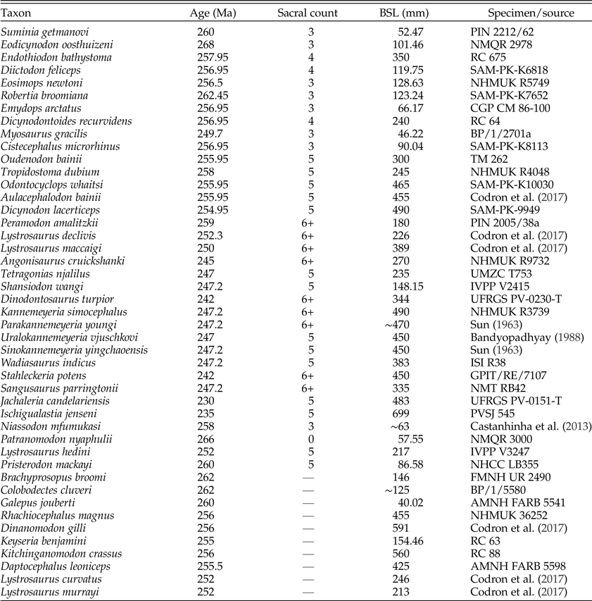
We used these morphological means of identifying the primordial sacral vertebrae of dicynodonts. The plesiomorphic dicynodont condition is to possess three sacral vertebrae inherited from the common therapsid ancestor (LeBlanc and Reisz Reference LeBlanc and Reisz2014; this study). This ancestral therapsid sacrum is composed of the two primordial sacral vertebrae present in the earliest synapsids, as well as an additional caudosacral posterior to these vertebrae, so the third plesiomorphic sacral in dicynodonts is actually a caudosacral when considered in a broader phylogenetic framework (LeBlanc and Reisz Reference LeBlanc and Reisz2014). Because the presence of three sacral vertebrae is the ancestral condition within the context of dicynodont sacral evolution, we will use the term “plesiomorphic” to describe all three of these vertebrae. The three primordial sacrals are always articulated dorsal to the medial wall of the acetabulum of the ilium. The sacral rib of the anteriormost primordial sacral vertebra—primordial sacral 1—possesses an anteriorly convex articular surface and is larger than the rib of plesiomorphic sacral 2, which is roughly columnar in shape (i.e., the articular surface of this rib resembles a column in lateral view). The rib of the third plesiomorphic sacral has a small, circular articular surface consistent with its history as a caudosacral, just as caudosacrals among earlier-diverging therapsids and relevant non-therapsid synapsids possess similar morphologies (LeBlanc and Reisz Reference LeBlanc and Reisz2014). Following the identification of the three plesiomorphic sacral vertebrae, we determined the number of sacral vertebrae anterior and posterior to these elements for those taxa with additional sacrals and used their locations and morphologies to hypothesize their origins. We used firsthand observations for nearly all taxa for which we describe or hypothesize sacral homologies. For the few taxa that we did not observe in person (Suminia, Peramodon, Parakannemeyeria, Uralokannemeyeria, Wadiasaurus, Niassodon), we used descriptions and figures of the sacrum and pelvis in published papers (Table 1) and scores from a recent phylogenetic data matrix (Olroyd et al. Reference Olroyd, Sidor and Angielczyk2018) to provide sacral count data and increase the sample size for our phylogenetic comparative analyses. This approach is based on our hypothesis that all dicynodonts with the same number of sacral vertebrae possess identically constructed sacra (see “Results”).
Taxa included in this study, with their estimated geological ages, numbers of sacral vertebrae, and basal skull lengths (BSL). Included are the sources of the BSL measurement, either the specimen or the study from which the measurement was taken. See text for institutional abbreviations for specimen/source.

Ancestral State Reconstructions
To track the changes in sacral count and body size during dicynodont evolution and to qualitatively assess any correlation between shifts in sacral count and body size, we reconstructed the ancestral states for these features across dicynodont phylogeny. For all these analyses, we used Olroyd et al.’s (Reference Olroyd, Sidor and Angielczyk2018) recent hypothesis of dicynodont relationships. We time-calibrated this tree by using dated terminal taxa as calibration points (Table 1), arbitrarily setting their branch lengths to 1 million years to prevent the existence of branches with a length of zero. Geological age ranges for terminal taxa were taken from the Paleobiology Database (www.paleobiodb.org), with dates assigned to taxa (Table 1) in the middle of these ranges. The only exception to this method was Galepus jouberti: “[T]he age of these localities [containing Galepus] is uncertain, but probably older than the Cistecephalus assemblage zone times, as suggested by Kitching (Reference Kitching1977; King, Reference King1992)” (Fröbisch and Reisz Reference Fröbisch and Reisz2011: p. 662). Then we estimated the age of unconstrained nodes by dividing the age of the closest calibrated node by the number of unconstrained internodes separating this calibrated node from the closest calibrated taxon, allowing for all branch lengths to be estimated. This avoids the calculation of infinite rates of evolution at unconstrained nodes by “smoothing” the time between divergences (Ruta et al. Reference Ruta, Wagner and Coates2006; Brusatte et al. Reference Brusatte, Benton, Ruta and Lloyd2008; Nesbitt et al. Reference Nesbitt, Smith, Irmis, Turner, Downs and Norell2009; Irmis Reference Irmis2011; Sookias et al. Reference Sookias, Butler and Benson2012; Turner and Nesbitt Reference Turner, Nesbitt, Nesbitt, Desojo and Irmis2013).
Because the actual processes by which new sacral vertebrae are incorporated into the sacrum are not well understood (Griffin et al. Reference Griffin, Stefanic, Parker, Hungerbühler and Stocker2017), determining which evolutionary models of character-state transition may accurately reflect biological reality is challenging. Intuitively, a standard meristic model, wherein only stepwise transitions are allowed to occur (e.g., 0→1 or 1→0 may occur, but not 0→2), and wherein these transitions possess equivalent rate parameters no matter the direction of change (e.g., 0→1 and 1→0 possess equivalent rates), makes sense for modeling transitions in sacral count if the sacrum evolves in a stepwise manner (e.g., a transition from three to five sacrals must proceed through an intermediate step of four sacrals). However, if some transitions in sacral count are more likely than their reverse, then a modified meristic model with all rates different (e.g., transitions are still stepwise, but 0→1 and 1→0 may not have equivalent rates) may more accurately reflect the evolution of this feature. Furthermore, the sacrum may not be constructed in a stepwise manner—it is at least plausible that two or more sacral vertebrae could be added simultaneously (e.g., via changes in Hox domains across more than one vertebra). Because of this, models that allow for all character transitions, such as an equal-rates model, an all-rates-different model, or a symmetrical-rates model (forward and reverse transitions possess the same rate parameter) may all be viable means of modeling the evolution of the dicynodont sacrum.
To select the model(s) with the best support, we used the fitDiscrete function in the package ‘geiger’ (Harmon et al. Reference Harmon, Weir, Brock, Glor and Challenger2008) within the statistical computing environment R (R Core Team 2016). Sacral states were scored following strategies of dicynodont phylogenetic matrices (e.g., Angielczyk Reference Angielczyk2001, Reference Angielczyk2007; Angielczyk and Kurkin Reference Angielczyk and Kurkin2003; Surkov et al. Reference Surkov, Kalandadze and Benton2005; Kammerer et al. Reference Kammerer, Angielczyk and Fröbisch2011, Reference Kammerer, Fröbisch and Angielczyk2013; Olroyd et al. Reference Olroyd, Sidor and Angielczyk2018): the plesiomorphic state of three sacral vertebrae as 0, four as 1, five as 2, and six or more sacral vertebrae as state 3. Because all dicynodonts with the same number of sacral vertebrae appear to construct their sacra in the same way (see “Results”), scoring based solely on sacral count is acceptable. We used the ‘geiger’ built-in rate index matrices for the standard meristic, equal-rates, all-rates-different, and symmetrical-rates models, as well as our user-supplied rate index matrix for the meristic all-rates-different model (see R code in Supplementary Material). To compare these five models, we used the corrected Akaike information criterion (AICc), log likelihood supplied by the function fitDiscrete, and ΔAICc and AICc weight supplied by the ‘geiger’ function aicw. We used AICc instead of AIC because this is recommended for smaller sample sizes and is an overall more robust comparative metric (Burnham and Anderson Reference Burnham and Anderson2002). The meristic all-rates-different model had the best overall fit, with the lowest AICc, highest log likelihood, and highest AICc weight (Table 2). The standard meristic model is the only other model with a large amount of support, with a ΔAICc of 1.21 (ΔAICc < 2 is considered to be high support for a model; Burnham and Anderson Reference Burnham and Anderson2002) and the second-highest AICc weight. All other models had negligible support (i.e., extremely low AICc weight and ΔAICc > 7; Burnham and Anderson Reference Burnham and Anderson2002). Therefore, we consider the two meristic models to be best for modeling sacral count evolution among dicynodonts. Rate parameters for transitions between character states are graphically reported for all models in the Supplementary Material (Supplementary Fig. 1).
The corrected Akaike information criteria (AICc), associated comparative metrics, and log likelihoods used in selecting models of discrete character evolution for the sacrum and continuous character evolution for body size. Models compared are an all-rates-different meristic model (Meristic ARD), a standard meristic model (Meristic), a symmetrical-rates model (SYM), an equal-rates model (ER), and an all-rates-different model (ARD), a Brownian motion model (BM), and an Ornstein-Uhlenbeck model (OU).

We used the function ace in the R package ‘ape’ (Paradis et al. Reference Paradis, Claude and Strimmer2004) to reconstruct the ancestral sacral counts, using maximum likelihood for both meristic models. Because there is no built-in rate index matrix for a meristic model or a meristic all-rates-different model in ace, we supplied our own that were identical to those used in the fitDiscrete analyses (see R code in Supplementary Material). To explore the influence of model selection on ancestral state reconstruction, we also conducted this analysis using equal-rates, symmetrical-rates, and all-rates-different models, which are built into ace. The results of these latter three analyses are reported in the Supplementary Material (Supplementary Figs. 2–4). Note that the rate parameters returned by fitDiscrete for the meristic all-rates-different and all-rates-different models are functionally identical, as are the rate parameters for the meristic and symmetrical-rates models. This results in identically reconstructed ancestral states for these models, even though the all-rates-different and symmetrical-rates models have much lower model support (Table 2). This also accounts for the identical log likelihoods of these models.
Because most dicynodont individuals are incompletely preserved, we used basal skull length (BSL; the distance between the occipital condyle and the snout tip) as a proxy for body size, a metric that is an accepted proxy for overall body size in paleobiological analyses of dicynodonts (e.g., Angielczyk and Walsh Reference Angielczyk and Walsh2008; Botha-Brink and Angielczyk Reference Botha-Brink and Angielczyk2010; Codron et al. Reference Codron, Botha-Brink, Codron, Huttenlocker and Angielczyk2017). We gathered the majority of basal skull lengths used in these analyses from direct measurements, with a few measurements taken from the literature (Table 1). For taxa with measurements of multiple individuals reported, we used the largest individual in an attempt to eliminate size bias caused by measuring skeletally immature individuals. We used the fitContinuous function in the package ‘geiger’ (Harmon et al. Reference Harmon, Weir, Brock, Glor and Challenger2008) to select a model, using the same criteria as for our sacrum state reconstruction model selection (Table 2), settling on a Brownian motion model. We used the anc.ML function in the R package ‘phytools’ (Revell Reference Revell2012) to reconstruct ancestral states for basal skull length under a Brownian motion model using likelihood and the function contMap to visualize ancestral states.
The R code and the data files (.csv files and .tre file) used in these analyses are presented in the online Supplementary Material.
Correlation between Sacrum Morphology and Body Size
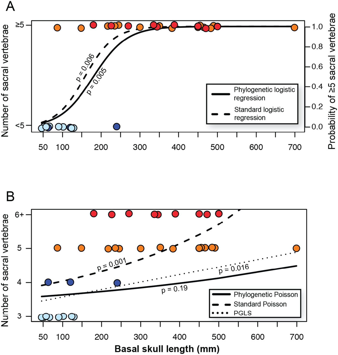
Although general trends of increasing body size (e.g., Keyser Reference Keyser1974; Kemp Reference Kemp2005; Angielczyk and Walsh Reference Angielczyk and Walsh2008) and increasing number of sacral vertebrae (Cruickshank Reference Cruickshank1967; Surkov et al. Reference Surkov, Kalandadze and Benton2005) through dicynodont evolution have been noted, no studies have statistically tested whether a correlative relationship exists between large body sizes and number of sacral vertebrae. Logistic regressions are appropriate when the independent variable is continuous (i.e., body size) and the dependent variable is discrete (i.e., number of sacral vertebrae). However, the only available phylogenetic logistic regression is constructed for use exclusively with a binary dependent variable (Ives and Garland Reference Ives and Garland2010), and our sacral data contain four character states. To circumvent this issue, we converted our sacral counts into binary states, with <5 sacral vertebrae coded as 0 and ≥ 5 sacrals coded as 1. This division is not arbitrary: the major shift in sacral count versus body size appears to be the shift between four and five sacral vertebrae (see “Results”), and this is supported by the rate parameters returned for the meristic and meristic all-rates-different models, in which there is a functional rate of zero for 2→1 but for no other reverse transition (see Supplementary Fig. 1). We used the function phyloglm in the R package ‘phylolm’ (Ho and Ane Reference Ho and Ane2014) to conduct this phylogenetic logistic regression, with the bound on the linear predictor that bounds the searching space set to 500 to ensure that the optimal solution was not missed by inadequate search space. Because Ives and Garland's (Reference Ives and Garland2010) α-statistic of phylogenetic signal returned by their phylogenetic logistic regression assumes an underlying model in which the probabilities of character-state transitions are constant, another measure of phylogenetic signal is beneficial. We used this same rescoring of sacral states to find Fritz and Purvis's (Reference Fritz and Purvis2010) D-statistic for the phylogenetic signal of binary traits, using the function phylo.d in the R package ‘caper’ (Orme et al. Reference Orme, Freckleton, Thomas, Petzoldt, Fritz and Isaac2013). We also used this statistic to determine the level of phylogenetic signal among bidentalian dicynodonts, which possess either state 2 or state 3 (see “Results”). We conducted a standard logistic regression using the function glm to explore the differences between phylogenetic logistic regression and standard logistic regression among our data.
Because our scoring scheme produces what are essentially count data, with different states scored for different numbers of sacral vertebrae, a phylogenetic Poisson regression (Paradis and Claude Reference Paradis and Claude2002) is also appropriate for determining whether a statistical relationship exists between body size and sacral count. Therefore, we used the phyloglm function again, this time with the argument for a phylogenetic Poisson regression included. We also performed a standard, nonphylogenetic Poisson regression to determine the effect phylogenetic signal had on obtaining a statistically significant result using the function glm. Finally, we fit a linear model using phylogenetic generalized least squares (PGLS) with the gls function in the package ‘nlme’ (Pinheiro et al. Reference Pinheiro, Bates, DebRoy and Sarkar2016), using Brownian motion as the within-group correlation structure. Although such an analysis is not strictly appropriate for noncontinuous data, it is useful for data exploration.
To independently test where in the tree major shifts in body size occurred, we implemented the R package ‘bayou’ (Uyeda and Harmon Reference Uyeda and Harmon2014). ‘bayou’ uses a multi-optima Ornstein-Uhlenbeck (OU) model in a Bayesian framework, incorporating reversible-jump Markov chain Monte Carlo (MCMC) simulations, to estimate the location, magnitude, and number of shifts in adaptive optima of a continuous trait across a phylogeny. This provides an independent, quantitative estimate of where major shifts in body size, if any, occurred across dicynodont phylogeny, using a “phylogenetic natural history” approach (Uyeda et al. Reference Uyeda, Zenil-Ferguson and Pennell2018) rather than strict hypothesis testing (as in the phylogenetic logistic regressions, see “Results, Correlation between Body Size and Sacral Morphology”). This method returns posterior probabilities of all parameters, integrating uncertainty in the estimated values of shifts in adaptive regimes. To conduct this analysis, we set distributions on our OU model priors from which the MCMC could draw. In our prior, the distributions for α (rate of adaptation) and σ2 (uncorrelated diffusion) were half-Cauchy distributions (scale = 0.1), a Poisson distribution for K (the number of shifts in adaptive optima; λ = 3, K max = 10), and a normal distribution for θ (adaptive optimum value; mean = mean of sampled basal skull lengths, SD = 1.5 times the SD of the sampled basal skull lengths). The measurement error factor was set at 0.01. We then ran our MCMC simulation with these priors on our OU model parameters for 1 million generations to ensure convergence. We also ran a second analysis for 1 million generations to generate another MCMC chain from a different starting point, and then compared the posterior probabilities of the branches of the two chains to ensure that convergence had been reached in both simulations.
Like the ancestral state reconstructions, the R code and the data files (.csv files and .tre file) used in these analyses are presented in the online Supplementary Material.
Results
Morphology and Homology of Sacral Vertebrae
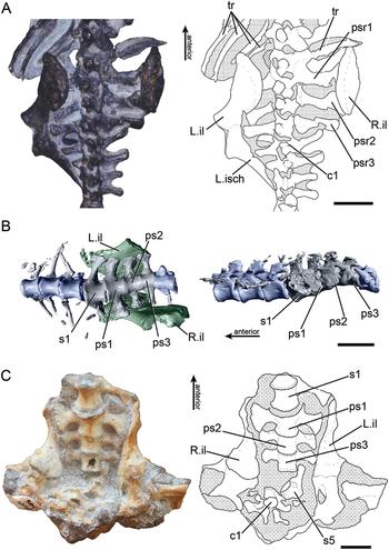
Based on our criteria for identifying the three plesiomorphic sacral vertebrae, we hypothesize that all dicynodonts with three sacral vertebrae possess the three plesiomorphic sacrals; this is even the case in taxa that have lost a sacral vertebra(e) to secondarily evolve the ancestral condition (e.g., Cistecephalus). In all taxa with four sacral vertebrae, the additional vertebra is anterior to the plesiomorphic sacrals. This anteriormost sacral possesses sacral rib morphology similar to plesiomorphic sacral 1, with a concave anterior margin and comparatively large area of articulation—larger than that of plesiomorphic sacrals 2 and 3, and of a similar size or larger than that of plesiomorphic sacral 1 (e.g., Niassodon; Fig. 1B). Therefore, we hypothesize that all dicynodont lineages that moved from three to four sacral vertebrae did so by adding a single vertebra anterior to the plesiomorphic three, and any that moved from four to three sacral vertebrae did so by losing this additional vertebra.
Axial morphology of dicynodonts possessing differing numbers of sacral vertebrae. A, Photograph (left) and line drawing (right) of the pelvic region of Eodicynodon oosthuizeni (NMQR 3155) in dorsal view. B, Reconstruction of the pelvic region of Niassodon mfumukasi (ML 1620) modified from Castanhinha et al. (Reference Castanhinha, Araújo, Júnior, Angielczyk, Martins, Martins, Chaouiya, Beckmann and Wilde2013: Fig. S1) in ventral (left) and left lateral (right) views. C, Photograph (left) and line drawing (right) of the pelvic region of Pristerodon mackayi (SAM-PK-10141) in ventral view; note that the fifth sacral and first caudal vertebrae are partly disarticulated and that the pelves have shifted dorsally and medially relative to the sacrum. Scale bars, 1 cm. Abbreviations: c1, first caudal vertebra; L.il, left ilium; L.isc, left ischium; ps1–3, first, second, and third plesiomorphic sacral vertebra; psr1–3, ribs of first, second, and third plesiomorphic sacrals; R.il, right ilium; s1, first sacral vertebra (non-plesiomorphic); s5, fifth sacral vertebra (non-plesiomorphic); tr, trunk rib.

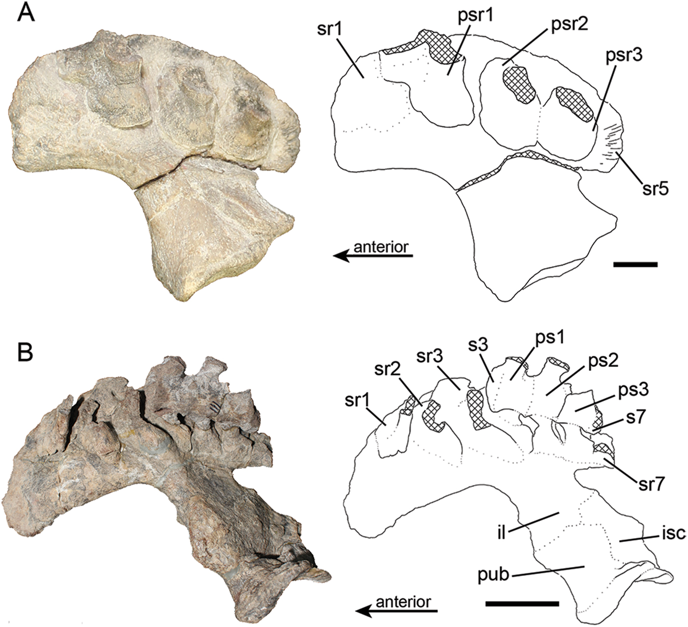
As in taxa with four sacrals, all dicynodonts with five sacral vertebrae appear to possess the same homologies for each element. In addition to the novel anterior vertebra that is present in taxa with four sacrals, dicynodonts with five sacral vertebrae also possess an additional sacral vertebra posterior to the three plesiomorphic sacrals. This vertebra possesses a small sacral rib with a rounded articular surface (Figs. 1, 2), similar to the caudosacrals of several early synapsid lineages and the plesiomorphic sacral 3 of dicynodonts, itself a caudosacral in the ancestral therapsid. Taxa with greater than five sacral vertebrae possess the same vertebrae as those with five—the three plesiomorphic sacrals, a large anterior sacral, and a small posterior sacral—in addition to one or more novel vertebrae anterior to these. These more anterior vertebrae possess sacral ribs similar in morphology to the first plesiomorphic sacral vertebra and the additional anterior vertebra, in that they possess large, anteriorly convex articular surfaces, and these anteriormost sacrals possess the largest ribs in the sacrum (Figs. 1, 2). Taken together, our observations indicate that all dicynodonts with a given sacral count appear to possess identically constructed sacra.
Morphology of the medial surface of the ilium is informative for sacral counts and homologies in dicynodonts. A, Photograph (left) and line drawing (right) of the right ilium of Oudenodon bainii (NMT RB37) in medial view, illustrating that scars from sacral ribs and fused sacral ribs can give information in the absence of a complete sacrum. B, Photograph (left) and line drawing (right) of the right innominate of Sangusaurus parringtonii (NMT RB42) in medial view. Scale bar, 1 cm (A); 10 cm (B). Abbreviations: il, ilium; isc, ischium; ps1–3, first, second, and third plesiomorphic sacral vertebra; psr1–3, ribs of first, second, and third plesiomorphic sacrals; pub, pubis; s3, s7, third and seventh sacral vertebrae (non-plesiomorphic); sr1–3, sr5, sr7, first through third, fifth, and seventh sacral ribs, or scars on the ilium marking the articulation of these ribs.

Ancestral State Reconstructions
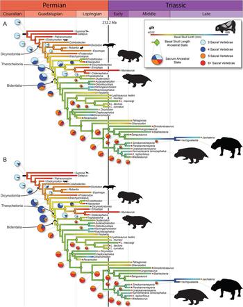
The ancestral state reconstructions indicate an increase in both body size and number of sacral vertebrae across dicynodont evolutionary history, and the general trend of increasing body size through evolution has previously been noted in this clade (Keyser Reference Keyser1974; Kemp Reference Kemp2005; Angielczyk and Walsh Reference Angielczyk and Walsh2008). The ancestral condition of Dicynodontia is small bodied (BSL = 107 mm, comparable to Eodicynodon), similar to most of their non-dicynodont anomodont outgroups, with only the three plesiomorphic sacral vertebrae (Fig. 3). Although there are occasional non-bidentalian taxa that evolve four sacral vertebrae (Diictodon, Dicynodontoides, Niassodon), and even two that evolve five (Pristerodon, Endothiodon), the major shift in sacral evolution appears to be near the base of Therochelonia, with changes from three sacrals to four, and finally to five at Bidentalia (Fig. 3). The ancestral condition at Bidentalia is reconstructed to be five sacral vertebrae, and the presence of five or more sacral vertebrae appears to become fixed at this point in the phylogeny (Fig. 3). A major shift in body size also occurs at this node, with a jump from a reconstructed BSL of 170 mm at the unnamed node Brachyprosopus + Wadiasaurus to 245–274 mm at the two earliest nodes of Bidentalia (Supplementary Table 1), concurrent with the increase in sacral count (Fig. 3). The small (86.58 mm BSL, NHCC LB355) non-bidentalian taxon Pristerodon, which possesses five sacral vertebrae, is an exception to this pattern—although the vertebrae have moved from life position and interpretation is somewhat ambiguous (Fig. 1C), we follow Angielczyk and Kurkin (Reference Angielczyk and Kurkin2003) in hypothesizing that this taxon possessed five sacral vertebrae, especially considering the anteriorly directed ribs of sacral 5 which are similar to an articulated specimen of Oudenodon (BP/1/749). The only other non-bidentalian taxon in our data set to evolve five sacral vertebrae independently is the extremely large Endothiodon (350 mm BSL, RC 675), which is further support for a correlation between the evolution of large body sizes and increased numbers of sacral vertebrae. Although the major shift in sacral vertebrae seems to be the addition of the fifth sacral, a smaller-scale shift may be present in Emydopoidea, which we have reconstructed as reducing their sacral count from four to three in parallel with a marked decrease in BSL (from 214 mm at the base of Therochelonia to 94 mm at Myosaurus + Cistecephalus). The same major shifts in sacral count are reconstructed at the same nodes no matter which evolutionary model is used, although the relative likelihood of different ancestral states changes depending on the model (Fig. 3, Supplementary Figs. 2–4). Among bidentalians, shifts between five sacral vertebrae and six or more appear to have no general pattern, either phylogenetically or with body size (Fig. 3). This apparent lack of phylogenetic signal is supported by Fritz and Purvis's (Reference Fritz and Purvis2010) D-statistic. In this statistic, D = 1 indicates no phylogenetic signal (a random pattern of character-state distribution), and D = 0 indicates a clumped, or Brownian, pattern of character evolution, indicating the presence of phylogenetic signal (note that the value of D is not restricted to a range between 0 and 1, and, e.g., a value greater than 1 is more likely to be significantly different from 0). Among bidentalians we found no significant difference between our returned D-statistic (D = 0.79) and that of no phylogenetic structure (D = 1; p = 0.29), but we also found no significant difference between our D-statistic and that indicating a strong phylogenetic structure (D = 0; p = 0.10). However, the difference between our observed value and 0 is closer to significance, and our recovered D is close to 1 in absolute terms, which suggests that the distribution of sacral character states among Bidentalia does lack phylogenetic structure, even if it cannot be distinguished statistically from a phylogenetically structured distribution.
Ancestral state reconstructions showing that both basal skull length and sacral count increase through anomodont evolution and appear to be correlated. A, Ancestral sacral counts reconstructed with a meristic all-rates-different model, with body size reconstructed using Brownian motion. B, Ancestral sacral counts reconstructed with a standard meristic model, with body size reconstructed using Brownian motion. Pie charts show the scaled likelihood of a given ancestral sacral count. Body silhouettes are to scale and either modified from Wikimedia Commons images or from PhyloPic (Suminia). Skull silhouettes modified from Ruta et al. (Reference Ruta, Angielczyk, Fröbisch and Benton2013).

Correlation between Body Size and Sacral Morphology
The phylogenetic logistic regression indicated a statistically significant relationship between dicynodont body size and the possession of five or more sacral vertebrae (p = 0.005; intercept = −4.19±1.6, β = 0.023±0.01; Fig. 4A), even given the strong phylogenetic signal in the data (α = 0.051, with α = 1 indicating no phylogenetic signal and phylogenetic signal increasing as α approaches 0). Fritz and Purvis's (Reference Fritz and Purvis2010) D-statistic is also indicative of phylogenetic signal in this data set (D = −1.55), significantly different from D = 1 (p = 0 < 0.001) but not from D = 0 (p = 0.99). The standard logistic regression was also statistically significant (p = 0.006; intercept = −4.19±1.6, β = 0.026±0.01) and possessed a slightly better fit (Fig. 4A; AIC = 19.9 compared with 22.7). Therefore, although there is good evidence for phylogenetic structure in the distribution of sacral character states in our data, there still appears to be a correlation between the presence of five or more sacral vertebrae and a larger body size.
Sacral count and body size are statistically correlated, even given phylogenetic signal. A, The phylogenetic logistic regression and standard logistic regression lines for basal skull length against sacral count, scored as a binary variable. B, The phylogenetic Poisson and normal Poisson regression lines, as well as the line for the phylogenetic generalized least squares (PGLS) regression, for basal skull length against sacral count. Note that the distribution of the sacral count data violate the assumptions of PGLS, so this is only included for data exploration. Color key follows Fig. 3; for grayscale, color key of A can be discerned by reference to B.

The phylogenetic Poisson regression, with each individual sacral count retained as its own character state, indicated that there was no statistical relationship between overall sacral count and body size among dicynodonts (p = 0.19; intercept = −0.58±1.0, β = 0.001±0.001). This lack of a statistical relationship may be because of a strong phylogenetic signal, but also because of the near-complete lack of a trend between body size and sacral count among dicynodonts with five or more sacral vertebrae (Fig. 4B). Our nonphylogenetic Poisson regression of the same data returned a statistically significant response (p = 0.001; intercept = −0.2±0.3, β = 0.002±0.001), indicating that phylogenetic signal may be influencing this result. The PGLS model indicated a statistically significant relationship between sacral number and body size as well (p = 0.016; intercept = 0.37±0.63, β = 0.002±0.001). Although PGLS is not strictly appropriate for count data as in our sacral states, it is useful as a method of data exploration to test the limits of the relationship between the increase in sacral count and increase in body size.
The analysis run using ‘bayou’ suggests that a major shift in body-size optimum occurred near the base of Bidentalia, at the same phylogenetic location as the transition from three or four to five sacral vertebrae. This analysis suggest a major shift from an original mean adaptive optimum (root θ) for BSL of 226 mm to a shift at Tropidostoma + Wadiasaurus (posterior probability = 0.301; θ = 422.5 mm) and a smaller probability shift at Aulacephalodon +Wadiasaurus (posterior probability = 0.156; θ = 431.0 mm) (Supplementary Figs. 5, 6). The traces returned by the MCMC simulations ensure that convergence had been reached (Supplementary Figs. 7, 8), and plotting the posterior probabilities of branches between the two MCMC chains results in points falling exactly along the y = x line, indicating that these two chains have converged on the same parameter values. This is quantitative, independent evidence that a major shift in body size occurred coincident with the evolution of five sacral vertebrae.
Discussion
Most novel sacral vertebrae are added anteriorly to the plesiomorphic sacrum in dicynodonts, with only one vertebra added posteriorly. Dorsosacrals—sacral vertebrae added from the trunk—are commonly identified not only by their anterior position in the sacrum, but also by the morphology of the vertebrae themselves, particularly the sacral ribs (e.g., Nesbitt Reference Nesbitt2011; Griffin et al. Reference Griffin, Stefanic, Parker, Hungerbühler and Stocker2017). In these cases, dorsosacral ribs are often relatively small and typically cylindrical, because they represent modified trunk ribs and articulating transverse processes. In contrast, the additional anterior sacral vertebrae of dicynodonts possess sacral ribs that are large and convex anteriorly, with the size of the rib and articular surface increasing in the anterior vertebrae. This anteriorly convex, C-shaped morphology is most similar to the rib of the first plesiomorphic sacral. Additionally, because dicynodonts, including taxa with these additional sacral vertebrae, consistently have 25–26 sacral vertebrae (e.g., Aulacephalodon) or more (e.g., Endothiodon), it is unlikely that the novel anterior sacrals are repatterned presacrals. Based on their location and morphological similarity, we hypothesize that these vertebrae are duplications of the first plesiomorphic sacral. Conversely, because of its small, peg-like sacral rib, which is similar to the transverse process of the anterior caudal vertebrae, we hypothesize that the sole sacral vertebra added posteriorly to the plesiomorphic sacrals is a caudosacral and represents a repatterning of the anteriormost caudal vertebra.
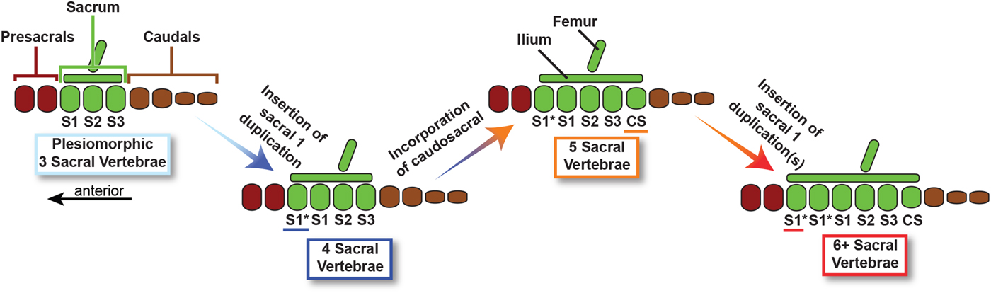
Given this interpretation of the dicynodont sacrum, and the meristic model suggested by the ancestral state reconstruction, we can hypothesize the sequence by which additional sacral vertebrae were acquired through evolutionary time (Fig. 5). The plesiomorphic three sacral vertebrae were first joined by a duplicated plesiomorphic sacral 1, resulting in a four-vertebra sacrum. Following this event, a caudosacral was added via repatterning, forming a five-vertebra sacrum. In some taxa the first plesiomorphic sacral was again duplicated one or more times, resulting in the most derived character state (6+ sacral vertebrae). The pre- and postacetabular processes of the ilium expanded concurrently with these changes, with the preacetabular process expanding much more than the postacetabular process for articulation with a greater number of large sacral ribs. Although it is conceivable that more than one sacral vertebra could be added in a single step, allowing for a shift from three to five sacrals as in the equal-rates model (Supplementary Fig. 3), we view this as unlikely for several reasons. For one, the statistical support for the meristic model of character transition is far greater than models that allow for the addition of multiple sacrals simultaneously, suggesting that sacrals were added in this stepwise manner. Additionally, it is likely that the mechanism by which the first sacral is duplicated is different from the mechanism that allows for the incorporation of a caudosacral, although the actual evolutionary developmental mechanisms by which sacrals are added are poorly constrained (Griffin et al. Reference Griffin, Stefanic, Parker, Hungerbühler and Stocker2017). Therefore, based on both the results of our evolutionary models and mechanistic considerations, we consider the stepwise addition of sacral vertebrae to be most consistent with the data.
Schematic of the hypothesized origins of additional sacral vertebrae among dicynodonts.

Both the ancestral state reconstructions and the statistical tests employed point toward increased body size being correlated with greater numbers of sacral vertebrae in dicynodonts. The main shift in body size seems to occur at roughly the same phylogenetic position as the shift from four to five sacral vertebrae. After five vertebrae evolve at the base of Bidentalia, concordant with a shift to greater body size (Fig. 3), neither the sacral count nor body size decreases in this clade. Moreover, the sole large-bodied, non-bidentalian dicynodont—Endothiodon—independently evolved five sacral vertebrae, further suggesting a relationship between five or more sacrals and the largest body sizes. Even when phylogenetic covariance is considered, there is statistical evidence of a relationship between body size and sacral count; notably, the evidence for this relationship is strongest at the same four- to five-sacral shift (Fig. 4). Based on this combined evidence, we are confident in hypothesizing that higher sacral counts are functionally related to larger body size in dicynodonts. Although the actual biomechanical consequences of the increase in sacral count are beyond the scope of this study, intuitively they would seem related to increased structural support. Testing this hypothesis represents an area for future research.
The developmental mechanisms by which vertebrae may be added to the sacrum have not been rigorously constrained (discussed by Griffin et al. Reference Griffin, Stefanic, Parker, Hungerbühler and Stocker2017), but the overall patterning of the vertebral column is well understood. The broad organization of the axial skeleton into distinct regions is controlled by the expression of Hox genes, and changes in the timing of Hox gene expression along the anteroposterior axis can result in shifts of vertebral identity and repatterning of the axial column (Casaca et al. Reference Casaca, Santos and Mallo2014). In particular, Hox11 paralogous genes are responsible for assigning identity to the sacral vertebrae (Burke et al. Reference Burke, Nelson, Morgan and Tabin1995; Wellik and Capecchi Reference Wellik and Capecchi2003; Wellik Reference Wellik2007), and changes in the timing of expression of Hoxd11 in mouse models have been shown to produce anterior (Gérard et al. Reference Gérard, Chen, Gronemeyer, Chambon, Duboule and Zákány1996, Reference Gérard, Zákány and Duboule1997; Boulet and Capecchi Reference Boulet and Capecchi2002) or posterior (Davis and Capecchi Reference Davis and Capecchi1994; Zákány et al. Reference Zákány, Gérard, Favier, Potter and Duboule1996, Reference Zákány, Gérard, Favier and Duboule1997) shifts in the sacrum, with the overall number of vertebrae remaining constant. Therefore, any changes in Hox11 paralogue expression along the axial column may be responsible in part for changes in the dicynodont sacrum. However, most changes in the dicynodont sacrum do not seem to be simple repatterning of the posteriormost presacral vertebrae, but instead consist of novel sacral vertebrae inserted anteriorly to the plesiomorphic three. Therefore, a change in Hox expression timing cannot be solely responsible for the trend of increasing sacral count. Additionally, because the sacral–hind limb unit shifts posteriorly as sacral vertebrae are inserted anteriorly (Fig. 5), changes in expression of Gdf11—which regulates the coordination of the hind limb with the sacral vertebrae (Matsubara et al. Reference Matsubara, Hirasawa, Egawa, Hattori, Suganuma, Kohara, Nagai, Tamura, Kuratani, Kuroiwa and Suzuki2018)—must also act in concert with these shifts in vertebral count.
If vertebrae are being added to the dicynodont sacrum as novel elements, and not repatterned from other vertebrae, this suggests that the overall number of precaudal vertebrae increased through dicynodont evolution. Vertebrae develop from somites along the anteroposterior axis of the embryo, so the number of vertebrae is directly connected to the number of somites present early in development (e.g., Dequéant and Pourquié Reference Dequéant and Pourquié2008). During somitogenesis, paraxial mesoderm is formed into somites in an oscillatory process (via the “clock and wavefront mechanism”; Cooke and Zeeman Reference Cooke and Zeeman1976; Palmeirim et al. Reference Palmeirim, Henrique, Ish-Horowicz and Pourquié1997; Dequéant et al. Reference Dequéant, Glynn, Gaudenz, Wahl, Chen, Mushegian and Pourquié2006), which results in one bilateral somite pair created each cycle, with somites forming in an anterior-to-posterior sequence (Dequéant and Pourquié Reference Dequéant and Pourquié2008). If the oscillation period decreases, the number of somites correspondingly increases, with the result that each somite is smaller at the onset of its formation than if the period was longer. For example, among squamates with similar developmental rates, a snake with a faster segmentation clock has a larger number of smaller somites, and a lizard with a slower clock has fewer, larger somites, with these differences eventually resulting in interspecific variation in vertebral count (Gomez et al. Reference Gomez, Özbudak, Wunderlich, Baumann, Lewis and Pourquié2008). Therefore, if there is an increase in the number of precaudal vertebrae in dicynodonts, it is reasonable to hypothesize that this change in the vertebral column was mediated at least in part by an increase in the oscillation rate of the segmentation clock (driven by the activation of Notch, Wnt, and fibroblast growth factor pathways; Cooke and Zeeman Reference Cooke and Zeeman1976; Palmeirim et al. Reference Palmeirim, Henrique, Ish-Horowicz and Pourquié1997; Dequéant et al. Reference Dequéant, Glynn, Gaudenz, Wahl, Chen, Mushegian and Pourquié2006). An increase in rate of somitogenesis would result in each somite being slightly smaller and could therefore influence the perceived location of Hox expression boundaries. If Hox boundaries remain spatially identical in the embryo, but somite size decreases with an increase in segmentation clock rate, this could induce a functional shift in Hox expression boundaries, because differing numbers of somites would be part of Hox expression regions. Therefore, changes in the segmentation clock rate in addition to changes in Hox expression boundaries may be responsible for the insertion of duplicates of the first plesiomorphic sacral. Indeed, experiments have shown that changes in the expression of genes involved in clock and wavefront mechanism pathways and changes in the segmentation clock effect both Hox expression boundaries and vertebral identity (Cordes et al. Reference Cordes, Schuster-Gossler, Serth and Gossler2004), in one case causing the duplication of the first sacral vertebra in a mouse model (Ikeya and Takada Reference Ikeya and Takada2001).
Vertebral counts among mammals and their nonmammalian synapsid relatives are highly conserved (Müller et al. Reference Müller, Scheyer, Head, Barrett, Werneburg, Ericson, Pol and Sánchez-Villagra2010), and the sacrum of most therapsids follows this trend, with majority of this clade retaining only the three ancestral sacral vertebrae (with a few exceptions discussed later; Romer Reference Romer1956; Oliveira et al. Reference Oliveira, Schultz and Soares2009; Martinelli et al. Reference Martinelli, Kammerer, Melo, Neto, Ribeiro, Da-Rosa and Soares2017). Even in therapsid taxa that approach the size of the largest dicynodonts, the conservative sacral morphology is retained. For example, the large dinocephalian therapsid Moschops (skull length = 381 mm; Barghusen Reference Barghusen1975) only possesses three sacral vertebrae (Gregory Reference Gregory1926), and this is the case in other extremely large (≥300 mm skull length) dinocephalians (Chudinov Reference Chudinov1965). Therocephalians possess three sacral vertebrae, regardless of size (Gebauer Reference Gebauer2007, Reference Gebauer, Kammerer, Angielczyk and Fröbisch2014), and gorgonopsians, which reached sizes comparable to the largest living terrestrial mammalian carnivores, also possessed this conservative sacral arrangement (Colbert Reference Colbert1948; Kemp Reference Kemp2005). Non-mammaliaform cynodonts have either three or four sacral vertebrae (e.g., Romer Reference Romer1956; Kielan-Jaworowska et al. Reference Kielan-Jaworowska, Cifelli and Luo2004; Oliveira et al. Reference Oliveira, Schultz and Soares2009; Martinelli et al. Reference Martinelli, Kammerer, Melo, Neto, Ribeiro, Da-Rosa and Soares2017), but the number of sacrals does not seem to be correlated with body size in this grade as it is in dicynodonts. The medium-sized Diademodon (290 mm skull length; Martinelli et al. Reference Martinelli, de la Fuente and Abdala2009) and Chiniquodon (~200 mm skull length) possess four sacrals (Gow and Grine Reference Gow and Grine1979; Oliveira et al. Reference Oliveira, Schultz and Soares2009) (although Romer and Lewis [Reference Romer and Lewis1973] reported five sacral vertebrae for Chiniquodon and von Huene [Reference Huene1936] reported only three), but the small Oligokyphus (~90 mm skull length) also possesses four sacrals (Kühne Reference Kühne1956), as may Therioherpeton (~30 mm skull length; Bonaparte and Barberena Reference Bonaparte and Barberena2001). Surprisingly, the medium-sized cynodont Exaeretodon (~200 mm skull length; Bonaparte Reference Bonaparte1962) possesses six to seven sacral vertebrae, the only non-dicynodont synapsid to do so (Bonaparte Reference Bonaparte1963). However, this sacrum possesses a very different morphology from the sacrum of the larger dicynodonts (Bonaparte Reference Bonaparte1963: Fig. 6) and was probably constructed in a different way from the derived dicynodont condition. The possession of three sacral vertebrae holds within mammaliaform cynodonts (e.g., Zhou et al. Reference Zhou, Wu, Martin and Luo2013) and the earliest mammals (e.g., Li and Luo Reference Li and Luo2006; Luo et al. Reference Luo, Chen and Chen2007a, Reference Luo, Ji and Yuan2007b; Ji et al. Reference Ji, Luo, Zhang and Xu2009; Chen and Luo Reference Chen and Luo2013), which are exclusively small-bodied. Therefore, the great increase in sacral count present in dicynodonts is completely unknown among all other synapsid lineages outside the crown. This conservativism cannot be explained by a retention of small body sizes in these lineages, because many (e.g., dinocephalians and gorgonopsians, as discussed earlier) reach the same sizes as large dicynodonts without increasing their sacral counts. The strong conservatism in sacral count within the synapsid lineage implies that strong functional, selective, and/or developmental constraints (discussed in Maynard Smith et al. Reference Maynard Smith, Burian, Kauffman, Alberch, Campbell, Goodwin, Lande, Raup and Wolpert1985; Alberch Reference Alberch1989; Schwenk Reference Schwenk1995; Arthur and Farrow Reference Arthur and Farrow1999; Arthur Reference Arthur2003) were present through the origin of mammals and largely persist to the present day. Although we can use observations from developmental studies to hypothesize a mechanism by which dicynodonts may have been able to escape this constraint (i.e., increase in segmentation clock rate with associated changes in Hox expression patterns), it is not clear why they were uniquely able to do this. Likewise, the source(s) of the general morphological conservativism of the synapsid sacrum and its relation to the origin and evolution of the mammalian body plan remain to be further explored.
Acknowledgments
We thank S. Nesbitt, M. Stocker, S. Xiao, S. Werning, Z. Morris, E. Bucholtz, K. Jones, A. Bhullar, C. Botelho, D. Smith, M. Fabbri, M. Hansen, J. Gauthier, and the VT Paleobiology group for helpful discussions. The collections staff at FMNH, YPM, BP, and SAM provided access to specimens for study by CTG. We thank K. Jones for photos of Lystrosaurus (MCZ T763), C. Kammerer and J. Codron for providing basal skull length data, and J. Uyeda and K. Langwig for discussion regarding the comparative phylogenetic analyses. This article was improved by input from S. Nesbitt and M. Stocker. We are grateful to C. Kammerer and J. Fröbisch, whose reviews improved the quality of the article. Funding was provided by National Science Foundation award EAR-1524938 (to KDA) and an NSF Graduate Research Fellowship (to CTG).