Introduction
Strongyloides stercoralis (Rhabditida: Strongyloididae), a soil-transmitted helminth, is the cause of strongyloidiasis in humans and dogs (Paradies et al., Reference Paradies, Buonfrate and Iatta2019). Infection in humans and dogs is widespread in tropical and subtropical areas, especially in poor communities with low sanitation standards (Beknazarova et al., Reference Beknazarova, Whiley and Ross2016). Strongyloides stercoralis has a unique life cycle consisting of free-living and parasitic stages. Parthenogenic female parasites multiply in the small intestine of the host. First-stage rhabditoid larvae are excreted in the faeces and through homogonic stage (sexual reproduction) and then moult via a second-stage rhabditoid larvae into infective filariform third-stage rhabditoid larvae (L3) stages in the environment. Infection usually occurs through penetration into the skin by L3 (Shoop et al., Reference Shoop, Michael, Eary and Haines2002; Jariwala et al., Reference Jariwala, Redding and Hewitt2017). Furthermore, internal and external autoinfection occurs in intestinal mucosa and/or in the perianal area of the hosts by L3, leading to hyperinfection, particularly in immunosuppressed individuals. Moreover, lactogenic transmission in dogs has been observed in several experimental studies (Shoop et al., Reference Shoop, Michael, Eary and Haines2002). The infection in dogs can be classified as an asymptomatic, self-limiting infection, with severe and life-threatening disease in immunodeficiency conditions (Basso et al., Reference Basso, Grandt, Magnenat, Gottstein and Campos2019).
Numerous studies have assessed the prevalence of S. stercoralis infection in the canine population (Ferreira et al., Reference Ferreira, Goncalves-Pires, Silva, Goncalves and Costa-Cruz2006). For decades, the zoonotic hypothesis of this disease has been debated by many researchers (Shoop et al., Reference Shoop, Michael, Eary and Haines2002; Dillard et al., Reference Dillard, Saari and Anttila2007). In this regard, different molecular studies have been carried out on S. stercoralis isolates from humans and dogs in endemic areas, which strongly support the cross-transmission between dogs and humans (Jaleta et al., Reference Jaleta, Zhou, Bemm, Schär, Khieu, Muth, Odermatt, Lok and Streit2017; Nagayasu et al., Reference Nagayasu, Aung and Hortiwakul2017; Sanpool et al., Reference Sanpool, Intapan, Rodpai, Laoraksawong, Sadaow, Tourtip, Piratae, Maleewong and Thanchomnang2019).
Since the use of companion animals especially dogs have become increasingly popular among different communities in recent years, the potential zoonotic aspect of S. stercoralis infection may be increased risk in humans. As such, it is essential to have comprehensive data about the global prevalence of S. stercoralis infection in canine populations. Hence, the present study performs the first systematic review and meta-analysis of existing data on the global prevalence of S. stercoralis in dogs.
Methods
Search scheme and selection criteria eligibility
This systematic review and meta-analysis study was performed in accordance with Predefined Protocol Items for Systematic Reviews and Meta-Analyses (PRISMA) statements (Munn et al., Reference Munn, Moola, Riitano and Lisy2014).
Only peer-reviewed original observational studies reporting the prevalence of S. stercoralis in canines were included. Studies with different designs including different case–control, cohort and cross-sectional studies reporting the prevalence of S. stercoralis infection in canines were included. Other types of studies such as experimental research, conference papers, reviews, case reports, case series, and letters or correspondences were excluded.
A comprehensive literature search of studies was preformed through six English language databases (PubMed, Web of Science, Science direct, Scopus, Google Scholar and SciElo) up to 10 February 2021. In addition, the reference lists of relevant articles were checked as grey literature. Our search was restricted to canine issues, but not limited to a publication period. In the next step, duplicate publications were removed and the initial title and abstract were screened by two independent researchers. The key search terms were applied alone or in combination as follows: (Strongyloides stercoralis OR S. stercoralis OR Strongyloidiasis OR gastrointestinal helminth OR intestinal parasites) AND (prevalence OR epidemiology) AND (dog OR puppy OR canine).
Data extraction and study quality assessment
A data extraction form in an Excel sheet was created by two independent individuals (T.G.F and S.S). The full-text of all selected papers was reviewed for inclusion and exclusion criteria by two independent authors (T.G.F and S.S.). Data were extracted and organized for the following variables: first author's last name; publication year; operation year; country; mean age or age range of the studied population; dog type (service dog, pet, stray, etc.); study design; total sample size; and number of infected subjects. In cases of dissimilarity in data extraction, an agreement was reached by the third researcher (N.K.). The Joanna Briggs Institute Prevalence Critical Appraisal Tool was used to evaluate the quality assessment (risk of bias) of the included studies. Nine questions were answered using this tool; then, the included studies were categorized based on the total score to show poor quality (high risk of bias), 0–3 points; moderate quality (moderate risk of bias), 4–6 points; and high quality (low risk of bias), 7–9 points (Wells et al., Reference Wells, Shea, O'Connell, Peterson, Welch, Losos and Tugwell2000; Munn et al., Reference Munn, Moola, Riitano and Lisy2014).
Data synthesis and statistical analysis
All statistical analysis was performed using Stata version 15 (College Station, Texas 77845 USA) and the significance level was considered as P- value < 0.05.
In the current meta-analysis, a random-effects model was used to calculate the pooled (weighted) proportion of S. stercoralis in canines, and the I 2 statistic was used to measure the inconsistency in the prevalence estimates across the studies. The sources of heterogeneity were assessed by subgroup analysis. Subgroup analysis was performed based on several variables including: World Health Organization (WHO) region; study period; type of dogs (service dog, pet, stray, etc.); sample size; diagnostic methods; gender; age; location (urban/rural); and Strongyloides species.
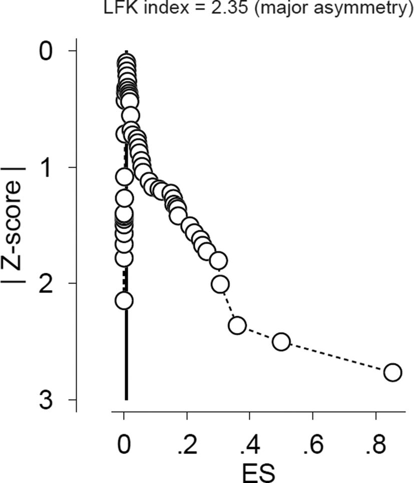
Publication bias was assessed using the Doi plot and Luis Furuya-Kanamori asymmetry index (LFK index). In the presence of symmetry, no publication bias and in the absence of symmetry, publication bias is expected. An LFK index within ±1, out of ±1 but within ±2 and > ± 2 means no asymmetry, minor asymmetry, and major asymmetry, respectively (Furuya-Kanamori et al., Reference Furuya-Kanamori, Barendregt and Doi2018).
Results
Main characteristics of the studies
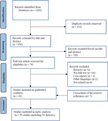
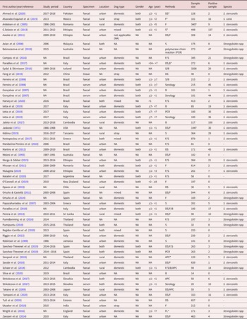
Figure 1 shows the process of study selection according to PRISMA. In brief, the preliminary search yielded 1202 studies. Subsequently, duplicated papers and unrelated studies conferring to title and abstract was removed. Seventy-four full texts of the remaining articles were evaluated, as a result of which 43 studies were eligible to be included in the quantitative analysis based on the inclusion and exclusion criteria. Also, seven articles, found through the reference list literature review, were included in quantitative analysis (fig. 1). Among 50 articles, 49, four and three studies evaluated S. stercoralis infection in canines by coprological, serological and molecular methods, respectively. Four studies used the coprological and serological methods (Ferreira et al., Reference Ferreira, Goncalves-Pires, Silva, Goncalves and Costa-Cruz2006; Gonçalves et al., Reference Gonçalves, Machado, Gonçalves-Pires, Ferreira-Júnior, Silva and Costa-Cruz2007; Štrkolcová et al., Reference Štrkolcová, Goldová, Bocková and Mojžišová2017; Iatta et al., Reference Iatta, Buonfrate and Paradies2019) and two reports applied coprological and molecular methods alongside each other (Iatta et al., Reference Iatta, Buonfrate and Paradies2019; Sanchez-Thevenet et al., Reference Sanchez-Thevenet, Carmena, Adell-Aledon, Dacal, Arias, Saugar, Rodriguez and Dea-Ayuela2019) (table 1). Only one study reported the prevalence based on molecular techniques (Beknazarova et al., Reference Beknazarova, Barratt, Bradbury, Lane, Whiley and Ross2019). Seven studies assessed the prevalence of S. stercoralis infection in dog owners together with their dogs (Takano et al., Reference Takano, Minakami, Kodama, Matsuo and Satozono2009; Schaer et al., Reference Schaer, Inpankaew and Traub2014; Jaleta et al., Reference Jaleta, Zhou, Bemm, Schär, Khieu, Muth, Odermatt, Lok and Streit2017; Mulugeta, Reference Mulugeta2019; Sanpool et al., Reference Sanpool, Intapan, Rodpai, Laoraksawong, Sadaow, Tourtip, Piratae, Maleewong and Thanchomnang2019; Harvey et al., Reference Harvey, Tang, da Paixao Sevá, Albano dos Santos, Santos Carvalho, Magalhães da Rocha, Oliveira and Albuquerque2020).
Predefined Protocol Items for Systematic Reviews and Meta-Analyses (PRISMA) flow chart of study identification and selection.

Main characteristics of included studies reporting prevalence of Strongyloides stercoralis infection in dogs.

DS*; direct smear, F*; flotation method, S*; sedimentation technique, B*; Baermann technique; APC*; Koga agar plate culture technique, FL*; FLOTAC technique.
In total, 56 datasets out of 50 studies were included in the current meta-analysis. These studies were carried out in 27 countries across five continents as follows: seven in America; two in Africa; eight in Asia; eight in Europe; and two in Oceania (table 1). These studies delivered data on 20,627 dogs from the 56 datasets across six WHO regions as follows: one dataset was accessible for the Eastern Mediterranean region (128 dogs); five for Africa (1783 dogs); five for the South-East Asian region (651 dogsdoges); 15 for America (3842 dogs); 23 for Europe (13,090 dogs); and eight for the Western Pacific region (1133 dogs). Among the Americas region, four studies were from North America (1938 dogs) and 11 studies were from South America (1904 dogs).
Overall prevalence of S. stercoralis infection in dogs
The global prevalence of S. stercoralis infection among dogs was 6% (95% confidence intervals (CI) 4–8%; 868/20,627) using pooled random effect with a very high heterogeneity (Q = 2152.1, df = 56, I2 = 97.4%, P < 0.001).
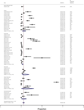
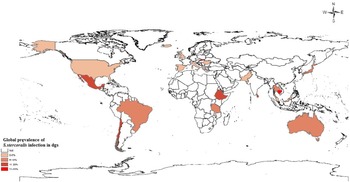
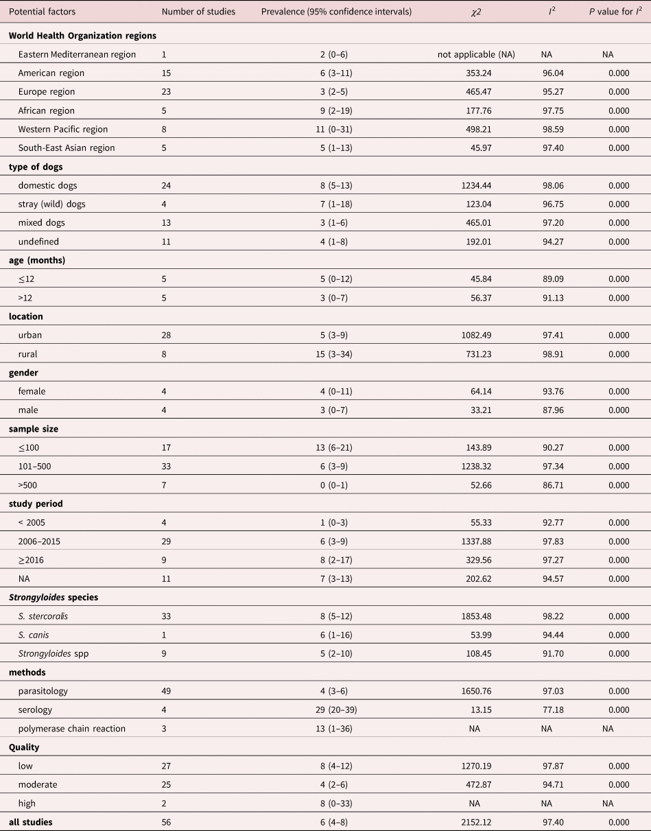
Subgroup analysis was performed to establish the source of heterogeneity with reference to the type of dogs, gender, age and area (urban vs. rural), sample size, methods and study period. The results showed very high heterogeneity among subgroups which decreased to 77.18% in the studies applied by serological assay (Q = 13.15, df = 4, I 2 = 77.18%, P < 0.001). The details of pooled prevalence of S. stercoralis infection obtained from subgroups analysis is presented in table 2. In accordance with WHO regions, the prevalence of this infection in dogs was highest in the Western Pacific region (11%, 0–31%) and lowest in Europe (3%, 2–5%) (Q = 953.054, P < 0.05). The pooled prevalence in other WHO regions was: African region (9%, 2–19%); America (6%, 3–11%); South-East Asia (5%, 1–13%); and Eastern Mediterranean (2%, 0–6%) (fig. 2). The global prevalence of S. stercoralis in dogs across countries is shown in fig. 3.
Forest plot of the prevalence of Strongyloides stercoralis infection in dogs based on World Health Organization region and globally.

Prevalence of Strongyloides stercoralis in dogs in different countries using a geographic information system (GIS).

Subgroup analysis of potential factors influencing the prevalence of Strongyloides stercoralis infection in dogs.

The results of the LFK index analysis showed that the publication bias was significant with major asymmetry (LFK index = 2.35) (fig. 4).
Publication bias using Doi plot and Luis Furuya-Kanamori asymmetry index (LFK index).

Prevalence of S. stercoralis infection in dog owners
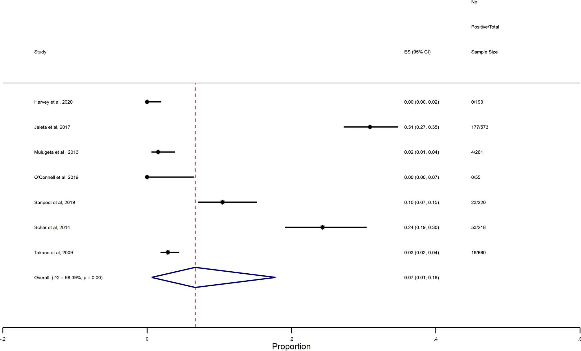
The pooled prevalence of S. stercoralis infection in dog owners was 7% (1–18%) with a very high heterogeneity (Q = 371.78, df = 6, I 2 = 98.39%, P = 0.00) (fig. 5).
Forest plot of the prevalence of Strongyloides stercoralis infection in dog owners.

Sensitivity analysis
A sensitivity analysis, in which a particular study was removed each time from the meta-analysis to calculate the stability of the results, was performed in the present study. The obtained results did not show any considerable changes in the heterogeneity of the studies (P < 0.0001).
Quality assessment
A summary of quality assessment for the included studies estimating the prevalence of S. stercoralis infection in dogs is shown in the Online Supplementary Table S1. Analysis of risk of bias showed that prevalence rates of S. stercoralis infection in studies with low (8%, 4–12%) and moderate (4%, 2–6%) risks of bias were considerable (table 2).
Discussion
The results of the present systematic review and meta-analysis study indicates the low global prevalence of S. stercoralis infection in dogs with a rate of 6% (95% CI 4–8%). It could be concluded that in most countries, it is not possible to estimate the precise prevalence of S. stercoralis, due to scarce and insufficient studies. A total of 20,627 dogs were analysed, from which 868 were infected. One possible explanation for the low prevalence rate may be related to the diagnostic methods applied. As was expected, most studies used stool examination for the diagnosis of S. stercoralis larvae which showed a lower prevalence rate. It is well documented that faecal examination using direct smear and different flotation/sedimentation techniques have very low-sensitivity for the diagnosis of strongyloidiasis (Requena-Méndez et al., Reference Requena-Méndez, Chiodini, Bisoffi, Buonfrate, Gotuzzo and Muñoz2013). Among parasitological techniques, flotation with saturated salt or Sheather's solution is not suitable for detecting larvae in faeces. However, flotation with zinc sulphate solution can yield detectable larvae in stool (Dillard et al., Reference Dillard, Saari and Anttila2007). In the current study, three and seven studies used the agar plate coproculture (APC) and the Baermann technique, respectively, which could moderately increase the sensitivity in comparison with the other parasitological methods (Schär et al., Reference Schär, Trostdorf, Giardina, Khieu, Muth, Marti, Vounatsou and Odermatt2013; Buonfrate et al., Reference Buonfrate, Formenti, Perandin and Bisoffi2015). However, most investigations on S. stercoralis and other soil-transmitted helminths were performed based on non-specific techniques. Therefore, the global and local prevalence of strongyloidiasis is extremely underestimated in human and canine populations (Buonfrate et al., Reference Buonfrate, Formenti, Perandin and Bisoffi2015). In this systematic review, only four and three studies were performed on the basis of serological and molecular techniques, respectively, which proved higher sensitivity compared to coprological techniques (Ferreira et al., Reference Ferreira, Goncalves-Pires, Silva, Goncalves and Costa-Cruz2006; Gonçalves et al., Reference Gonçalves, Machado, Gonçalves-Pires, Ferreira-Júnior, Silva and Costa-Cruz2007; Beknazarova et al., Reference Beknazarova, Whiley and Ross2016; Štrkolcová et al., Reference Štrkolcová, Goldová, Bocková and Mojžišová2017; Iatta et al., Reference Iatta, Buonfrate and Paradies2019; Sanchez-Thevenet et al., Reference Sanchez-Thevenet, Carmena, Adell-Aledon, Dacal, Arias, Saugar, Rodriguez and Dea-Ayuela2019). In recent years, the use of serological assays along with molecular techniques has increased true positive diagnosis of S. stercoralis infection in humans (Buonfrate et al., Reference Buonfrate, Requena-Mendez and Angheben2018; Kalantari et al., Reference Kalantari, Chehrazi, Ghaffari and Gorgani-Firouzjaee2020). According to geographical regions, the Western Pacific region has the significantly highest prevalence rate compared to European and Eastern Mediterranean regions. The main reasons for these differences may be associated to climate conditions. This is evidence that humid and warm weathers provide ideal conditions for the reproduction of S. stercoralis (Štrkolcová et al., Reference Štrkolcová, Goldová, Bocková and Mojžišová2017). Countries which are located in Western Pacific region have such a climate. Also, several studies conducted in Europe indicated that the rate of infection in dogs in warmer regions such as France, Italy and Spain was higher than in cold regions (Ortuño & Castellà, Reference Ortuño and Castellà2011; Paradies et al., Reference Paradies, Iarussi, Sasanelli, Capogna, Lia, Zucca, Greco, Cantacessi and Otranto2017; Sanchez-Thevenet et al., Reference Sanchez-Thevenet, Carmena, Adell-Aledon, Dacal, Arias, Saugar, Rodriguez and Dea-Ayuela2019; Tull et al., Reference Tull, Moks, Laurimaa, Keis and Suld2020). According to the studies conducted on the prevalence of S. stercoralis in humans, the rate of infection is high in Thailand and Brazil with the prevalence ranging from 23 to 43%, with those countries considered as endemic countries for strongyloidiasis (Schär et al., Reference Schär, Trostdorf, Giardina, Khieu, Muth, Marti, Vounatsou and Odermatt2013; Salvador et al., Reference Salvador, Treviño and Bosch-Nicolau2020). As mentioned in the present work, three studies investigated the prevalence of strongyloidiasis using simple parasitological methods in the canine population in Thailand (Pumidonming et al., Reference Pumidonming, Salman, Gronsang, Abdelbaset, Sangkaeo, Kawazu and Igarashi2016; Pumipuntu, Reference Pumipuntu2018; Sanpool et al., Reference Sanpool, Intapan, Rodpai, Laoraksawong, Sadaow, Tourtip, Piratae, Maleewong and Thanchomnang2019). Moreover, seven studies evaluated strongyloidiasis in the dog population in Brazil, where only two studies showed high prevalence rates when diagnosed with serological techniques (table 1) (Ferreira et al., Reference Ferreira, Goncalves-Pires, Silva, Goncalves and Costa-Cruz2006; Gonçalves et al., Reference Gonçalves, Machado, Gonçalves-Pires, Ferreira-Júnior, Silva and Costa-Cruz2007).
Dogs with an age of <1 year have a higher prevalence of S. stercoralis infection than their older counterparts (>1 year of age). This finding is consistent with most of the previous studies (Jaskoski, Reference Jaskoski1971; Alvarado-Esquivel et al., Reference Alvarado-Esquivel, Romero-Salas, Aguilar-Domínguez, Cruz-Romero, Ibarra-Priego and Pérez-de-León2015). This difference might be associated with an under-developed immune system in younger dogs than fully grown subjects (Fontanarrosa et al., Reference Fontanarrosa, Vezzani, Basabe and Eiras2006). In this review, the distribution of strongyloidiasis in dogs from rural areas was higher than urban areas. Factors such as poor preventive veterinary care, health conditions, and treatment programmes can affect the prevalence of infections in rural areas (Jaleta et al., Reference Jaleta, Zhou, Bemm, Schär, Khieu, Muth, Odermatt, Lok and Streit2017). The highest infection rate was reported in rural regions in the study conducted by Jaleta et al. (Reference Jaleta, Zhou, Bemm, Schär, Khieu, Muth, Odermatt, Lok and Streit2017). Also, the infection rate in dog populations was high in this study.
Seven studies in the current review evaluated strongyloidiasis prevalence in dog owners along with their animals with a rate of 7%. This may strongly confirm the cross-transmission between dogs and humans especially in rural conditions, where people share their space closely with dogs (Schaer et al., Reference Schaer, Inpankaew and Traub2014; Jaleta et al., Reference Jaleta, Zhou, Bemm, Schär, Khieu, Muth, Odermatt, Lok and Streit2017; Nagayasu et al., Reference Nagayasu, Aung and Hortiwakul2017).
The limitations of the present study are as follows: (i) epidemiological data were not available for a number of countries in the WHO regions; (ii) some potentially eligible papers, which were selected for the current systematic review, were not available in full text; and (iii) a number of potentially relevant studies selected in our review could not be assessed, as they were not in English. Therefore, a potential selection bias may be increased to some extent in this study due to the exclusion of some appropriate data.
Conclusions
This investigation concludes that the low global prevalence of S. stercoralis in dogs may be strongly associated with the lack of improved and sensitive diagnostic methods, along with the low consideration given to S. stercoralis infection compared to other species of soil-transmitted helminths. Therefore, for the accurate evaluation of strongyloidiasis, the use of appropriate diagnostic methods with high sensitivity is recommended.
Supplementary material
To view supplementary material for this article, please visit https://doi.org/10.1017/S0022149X21000808
Acknowledgements
The authors would like to thank to Taraneh Ghaffari for English editing and the research council of Babol University of Medical Sciences for their supports.
Financial supports
The present work was funded by the research council of Babol University of Medical Sciences, Babol, Iran (Grant No: 14012341).
Conflict of interest
None.
Ethical standards
This work was approved by the ethical committee of Babol University of Medical Sciences, Babol, Iran (IR.MUBABOL.REC. 1400.013).
Author contributions
T.G.F. designed the study. T.G.F. and S.S. conducted the search study. T.G.F. and S.S. contributed to data extraction. M.C. performed data analysis. T.G.F., N.K. and S.G. drafted and edited the manuscript. We confirm that all authors observed the drafts of the manuscript and approved the final version of the paper.