Introduction
Myostatin, a muscle-specific member of the transforming growth factor beta (TGFβ) family, negatively regulates muscle development (Ríos et al., Reference Ríos, Carneiro, Arce and Devesa2002). It acts by blocking cell proliferation, modifying expression of cell cycle proteins. An increase in p21cip1 and reduction in Cdk2 expression precede myoblast fusion and muscle fibre formation (Oldham et al., Reference Oldham, Martyn, Sharma, Jeanplong, Kambadur and Bass2001).
Cattle that showed a significant increase in muscle mass were found to carry natural mutations in the myostatin gene, which resulted in changes in the myoblast cell cycle and differentiation (Kambadur et al., Reference Kambadur, Sharma, Smith and Bass1997; McPherron & Lee, Reference McPherron and Lee1997; Grobet et al., Reference Grobet, Royo, Poncelet, Dimitri, Brouwers, Riquet, Schoeberlein, Dunner, Ménissier, Massabanda, Fries, Hanset and Georges1997, Reference Grobet, Poncelet, Royo, Brouwers, Pirottin, Michaux, Menissier, Zanotti, Dunner and Georges1998). These animals presented an increase in muscle mass of around 25% and were still viable and fertile, demonstrating the potential value of producing an increase in muscle mass by disrupting myostatin function, even in other breeds and species (McPherron & Lee, Reference McPherron and Lee1997). In the mouse, it was possible to create mutations in the myostatin gene through homologous recombination, in order to increase muscle mass (McPherron et al., Reference McPherron, Lawler and Lee1997). Unfortunately, this process is expensive and time consuming. At this time, it is not applicable to farm animals.
RNA interference (RNAi) is a process in which a double-stranded DNA (dsRNA) silences gene expression, either by inducing the degradation of complementary mRNA or by inhibiting translation. The silencing can be stably induced by short hairpin RNAs (shRNAs), produced by DNA expression vectors introduced into mammalian cells (Scherr and Eder Reference Scherr and Eder2007).
The aims of the present experiments were to test the efficiency of shRNA targeting myostatin delivered by a lentiviral vector in murine myoblasts and verify the feasibility of lentiviral-mediated delivery of shRNA into in vitro produced transgenic bovine embryos.
Material and methods
Production of shRNA
The bovine myostatin mRNA (GenBank NM_001001525) was processed through a computer algorithm Strand Analysis (http://lgm.fcm.unicamp.br:9001/cgi-bin/SA/SA.cgi) that predicted a best siRNA design for the myostatin gene sequence (>90% degradation). The siRNA was designed to map to a conserved region between the murine and bovine myostatin genes. Individual shRNA were ordered as sense and antisense oligonucleotides (Invitrogen). They were annealed and ligated into a pSilencertm 2.1-U6 vector (Ambion) that contained a U6 promoter directly upstream of the predicted shRNA site.
Lentiviral vector production
For the lentiviral packaging plasmid, envelope and transference plasmids were used to generate a lentiviral vector as described by Lois et al., Reference Lois, Hong, Pease, Brown and Baltimore2002. Myostatin shRNA with a U6 promoter were cloned into the PacI restriction site of the transference plasmid, followed by a ubiquitin promoter and a green fluorescent protein (GFP) cassette. As a control, a lentiviral vector was produced that carried only the ubiquitin promoter and GFP cassette.
The human embryonic kidney cell line A293T was treated with 25 μM chloroquine and all plasmids were added to culture medium that contained 2 M calcium chloride. Medium was changed after 22 h and cells were cultured for an additional 24 h, after which medium was collected and centrifuged at 5000 g for 5 min. The supernatant (viral suspension) was collected and frozen at –80°C. Viral titration was performed by use of serial dilutions of viral suspension in the NIH 3T3 cultured cell line and determination of GFP-positive colonies.
Murine myoblast (C2C12) cell culture
C2C12 myoblasts were cultured in Dulbecco's Modified Eagle Medium (DMEM; Gibco) supplemented with 10% fetal calf serum (FCS; Gibco) and gentamycin (250 μg/ml; Sigma-Aldrich) at 37°C and 5% CO2. Cells were cultured until 80% confluence and then were passaged by incubation with trypsin solution (0.1% trypsin; Gibco) and 0.03% EDTA (Sigma-Aldrich) at 37°C. Myoblasts were centrifuged and resuspended in DMEM supplemented with 20% FCS and 10% dimethyl sulfoxide (DMSO; Sigma-Aldrich), then cells were frozen in 1 ml aliquots using the freezing container ‘Mr Frosty’ (Nalgene) and stored in liquid nitrogen.
Cell transduction and flow cytometry
Myoblasts were seeded in 6-well plates and cultured until 50% confluent. Medium was then removed and 2.5 × 10⁴ viral particles were added together with 8 μg/ml hexadimethrine bromide (Sigma-Aldrich). The medium volume was made up to 500 μl with DMEM and then incubated at 37°C and 5% CO2 for 8 h, at which time the DMEM plus viral particles was changed to DMEM with 10% FCS. Two cell lines were produced according to the lentiviral vector used: (1) GFP: containing only the GFP cassette; and (2) GFP-shRNA: containing the GFP cassette and the shRNA targeting myostatin. Transduced cells were cultured for an additional 24 h and incubated with trypsin. Myoblasts were resuspended in phosphate buffered saline (PBS) with no calcium or magnesium and submitted to flow cytometric sorting. GFP-positive myoblasts were sorted by FACS Aria (Becton Dickinson).
Cell differentiation
Cell differentiation was performed to induce myostatin expression. To induce myotube formation, myoblasts were cultured until 80% confluent and medium was changed using DMEM with 1% FCS. Cells were cultured for an additional 72 h.
Real time PCR
Real-time PCR was conducted in order to verify shRNA efficiency in myostatin degradation. Primers were designed using the Primer3 online software (Whitehead Institute for Biomedical Research). Primer sequences were MSTNf 5′-GAGGGCAGTGAGAGAGAAGA-3′ and MSTNr 5′-GTCAGACTCTGTAGGCATGG-3′ for myostatin amplification and GAPDHf 5′-TCGGAGTGAACGGATTTG-3′ and GAPDHr 5′-CCTGGAAGATGGTGATGG-3′, for glyceraldehyde 3-phosphate dehydrogenase (GAPDH) amplification (used as a housekeeping gene). Transduced or non-transduced myoblasts that had been induced to differentiate for 72 h had their total RNA isolated with the Illustra RNAspin MiniRNA Isolation Kit® (GE Healthcare). From purified RNA, RT-PCR was performed to synthesize cDNA using the Superscript First-Strand Synthesis System for RT-PCR® (Invitrogen) according to manufacturer's protocols. Real-time PCR was carried out using Platinum SYBR Greener qPCR SuperMix (Invitrogen) and a thermal cycling profile of 40 cycles of 15s at 95°C and 45s at 60°C for myostatin amplification and 40 cycles of 15s at 95°C, 20s at 55°C and 20s at 68°C for GAPDH amplification (Mastercycler EP).
Cell differentiation kinetics
Transduced or non-transduced myoblasts were induced to form myotubes after 72 h of differentiation. Cell differentiation kinetics was assessed as described previously (Minotti et al., Reference Minotti, Scicchitano, Nervi, Scarpa, Lucarelli, Mollnaro and Adamo1998) with minor modifications. Cells were fixed in ethanol, stained with Giemsa for 3 min and observed under optical microscopy. Cells that presented continuous cytoplasm and had at least three nuclei were considered myotubes. Total myotube numbers were divided by total number of nuclei and expressed as percentage of fusion. At least seven random fields and more than 500 total nuclei were counted for each sample. The results are presented as mean ± SE of the counts from three independent experiments.
In vitro transgenic embryo production
Bovine ovaries were obtained from a local abattoir and transported to the laboratory in 0.9% NaCl solution at 30°C. Follicles 2–8 mm in diameter were aspirated with a 21-gauge needle attached to a 5 ml syringe and fluid placed in 15 ml conical tube. Cumulus–oocyte complexes (COCs) were allowed to settle for 10 min and COCs were selected according to cytoplasm aspect and number of granulosa cell layer.
Groups of 30 COCs were matured in 90 μl drops of TCM-199 bicarbonate supplemented with 10% FCS and 0.5 μg/ml FSH (Follitropin-V), 100 IU/ml hCG (Chorulon, Intervet) and 1.0 μg/ml estradiol under mineral oil for 18 h at 38.5°C and 5% CO2 in air and high humidity. Cumulus cells were enzymatically removed using 0.2% hyaluronidase in calcium-free PBS for 10 min and repeated pipetting for 2 min.
Oocytes that extruded the first polar body were selected and microinjected with a solution containing lentiviral particles (2.5 × 10⁶ UI/ml) using FemtoJet (Eppendorf). In vitro fertilization was performed after centrifugation of frozen–thawed semen on discontinuous Percoll density gradient (45% Percoll upper and 90% lower layer) in TALP stock medium supplemented with 0.6% fatty acid-free BSA, 3.0 μg/ml heparin, 0.2 mM sodium pyruvate, 50 μg/ml gentamicin, 3.0 μg/ml penicillamine, 1.1 μg/ml hypotaurine, 0.3 μg/ml epinephrine (Parrish et al., Reference Parrish, Susko-Parrish, Handrow, Sims and First1989).
Presumptive zygotes were in vitro cultured (IVC) on a granulosa cell monolayer in SOFaa medium (Tervit et al., Reference Tervit, Whittingham and Rowson1972) supplemented with 10% FCS, 0.02% essential, and 0.01% non-essential amino acids. After 2 days of IVC, 90 μl of SOFaa medium was added to the culture drops and cleavage was evaluated after 4 days. Blastocyst rate was evaluated at day 7 of IVC. Embryos were assessed for GFP expression under epifluorescence microscopy (Olympus) using 610 nm emission filter and 520 nm excitation filter at day 7–9. Cleavage, blastocyst and hatching rates and also GFP-positive embryos were presented as mean ± SE of three independent replicates.
Statistical analysis
Quantitative PCR data were analysed using REST MCS beta software by Pairwise Fixed Reallocation Randomization Test (Pfaffl et al., 2005). Reactions were normalized by expression of the housekeeping gene GAPDH. Differentiation kinetics data were analysed by ANOVA using SAS System for Windows (SAS Institute Inc.). Dependent variable was the number of myotubes and independent variable was cell type. Comparison of mean values was performed using Tukey's test with 5% of significance level. Cleavage and blastocyst rates and also GFP positive embryos were presented as LS means ± SEM of three independent replicates and analysed by ANOVA using MINITAB Release 14 Statistical Software and Tukey's test as described above.
Results
Myostatin shRNA efficiency in myoblasts
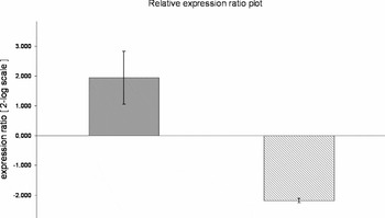
Relative expression of myostatin in non-transduced myoblasts, myoblasts with GFP only (GFP) and GFP plus shRNA targeting myostatin, differentiated for 72 h, was verified by real time PCR. Myostatin expression was upregulated in GFP group and downregulated in GFP shRNA group (p < 0.05) when compared to non-transduced cells (Fig. 1).
Changes in myostatin gene expression in non-transduced, GFP and GFP-shRNA by real-time PCR. Myostatin mRNA expression was normalized with GAPDH mRNA expression. Data represent means ± SEM for three replicates. Myostatin expression was upregulated in the GFP (grey bar) group and downregulated in the GFP-shRNA (grey hatched bar) group (p < 0.05) when compared with non-transduced cells after 72 h differentiation.

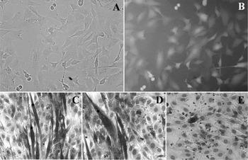
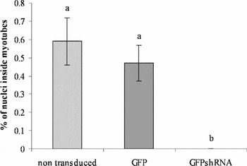
Transduced myoblasts were photographed under epifluorescence microscopy in order to verify GFP expression. After cell sorting and differentiation myotube formation was comparatively evaluated between non-transduced and transduced cells. The number of nuclei inside myotube did not differ between GFP and non-transduced cells. However, GFP shRNA presented lower formation of myotubes (Figs 2 and 3).
Evaluation of myotubes differentiation kinetic. (A) Transduced myoblasts, after cell sorting, photographed under optical microscopy. (B) Transduced myoblasts, after cell sorting, photographed under epifluorescence microscopy in order to verify GFP expression. (C) Control (non-transduced) myoblasts after 72 h differentiation and Giemsa stained, observed under optical microscope. (D) GFP cell line after 72 h differentiation and Giemsa stained, observed under optical microscopy. (E) GFP-shRNA cell line after 72 h differentiation and Giemsa stained, observed under optical microscopy. *, myotube.

Evaluation of myotubes differentiation kinetic. The percentage of nuclei inside the myotube was comparatively evaluated in non-transduced, GFP and GFP-shRNA cell lines. Different letters are significantly different (p < 0.05).

Transgenic embryo production
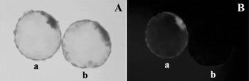
Microinjected or non-microinjected embryos had cleavage rates assessed at day 4 and blastocyst rates assessed at day 7. No difference (p < 0.05) was observed between groups (Table 1). GFP-positive embryos were present only in microinjected group (Fig. 4). After hatching, 3.07% of embryos were GFP positive (Table 1).
Embryo development and GFP expression. Control (non-microinjected) and microinjected embryos were evaluated at day 4 (cleavage) and day 7 (blastocyst). GFP expression was assessed at days 7–9 (after hatching).

Different letters are significantly different (p < 0.05).
Embryo development and GFP expression. GFP-positive embryos were present only in the microinjected group. (A) Hatched blastocysts from the microinjected (a) and control (b, non-microinjected) groups, photographed under optical microscopy. (B) Hatched blastocysts from microinjected (a) and control (b, non-microinjected) groups photographed under epifluorescence microscopy.

Discussion
Applying gene knockout technologies in farm animals is limited at the present time. Therefore, post-transcriptional gene silencing using RNAi is an alternative for production of transgenic livestock. Generation of double muscle animals would useful for the beef cattle industry. Thus, this study verified the reduction of myostatin expression in cultured cells and the feasibility of transgenic embryo production by means of lentiviral-mediated shRNA delivery.
A lentiviral vector containing myostatin shRNA was constructed and proved itself to efficiently deliver foreign DNA into cultured cells. Compared with non-viral transfection methods, such as cationic lipids and electroporation, lentiviral vectors showed higher genome integration and the possibility of transducing non-dividing cells (Naldini et al., Reference Naldini, Blomer, Gallay, Ory, Mulligan, Gage, Verma and Trono1996).
Lentivirus-delivered shRNA reduced expression of myostatin efficiently, as expected, demonstrating the efficiency of the designed shRNA in promoting gene silencing. Sorted GFP shRNA cells submitted to differentiation showed reduction of myotube formation in accordance with previous studies (Wagner et al., Reference Wagner, Liu, Chang and Allen2005, Sato et al., Reference Sato, Kurokawa, Yamauchi and Hattori2006). This reduction, however, did not compromise muscle development, as myostatin mutant mice and cattle still presented skeletal muscle differentiated from myoblasts (McPherron & Lee, Reference McPherron and Lee1997; McPherron et al., Reference McPherron, Lawler and Lee1997). Surprisingly, the GFP cell line exhibited an upregulation of myostatin expression. This may be due to vector integration near to the myostatin gene transcriptional control regions or some other gene related to its pathway.
After efficiency verification, the lentiviral vector was used to produce in vitro transgenic embryos as previously reported (Hofmann et al., Reference Hofmann, Zakhatchenko, Weppert, Sebald, Wenigerkind, Brem, Wolf and Pfeifer2004, Golding et al., Reference Golding, Long, Carmell, Hannon and Westhusin2006). After hatching, 3.07% of microinjected embryos were GFP positive. Although other groups have obtained better results using the lentiviral delivery system, our lower results may be caused by a very low viral titration compared with other groups (Hofmann et al., Reference Hofmann, Zakhatchenko, Weppert, Sebald, Wenigerkind, Brem, Wolf and Pfeifer2004; Golding et al., Reference Golding, Long, Carmell, Hannon and Westhusin2006). In mice, a single injection with a low viral titration of 2.4 × 10⁷ resulted in 1% positive embryos (Ritchie et al., Reference Ritchie, Neil, King and Whitelaw2007). In spite of the fact that nuclear transfer may generate 100% positive embryos, microinjection technique may result in higher blastocyst rates due to the current inefficiency of nuclear reprogramming (Wilmut et al., Reference Wilmut, Beaujean, de Sousa, Dinnyes, King, Paterson, Wells and Young2002).
In conclusion, lentiviral shRNA delivery was efficiently performed in C2C12 myoblast cell culture; downregulated myostatin gene expression; and reduced myotube formation in vitro. Also, our lentiviral vector carrying myostatin shRNA was able to produce in vitro transgenic bovine embryos by microinjection. The results herein described may be suitable to enhance meat production in the beef cattle industry and as models for muscle disorders in other species.
Disclosure statement
There are no competing financial interests exist.
Acknowledgements
The authors would like to thanks São Paulo Research Foundation Support (FAPESP) for the financial support.