Case story
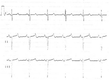
The patient was born at a gestational age of 35 weeks and 6 days, with a birth weight of 2.393 g, as the second child of healthy second-degree consanguineous parents. At birth, electrocardiogram demonstrated congenital complete atrioventricular block with a heart rate between 58 and 63 bpm and quantum tunnelling composite up to 0.504 s (Fig 1).
Section of electrocardiogram from day 2 of life. Quantum tunnelling composite is 504 ms.

Echocardiography demonstrated a structurally normal heart apart from two haemodynamic non-significant muscular ventricular septal defects, later spontaneously closing. The mother was anti-SSA/Ro and anti-SSB/La negative. Genetic testing of the child for long-QT syndrome mutations in the SCN5A gene was negative.
Pacemaker implantation was indicated because of congenital complete atrioventricular block and long-QT syndrome. A VVI-pacemaker (Identity Model 5172; St. Jude Medical) implanted via sternotomy with the leads positioned epicardial on the right ventricle outflow tract was implanted at 31 days of age. Propranolol 1.5 mg/day was initiated and later increased to 3 mg/day. It is standard policy in our hospital to start propranolol treatment at a very low dosage because of the risk of deterioration in pre-existing heart failure. Initial pace rate was set to 80, and increased within 6 days of implantation to 120 bpm.
At age 7 months, the patient was admitted to a local hospital with confirmed respiratory syncytial virus infection.
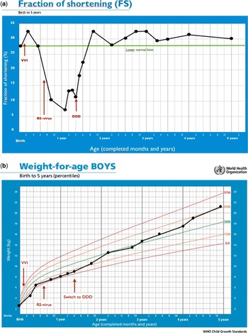
The patient was diagnosed with dilated cardiomyopathy and a 10% fraction of shortening at the age of 8.5 months (Fig 2a). Treatment with furosemide, spironolactone, and captopril was started but, in spite of this, the patient's clinical condition deteriorated without any improvement of left ventricle function during the following 6 months. He was treated on a monthly basis with antibiotics for recurrent pneumonia; he was sleeping 16 hours daily and had poor weight gain (3rd percentile see Fig 2b).
(a) Rapid decline in fraction of shortening (FS) after implantation of VVI pacemaker. The time of respiratory syncytial virus (RSV) infection and switch to DDD system is marked and shows normalisation of left ventricular fraction of shortening. (b) Growth retardation in the first year of life, markedly improved after switch to a DDD system.

At age 15 months, the patient was considered for heart transplantation and had a cardiac catheterisation. Using thermodilution, an immediate 63% increase in cardiac output (0.9–1.5 l/min) was seen by temporary pacing in the right ventricular outflow tract obtaining narrow QRS complexes. Furthermore, the central venous O2 saturation increased from 42% to 56%. Other pacing sites were tested, but could not produce a similar increase.
Consequently, at age 16 months, a biventricular epicardial DDD pacemaker (InSync III Model 8042; Medtronic) was implanted. Bipolar leads were positioned on the right atrium and on the right ventricular outflow tract. On the basis of the heart catheterisation experience, a unipolar lead was placed on the left ventricle close to the posterior descending artery, and as close to the atrioventricular groove as possible. This resulted in an instant arterial blood pressure and venous saturation increase, and decreased central venous pressure.
During the subsequent 9 months, the left ventricle diastolic dimension and fraction of shortening normalised. Furosemide, spironolactone, and captopril were reduced during the following 12 months. Furthermore, he gained weight and caught up with his age group (50th percentile). In the follow-up period, he was diagnosed with mannan-binding lectin deficiency treated with Spectramox, congenital muscular dystrophy – probably limb-girdle dystrophy – and attention deficit hyperactivity disorder treated with Ritalin. He has been followed up in the outpatient clinic every 6 months for the last 10 years, where no arrhythmias have been detected (since birth) and fraction of shortening continues to be normal (Fig 2a). At the end of the follow-up period (2011), he was treated with Ritalin, Concerta, Atenolol, and Zithromax. The patient is currently being assessed for Smith–Magenis syndrome.
Discussion
Up to 30% of children with congenital complete atrioventricular block develop dilated cardiomyopathy.Reference Silvetti, Drago and Rava 1 The aetiology has often been attributed to anti-SSA/Ro and anti-SSB/La antibody-induced autoimmune myocarditis. However, children negative of the specific antibodies, like the patient described, may also develop dilated cardiomyopathy.Reference Silvetti, Drago and Rava 1 Before the diagnosis of dilated cardiomyopathy, our patient had a confirmed respiratory syncytial virus infection, which can induce myocarditis and dilated cardiomyopathy. No other cause of dilated cardiomyopathy was identified during examinations – metabolic disorders, familial cardiomyopathies, coronary artery disease, and myocarditis. One possible cause is limb-girdle dystrophy, as cardiac disturbances are common in these patients.Reference van der Kooi, de Voogt and Barth 2 It is caused by various degrees of muscle wasting with fibrosis and fat replacement.Reference Groh 3 Conduction disturbances and heart malformations such as ventricular septum defect is a known phenotype in Smith–Magenis syndrome, as well as upper airway infections and behavioural disorders, but there has been no reported development of dilated cardiomyopathy.Reference Edelman, Girirajan and Finucane 4
Some studies suggest that right ventricle pacing can induce dilated cardiomyopathy.Reference Moak, Hasbani and Ramwell 5 , Reference Janousek 6 It has also been proposed that cardiac resynchronisation therapy can have clinical benefits for patients with heart failure.Reference Moak, Hasbani and Ramwell 5 , Reference Janousek 6 – Reference Dubin, Janousek and Rhee 8
The potential beneficial effect of biventricular DDD pacing was discussed at a time point, where heart transplant was considered. On the basis of information from a catheter intervention, the VVI pacemaker was changed to a biventricular DDD pacemaker. During the following 9 months, the patient improved clinically, regained normal weight, and reverted to normal left ventricle dimensions and normal left ventricle systolic function.
We cannot rule out that the heart failure in this patient was caused by suboptimal lead placement in the initial pacemaker system. A simple adjustment of lead placement might have produced a similar effect in cardiac output. However, based on the existing knowledge, the physicians on the case decided that the safest treatment was to switch to a biventricular system.
Echocardiographic measurements of left ventricle function have remained normal throughout the follow-up period (10 years), making us believe that his dilated cardiomyopathy was caused by single-chamber pacing.
On the basis of this case, we propose that cardiac catheterisation can be used to evaluate the potential beneficial effect of replacing a single-ventricle chamber pacing with a biventricular chamber system including patients with multi-system disorders.
Conclusion
Biventricular DDD pacing can normalise left ventricle systolic function and left ventricular diastolic dimensions in a child with congenital complete atrioventricular block, long-QT syndrome, congenital muscular dystrophy, and dilated cardiomyopathy after VVI pacing even in a multi-system syndrome patient like ours. Consequently, biventricular pacing may be the method of choice in these patients to preserve ventricular function.
Acknowledgements
None.
Financial Support
This research received no specific grant from any funding agency, commercial or not-for-profit sectors.
Conflicts of Interest
None.
Ethical Standards
None.