Although primarily sporadic, pneumococcal pneumonia has been identified in outbreaks within closed settings, such as hospitals, nursing homes, military camps, and shelters. Reference Mercat, Nguyen and Dautzenberg1–Reference Millar, Brown, Tobin, Murphy, Windsor and Speller5 An outbreak of serotype 19A pneumococcal pneumonia occurred in a relief facility in Japan. The facility was a 2-story building with a capacity of 100 individuals; 99 long-term residents were present in the facility at that time. The facility offers daily-life activity assistance to people with physical, mental, or intellectual disabilities. The residents live in separate rooms, with 1 or 2 individuals in each room, but they gather for meals in the dining room (Fig. S1). Other shared areas include a courtyard and restrooms.
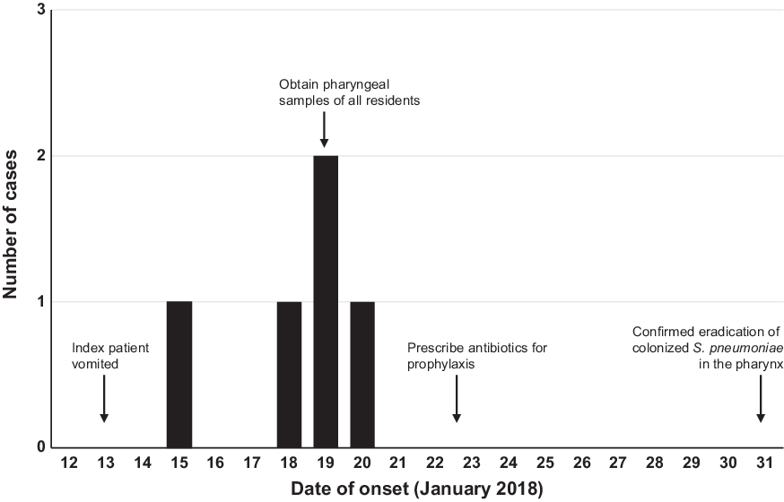
In January 2018, 1 resident (case 1) vomited in the dining room of the relief facility and developed a fever. The resident was hospitalized and diagnosed with pneumococcal pneumonia 2 days later. In addition, 4 residents (cases 2–5) were hospitalized with similar symptoms within a period of 6 days (Fig. 1). The other 4 patients were seated close to the index cases (case 1) at the time of the vomiting incident in the dining room. The first and second patients developed invasive pneumococcal disease (IPD), and the index case died from respiratory failure.
Number of pneumococcal pneumonia cases among the residents of the relief facility and timeline.

Because a pneumococcal pneumonia outbreak was strongly suspected, pharyngeal swabs from 93 non-onset residents were obtained, except from 1 resident who did not provide consent. Streptococcus pneumoniae was isolated in 6 of these samples. Standard precautions were implemented, and the facility was ventilated and cleaned. The staff were instructed to remain aware of symptoms among the residents, such as body temperature changes. On January 23, all residents and staff who provided consent were prescribed amoxicillin or amoxicillin/clavulanate for 7 days. Thereafter, no further cases of pneumococcal infection were observed. Pharyngeal swabs were taken after prophylaxis in 6 asymptomatic carriers; no S. pneumoniae was identified in the cultures.
We performed microbiological examinations of the isolated pneumococcal strains using pulsed-field gel electrophoresis and the Quellung reaction with pneumococcal antisera. Whole-genome sequencing (WGS) and phylogenetic analysis were performed in our previous study, modifying the removal of single-nucleotide variations (SNVs) in recombinogenic regions. Reference Chang, Morita, Lee and Ohnishi6 To exclude SNVs in recombinogenic regions, SNV clusters with >2 single-nucleotide polymorphisms (SNPs) within 100 base pairs were removed. Sequence type (ST) assignment of the S. pneumoniae strains was performed using the pneumococcal multilocus sequence typing website (https://pubmlst.org/) based on analysis of the 7 housekeeping genes aroE, gdh, gki, recP, spi, xpt, and ddl.
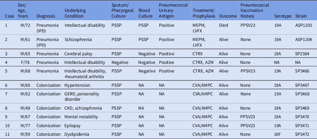
Among the 99 residents in the facility, 56% were male (average age, 66.4 years); 44% of the residents had been vaccinated with the 23-valent pneumococcal polysaccharide vaccine (PPSV23). No residents had been vaccinated with the 13-valent pneumococcal conjugate vaccine (PCV13). Among the residents, 5 developed pneumonia, and S. pneumoniae was isolated from sputum cultures in 4 of these cases (cases 1, 2, 3, and 5). The urinary pneumococcal antigen test was positive in all 5 cases. Also, 2 cases (cases 1 and 2) were diagnosed with IPD based on positive blood cultures, 1 of whom died (case 1). The characteristics of the patients are shown in Table 1. In 6 of 93 non-onset residents, S. pneumoniae was isolated from pharyngeal swabs, and these 6 were diagnosed as asymptomatic carriers (cases 6–11). In summary, the attack rate was 5.1% (4.5% of vaccinated residents and 5.8% of unvaccinated residents), and the colonization rate in non-onset residents was 6.4% (4.8% of the vaccinated residents and 8.3% of the unvaccinated residents). The proportions of residents and staff who had received the influenza vaccine were 98% and 71%, respectively. No other infectious diseases were observed during the outbreak.
Clinical and Microbiological Characteristics of Residents Who Developed Pneumococcal Pneumonia and of Nasopharyngeal Carriers of Streptococcus pneumoniae

Note. IPD; invasive pneumococcal disease, GERD; gastroesophageal reflex disease, CKD; chronic kidney disease, PSSP; penicillin-susceptible Streptococcus pneumoniae, MEPM; meropenem, LVFX; levofloxacin, CTRX; ceftriaxone, AZM; azithromycin, CVA/AMPC; clavulanic acid/amoxicillin, NA; not available.
The serotypes of the S. pneumoniae strains ASP1203, ASP1204, SP2364, and SP3466 from 4 of 5 patients (cases 1, 2, 3, and 5) with pneumonia and the strains SP3467, SP3468, SP3469, SP3470, and SP3471 from 5 of the 6 carrier cases (cases 6–10) were serotype 19A. The strain SP3472 from the remaining case (case 11) was serotype 16F (Table 1). The genotype of S. pneumoniae isolated from culture-positive cases (all cases except for case 4) was examined by multilocus sequence typing and WGS. The ST type of the 9 strains belonging to the 19A serotype was ST3111, whereas that of the 16F strain was ST3117. According to the WGS results, nine 19A strains formed a cluster in the SNV-based phylogenetic tree, and no evidence of polymorphism was observed among the sequences. However, they differed from the 16F isolate SP3472 in that cluster by 5,808 SNPs (Fig. S2).
To the best of our knowledge, an outbreak of serotype 19A pneumococcal has not been reported previously. According to a surveillance data, 19A remains the most frequently isolated serotype among adults in Japan, where the introduction of PCV13 was relatively late. Reference Ubukata, Takata and Morozumi7 Regarding pathogenicity, a systematic review on the risk of death from pneumococcal pneumonia demonstrated that serotype 19A conferred a moderate risk. Reference Weinberger, Harboe and Sanders8 Additionally, serotype 19A has shown increased potential to cause invasive disease relative to other serotypes. Reference Lai, Huang, Lee, Lu, Shao and Chang9 On the other hand, previous reports have described the risk of pneumonia in individuals with physical, mental, or intellectual disabilities. Reference Seminog and Goldacre10 Taken together, multiple factors, including local demographic background, potential virulence of the strain, a closed environment, and host vulnerability, may have contributed to the current outbreak due to serotype 19A S. pneumoniae.
Interestingly, serotype 19A predominated among the carriers, and no SNPs were found among the sequences of all the 19A strains isolated from cases with or without pneumonia. These results indicated that this outbreak was caused by a single S. pneumoniae strain. The opportunity for direct contact between the index case and the remaining residents was limited; the bacteria may have spread in the dining room, through droplets from coughing or vomiting. Because many residents require assistance for daily activities, pathogen transmission may be mediated by staff members with pneumococcal colonization.
The colonization of the nasopharynx by pneumococci is a precursor of various pneumococcal diseases, resulting in a horizontal propagation of pneumococci. Thus, antibiotic prophylaxis is an acceptable strategy to eradicate colonization and prevent the spread of pneumococcal infection. In this case, amoxicillin or amoxicillin/clavulanate was prescribed for 7 days based on the initial susceptibility results and the previous reports. Reference Nuorti, Butler and Crutcher2 The detected serotype 19A S. pneumoniae strain was penicillin sensitive, and the organism was not recovered from nasopharyngeal cultures after completed prophylaxis. The vaccination rate of PPSV23, which covers the serotype 19A, was higher than that observed in previous reports of outbreaks. Reference Mercat, Nguyen and Dautzenberg1–Reference Kuroki, Ishida and Suzuki4 In fact, the attack rate and the colonization rate were lower in residents vaccinated with PPSV23 in the present case.
In conclusion, although serotype 19A is not normally considered an outbreak strain, it can cause rapidly spreading pneumonia in closed settings, especially among vulnerable individuals with predisposing factors. In this case, relatively high coverage rate of pneumococcal vaccination and prophylaxis with penicillin may have prevented the spread of infection. Further studies on larger and more diverse cohorts are needed to validate our findings.
Supplementary material
To view supplementary material for this article, please visit https://doi.org/10.1017/ice.2021.328
Acknowledgments
The authors thank the employees of the relief facility for assistance with data collection. The authors also thank Enago (www.enago.jp) for the English language review. This study was in compliance with the principles of the Helsinki Declaration as well as current ethical guidelines and was approved by the ethics committee of Niigata University Medical and Dental Hospital (no. 2020-0359).
Financial support
No financial support was provided relevant to this article.
Conflicts of interest
All authors report no conflicts of interest relevant to this article.