INTRODUCTION
Cryptosporidiosis is an emerging protozoan disease associated with severe acute diarrhoea in humans and animals. Cryptosporidium parasite is composed of multiple species and genotypes, and thus far 8 species and genotypes (C. hominis, C. parvum, C. meleagridis, C. felis, C. canis, C. muris, C. suis and Cryptosporidium cervine genotype) are known to be transmissible between humans and animals (Xiao and Ryan, 2004). Among them, C. parvum (previously known as genotype II or cattle genotype) is the zoonotic species most commonly found in domestic and wild ruminants (Xiao and Ryan, 2004; Appelbee et al. 2005; Hunter and Thompson, 2005). Recent molecular studies show genetic heterogeneity among C. parvum isolates from humans and animals, and especially DNA sequencing analysis of the C. parvum 60 kDa glycoprotein (GP60) gene have identified at least 9 subtype allele families (IIa, IIb, IIc, IId, IIe, IIf, IIg, IIh and IIi) in this species (Peng et al. 2001; Sulaiman et al. 2001; Alves et al. 2003; Peng et al. 2003a,b; Sulaiman et al. 2005; Abe et al. 2006; Akiyoshi and Tzipori, 2006; Alves et al. 2006; Trotz-Williams et al. 2006). Although the data about subtyping of C. parvum isolates from animals have been extremely limited, only 2 subtype families IIa and IId have been found in the isolates from cattle (Alves et al. 2003; Peng et al. 2003b; Abe et al. 2006; Alves et al. 2006; Trotz-Williams et al. 2006). These findings suggest that some subtype families in C. parvum are zoonotic and some are anthroponotic. Therefore, it is necessary to identify isolates at the subtype level in order to control zoonotic transmission among humans and animals and understand the molecular epidemiology of C. parvum infection.
In Serbia and Montenegro, Cryptosporidium infection has been found in domestic animals such as cattle, pigs, sheep and goats, but the genetic information of the isolates is quite unclear because the identification was performed only by light microscopy (Misic et al. 2001, 2002, 2003, 2006). In the present study, we collected fecal samples from 103 calves, which are recognized as the main reservoir of C. parvum, on 10 farms in the vicinity of Belgrade and identified the isolates at the molecular level. Consequently, all isolates detected by PCR were found to be C. parvum. It was confirmed by molecular phylogenetic analysis of the hyper-polymorphic gene sequences that at least 6 subtypes including 4 new subtypes were present in calves in that area of Serbia and Montenegro.
MATERIALS AND METHODS
Fecal samples and purification of Cryptosporidium oocysts
We collected fecal samples from 103 dairy calves aged up to 1 month (42 calves aged 0–10 days, 46 aged 11–20 days and 15 aged 21–30 days) on 10 farms around Belgrade, Serbia and Montenegro, during April–May 2003 (Table 1). The farms A, B, E, F, I and J are situated in the north area approximately 18 to 34 km away from downtown Belgrade. The farms C, D and H, G are in the southwestern and western areas approximately 35, 45, 25 and 15 km, respectively, from the centre of Belgrade. The purification of Cryptosporidium oocysts from fecal samples was done by the sucrose flotation method (Abe et al. 2002), and the purified oocysts were stored in 2·5% potassium dichromate solution at 4 °C.
Table 1. Prevalence of Cryptosporidium infection in dairy calves around Belgrade by PCR and molecular typing of the isolates examined in the present study (Two isolates identified as C. parvum were randomly selected on every farm for subtyping by GP60 gene sequences. (*))

DNA purification
Purified oocysts were washed with phosphate-buffered saline (PBS) by centrifugation to remove preservatives. After centrifugation, the supernatant was discarded and the sediment was suspended in 200 μl of PBS and subjected to 3 freezing and thawing cycles. Subsequently, the suspension was boiled for 10 min and centrifuged. The supernatant was purified using a QIAamp DNA Mini Kit (Qiagen GmbH, Hilden, Germany) according to the manufacturer's instructions.
Detection of Cryptosporidium-specific fragment by PCR and identification of species or genotypes of the isolates by PCR-restriction fragment length polymorphism (RFLP) analysis
Two PCR protocols targeting the different loci, Cryptosporidium small subunit ribosomal RNA (SSUrRNA) and Cryptosporidium oocyst wall protein (COWP) genes, were performed to diagnose the Cryptosporidium infection, positive samples were identified to the species or genotype level by PCR-RFLP analysis of both products. The regions of the Cryptosporidium SSUrRNA and COWP genes were amplified using the primer sets reported previously (Spano et al. 1997; Xiao et al. 1999, 2000). The PCR amplification was performed in a 50 μl volume containing 10X PCR buffer, 2 mM MgCl2, 250 μM of each dNTP, 0·5 μM of each primer, 1·25 units of Ex Taq DNA polymerase (TAKARA Shuzo Co., Ltd, Otsu, Japan), and 5 μl of DNA sample. We used the PCR buffer and dNTP mixture supplied with Ex Taq DNA polymerase. Cryptosporidium HNJ-1 strain, which has already been found to be C. parvum (Abe et al. 2002), was used as the positive control for the PCR and the PCR-RFLP. The reaction was performed on a PE9700 thermal cycler (Applied Biosystem). The samples were denatured at 94 °C for 5 min, and then subjected to 35 cycles of 94 °C for 30 sec, 55 °C for 30 sec, and 72 °C for 1 min, followed by a final extension at 72 °C for 7 min. The amplification products were subjected to electrophoretic separation using 3% agarose gels, stained with ethidium bromide, and visualized on a UV transilluminator. The PCR-RFLP analyses were done using SspI and VspI restriction enzymes for SSUrRNA gene (TAKARA Shuzo Co., Ltd, Otsu, Japan), and RsaI enzyme (TAKARA Shuzo Co., Ltd, Otsu, Japan) for COWP gene at 37 °C for 1 h according to the manufacturer's instructions. The amplification products were subjected to the electrophoretic separation using 3% agarose gels, stained with ethidium bromide, and visualized on a UV transilluminator.
Identification of subtypes of the isolates based on the sequencing analysis of the 60 kDa glycoprotein (GP60) gene
For the subtyping analysis, 2 isolates identified as C. parvum were selected randomly from each farm, and the following 18 isolates from 9 farms were analysed: isolate nos. 6 and 8 from farm A, 24 and 28 from B, 42 and 43 from D, 58 and 61 from E, 69 and 71 from F, 80 and 83 from G, 89 and 90 from H, 102 and 110 from I, 112 and 113 from J. Among these isolates, only 1 (no. 113) of them originated from an asymptomatic calf, and all others were from calves with diarrhoea. The GP60 gene was amplified by the nested PCR with the primer sets reported previously (Peng et al. 2001). The secondary PCR products were sequenced in both directions on an automated sequencer (ABI PRISM 310 model; Perkin-Elmer, USA), and the nucleotide sequences excluding forward and reverse primer regions (approximately 400 base pairs) were aligned with available GP60 gene sequences of subtypes in C. parvum (subtype families IIa–IIi) and C. hominis (subtype families Ia–If) (Morgan-Ryan et al. 2002) (previously known as genotype I or C. parvum human genotype) reported previously using ClustalW with initial fixed parameter values at the DNA Data Bank of Japan (DDBJ, http://www.ddbj.nig.ac.jp/search/clustalw-j.html). A tree was constructed using the neighbour-joining algorithm based on evolutionary distances calculated by the Kimura two-parameter model with 1000 bootstrap sampling, and was drawn using the Njplot program. The partial sequences of GP60 gene of the isolate examined in the present study were deposited in the DDBJ (http://www.ddbj.nig.ac.jp/) under Accession numbers AB242224–AB242229.
RESULTS
The diagnostic fragments, SSUrRNA and COWP genes, were successfully amplified in 62 (60·2%) samples from 103 calves. The calves positive for Cryptosporidium infection were found on 9 of 10 examined farms, but the prevalence of Cryptosporidium infection varied (33·3–90%) (Table 1). The prevalence of this parasite also varied in every age group as follows: 82·6% (38/46) in 11–20 day group, 50% (21/42) in 0–10 day group and 20·0% (3/15) in 21–30 day group. Among the calves positive for Cryptosporidium infection, 40 calves (64·5%) had diarrhoea, but the other 22 (35·5%) were asymptomatic.
The PCR-RFLP profiles of both loci amplified were identical among the 62 isolates and were correspondent with those of HNJ-1 that were identified previously as C. parvum (data not shown). Therefore, all 62 isolates from calves were identified as C. parvum.
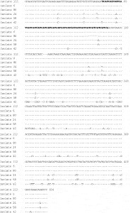
The GP60 gene sequences of the 18 isolates from 9 farms were classified into 6 types of sequences, and interestingly the sequences of the following isolates from different farms were identical to each other. Namely, the sequences of 6 isolates (nos. 6, 8, 69, 71, 89 and 90) from the farms A, F and H were identical to each other, and of 4 isolates (nos. 24, 28, 102 and 110) from the farms B and I were also identical to each other. On the other hand, the sequences of the isolates from the farm G (nos. 80 and 83) and J (nos. 112 and 113) were identical within each farm. As shown in Fig. 1, the GP60 gene sequences among the isolates examined differed from each other in the number of nucleotide repeat on the TCA or TCG repeat region (nucleotide position 49–111), and they showed the nucleotide polymorphisms mainly at the non-repeat regions. The sequences of the isolates (nos. 112 and 113) from the farm J had 20 copies of the TCA repeat, 1 copy of the TCG repeat and 1 copy of the sequence ACATCA immediately after the tri-nucleotide repeat. As compared with the sequence of the isolate 112, the sequences of the isolates from other farms were different as follows (Fig. 1): the 6 isolates (nos. 6, 8, 69, 71, 89 and 90) from the farms A, F and H had 16 copies of the TCA repeat; the 4 isolates (nos. 24, 28, 102 and 110) from the farms B and I had 16 copies of the TCA repeat with no copy of the TCG repeat, 2 copies of the sequence ACATCA and 16 nucleotide substitution (3 substitution were found before the TCA repeat region, 2 in the repeat region, and 11 in after the repeat region); the 2 isolates (nos. 58 and 61) from the farm E had 17 copies of the TCA repeat with no copy of the TCG repeat, 2 copies of the sequence ACATCA and the same nucleotide polymorphism with those of the isolates from the farms B and I; the 2 isolates (nos. 80 and 83) from the farm G had 18 copies of the TCA repeat; the last 2 isolates (nos. 42 and 43) from the farm D had 18 copies of the TCA repeat, 1 copy of the TCG but its position (nucleotide position 85–87) was different from that (nucleotide position 55–57) of the isolate 112, and showed high nucleotide polymorphism especially at the region after the TCA or TCG repeat region.

Fig. 1. Diversity in the GP60 gene sequences among the Cryptosporidium parvum isolates detected in the present study. The dots indicate the nucleotides that are identical to the isolate 112, and the dashes denote deletions. TCA or TCG repeat regions are boldfaced. The subtype names of the isolates examined are as follows: IIaA20G1R1 (isolate 112), IIaA16G1R1b (isolate 6), IIaA18G1R1 (isolate 80), IIjA16R2 (isolate 24), IIjA17R2 (isolate 58), IIdA18G1b (isolate 42).
The homology searching of the partial GP60 gene sequence obtained by using the FASTA program at the DDBJ (http://www.ddbj.nig.ac.jp/search/fasta-j.html) showed that the sequences of the isolates from the farms A, F, G, H and J had identities over 99% compared with those of the following C. parvum isolates belonging to the subtype family IIa (Peng et al. 2003b; Sulaiman et al. 2005): B-39 (IIaA16G1R1), B-254 (IIaA16G3R2), B-255 (IIaA16G2R1), B-194 (IIaA15G2R2), B-36 (IIaA16G3R1), H-A2C (IIaA16G2R2), 7499 (IIaA15G2R1) and 7503 (IIaA15G1R1). Similarly, the sequence of isolates from the farm D also had identities over 99% with those of the both isolates in subtype family IId (Sulaiman et al. 2005): 8025 (IIdA20G1) and 8897 (IIdA18G1). On the other hand, the highest sequence identities of the isolates from the farms B, I and E were 96·21% and 96·47% with those of the isolate B-255 (IIaA16G2R1) and B-194 (IIaA15G2R2) mentioned above, respectively.
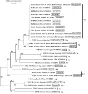
The phylogenetic analysis of the GP60 gene sequence also showed a close relatedness among the isolates examined in the present study and the ones identified previously as subtype allele family IIa or IId: the isolates from the farms A (no. 6), G (no. 80) and J (no. 112) were grouped into the subtype family IIa, and the isolate from the farm D (no. 42) into the subtype family IId (Fig. 2). However, the isolates from the farms B (no. 24) and E (no. 58) were in the same group, but were clearly separated from the subtype families IIa and IIg groups with high bootstrap values (Fig. 2). On the basis of the results obtained from the GP60 gene sequencing analyses, the 18 isolates were found to be subtypes belonging to the allele family IIa, IId or a new family, and in the present study we assigned the subtype names to the isolates examined following the nomenclature for the C. parvum and C. hominis subtypes reported previously (Sulaiman et al. 2005). Namely, the subtypes composed of 6 isolates (nos. 6, 8, 69, 71, 89 and 90) from the farms A, F and H, 2 isolates (nos. 112 and 113) from the farm J, 2 isolates (nos. 80 and 83) from the farm G and 2 isolates (nos. 42 and 43) from the farm D were designated IIaA16G1R1b, IIaA20G1R1, IIaA18G1R1 and IIdA18G1b, respectively (Fig. 2). In addition, we proposed a new subtype family IIj for the isolates from the farms B, I, E, and assigned the subtype names IIjA16R2 and IIjA17R2 to the isolates from the farms B (nos. 24 and 28), I (nos. 102 and 110) and E (nos. 58 and 61), respectively (Fig. 2).

Fig. 2. Phylogenetic relationships of the isolates from calves examined in the present study to multiple subtypes in Cryptosporidium parvum and C. hominis as inferred by neighbour-joining analysis based on the nucleotide sequences of GP60 gene. Original hosts, localities (also farms in the present isolates), and Accession numbers in GenBank are shown in parentheses after the names of the isolates. The names of subtypes in C. parvum isolates analysed are boxed. The bootstrap proportions (%) greater than 50% are shown at each branch except for the very shorts ones within each group.
DISCUSSION
In the present study, we revealed the high prevalence of C. parvum infection in dairy calves kept on farms around Belgrade, and also revealed by sequencing analysis of the GP60 gene that this species was genetically diverse in only a limited area.
In Serbia and Montenegro, by using light microscopy methods, the prevalence of Cryptosporidium infection was reported in 54% (68/126) of calves aged up to 3 months and 42·5% (34/80) of calves aged 3–12 months in the Belgrade area (Misic et al. 2001, 2002). Based on the size of oocysts, most isolates were suspected to be C. parvum. However, cattle also have Cryptosporidium bovis (previously known as Cryptosporidium bovine genotype B) or Cryptosporidium deer-like genotype, which is morphologically indistinguishable from C. parvum (Santin et al. 2004; Fayer et al. 2005), and Cryptosporidium parvum cannot be easily differentiated from both species and genotype by PCR-RFLP of SSUrRNA gene, but the COWP gene used in the present study cannot amplify in C. bovis and the deer-like genotype (Jiang et al. 2005; Trotz-Williams et al. 2006). Although we cannot totally exclude the possible presence of both species and genotype in calves studied, we certified, for the first time, the presence of C. parvum in calves kept on farms around Belgrade, because the COWP, as well as SSUrRNA genes were amplified in 62 samples and their RFLP profiles were quite correspondent with that of C. parvum.
The prevalence of C. parvum infection in the 0–20 day group was considerably higher (67%: 59/88) than that (20%: 3/15) in the 21–30 day group. This peak among the calves aged 0–20 days was also found in calves in other countries (Becher et al. 2004; Santin et al. 2004). It has been shown that calves aged over 7–8weeks harbour mostly C. bovis and C. andersoni and not C. parvum (Santin et al. 2004; Olson et al. 2004). Our previous studies have identified suspected C. andersoni isolates by microscopy in calves aged over 3 months (Misic et al. 2002). A continuous survey at the farms is required to clarify Cryptosporidium infection dynamics in calves around Belgrade.
The previous reports identified at least 9 subtype allele families (IIa, IIb, IIc, IId, IIe, IIf, IIg, IIh and IIi) among the C. parvum isolates from humans and cattle (Peng et al. 2001, 2003a,b; Alves et al. 2003, 2006; Sulaiman et al. 2005; Abe et al. 2006; Akiyoshi and Tzipori, 2006; Trotz-Williams et al. 2006). Among these subtype families, most isolates from cattle belong to the IIa, and thus far following 11 subtypes, IIaA13G2R1, IIaA15G1R1, IIaA15G2R1, IIaA15G2R2, IIaA16G1R1, IIaA16G2R1, IIaA16G2R2, IIaA16G3R1, IIaA16G3R2, IIaA17G2R1 and IIaA18G3R1, have been identified. In the present study, 3 subtypes in subtype family IIa, IIaA16G1R1, IIaA18G1R1 and IIaA20G1R1 were identified, and therefore 2 subtypes excluding IIaA16G1R1 were found to be new subtypes. The GP60 gene sequences analysed in the 6 isolates from farms A, F and H were subtyped as IIaA16G1R1, but its sequences showed a single nucleotide polymorphism compared with that of the isolate B-39 (IIaA16G1R1) from cattle in Michigan State (Peng et al. 2003b). We designated the B-39 and our isolates as IIaA16G1R1a and IIaA16G1R1b, respectively, following the nomenclature for the C. parvum and C. hominis subtypes reported previously (Sulaiman et al. 2005). The other subtype IIdA18G1 in the subtype family IId was also identified in the isolates examined in the present study. As shown in Fig. 2, this subtype had already been identified in the human isolate (8897) from Kuwait (Sulaiman et al. 2005), but the isolates from the farm D examined and the isolate 8897 differed from each other in single nucleotide polymorphism in the region after TCA or TCG repeat. Therefore, we designated the isolate 8897 and the isolates from the farm D as IIdA18G1a and IIdA18G1b, respectively, following the nomenclature mentioned above. The 2 subtypes belonging to the new subtype family IIj, IIjA16R2 found in the isolates from the farms B and I, IIjA17R2 in the isolates from the farm E, were unique subtypes because both subtypes had no TCG repeat the same as subtype IIgA9R1 in spite of the fact that all the subtypes in IIa identified previously have 1-3 TCG repeats.
Although only 2 isolates from each farm were selected randomly for subtyping analysis, the GP60 gene sequences of the isolates examined were identical to each other on every farm. This result suggests that a single subtype is circulating in calves on each farm. In contrast, the presence of plural subtypes on several farms was shown on the farms in USA (Michigan State) and Canada (Ontario State) (Peng et al. 2003b; Trotz-Williams et al. 2006), and therefore it is necessary to analyse more isolates on each positive farm in order to confirm whether only a single subtype circulates in calves on a farm or not.
To our knowledge, 145 cattle isolates from Portugal (72 isolates), USA (34), Canada (36) and Japan (3) have been subtyped, and the 141 isolates were classified into subtype family IIa and only 4 isolates from Portugal into IId (Peng et al. 2003b; Alves et al. 2003, 2006; Abe et al. 2006; Trotz-Williams et al. 2006). The 12 isolates from the 9 farms examined in the present study were also classified into only IIa and IId. Although molecular typing data at subtype level for the C. parvum isolates from animals is limited, only IIa and IId among 9 subtype families in C. parvum appeared to be zoonotic and the other subtype families (IIb, IIc, IIe, IIf, IIg, IIh and IIi) might be anthroponotic. A recent molecular study shows that a raccoon dog isolate (CRD-154) identified as C. parvum was speculated to be a novel subtype family quite different from other subtype families already known (Abe et al. 2006), and in the present study new subtype family IIj was also found in the isolates from calves. These new findings also support the speculation that C. parvum might be composed of zoonotic and anthroponotic multiple subtypes. Further molecular epidemiological studies, including the typing at the subtype level, are required to clarify the zoonotic potential and host specificity of the subtypes in C. parvum.
The raccoon dog isolate mentioned above had the unique GP60 gene sequence with no TCG repeat (A14R1), and its highest sequence identity was 77·81% with that of the isolate G1946 (IIbA14) in subtype family IIb, and as shown in Fig. 2 this isolate was clearly separated from the subtype family IIb. Therefore, we designated this isolate as subtype IIkA14R1 in additional new subtype family IIk following the nomenclature (Sulaiman et al. 2005).
Molecular methodologies have been used to identify the infection sources of the outbreaks or to track the spread of infections. Despite the extensive earlier studies, it is still unclear whether the pathogenesis or infectivity of the Cryptosporidium organism is related to some specific subtypes or not. In the present study, the calves that harboured the following 6 subtypes, IIaA16G1R1b, IIaA18G1R1, IIaA20G1R1, IIjA16R2, IIjA17R2 and IIdA18G1b, showed diarrhoea, and therefore these subtypes appear to be pathogenic to calves. In Serbia and Montenegro, human cryptosporidiosis occurs in AIDS patients (Misic et al. unpublished observations). It is necessary to analyse the human isolates molecularly for probing the possibilities whether the several subtypes found in calves are zoonotic and pathogenic to humans or not.
This study was financially supported by the Ministry of Science and Environment Protection of Serbia (Project No. 143022: Ecophysiological and genetic investigations of domestic animals and bees for the purpose of increasing reproductive traits and disease resistance).