Introduction
The presence of a nonprogressive, conductive hearing loss in the 40–60 dB range, with a normal tympanic membrane and no history of trauma or infection, is highly suggestive of congenital ossicular malformation. A stapes anomaly is the most common such malformation; this condition is clinically important because hearing can be restored with appropriate surgery.
The exact origin of the ossicles remains controversial, although the main source is known to be the neural crest mesenchyme of the first and second branchial arches. In addition, the otic capsule has a role in forming the stapes footplate, and the origin of the malleus handle and incus long process is known to be either the first or second branchial arch.Reference Bailey1
Many different congenital ossicular anomalies exist. However, there have been no previous reports of stapedial suprastructure fixation with normal footplate mobility, incus agenesis, deformity of the malleus handle and facial nerve exposure. We report such a case, which was diagnosed after clinical examination, radiological evaluation and surgical exploration.
Case report
A 50-year-old woman had suffered hearing loss in both ears since early childhood. She had no history of otalgia, otorrhoea, vertigo, ear surgery or head trauma. She was born at term via a normal delivery, with no perinatal problems.
Physical examination revealed that both tympanic membranes were intact and mobile, with a minimal anomalous shadow of the malleus handle bilaterally (Figure 1b).
Operating microscope photographs of middle-ear structures, with explanatory diagrams. Note the bony bridge between the fallopian canal and stapedial suprastructure. The incus is missing, while the malleus handle is minimally deformed. The head of the stapes was separated using a right-angled pick, and covered with a partial ossicular replacement prosthesis (a; right ear; black arrow = tympanic portion of the facial canal; white arrow = suprastructure fixed to the facial canal). The shape of the manubrium was atypical, being shorter and more anterolateral than normal (b). The tympanic portion of the facial nerve was exposed completely. The incus was missing and the malleus handle was minimally deformed. A bony bridge connected the stapes suprastructure with the medial bony portion of the epitympanum (c; left ear; black arrow = exposed facial nerve tympanic portion; white arrow = bony bridge between the stapes suprastructure and epitympanic space medial wall). While attempting to separate the bony bridge, the stapes footplate was disconnected from the oval window (d; white arrow).

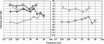
Audiometric analysis demonstrated moderate conductive hearing loss bilaterally (Figure 2). Temporal bone computed tomography (CT) detected the malleus and stapes but not the incus (Figure 3).
Composite audiogram showing pre-operative bilateral conductive hearing loss (dotted line). One year after the final operation, an improved right ear air conduction threshold was seen (solid line). Hearing on the left remained unchanged due to subluxation of the footplate.  = right side bone conduction level;
 = left side bone conduction level;
 = right side air conduction level;
 = left side air conduction level

Pre-operative computed tomography findings. (a) & (b) In the axial plane, the ‘ice cream cone’ configuration of the malleus head is well seen, but the incus (the cone) is not observed. (c) & (d) In the coronal plane, the incus is not seen and only part of the stapes is seen. R = right

An exploratory tympanotomy was conducted on the right ear under local anaesthesia. This revealed that the long process of the incus was absent, and that the stapes suprastructure was connected to the tympanic portion of the bony fallopian canal via a bony bridge (Figure 1a). When this bony bridge was disconnected using a right-angled pick, the stapes moved normally. A partial ossicular chain replacement prosthesis (PORP) was placed under the drum and on top of the suprastructure of the stapes. This prosthesis was extruded after one month, so the tympanic membrane was grafted with perichondrium with a thin layer of attached cartilage, and a new PORP was repositioned.
Three months after this latter procedure, the patient had made a good recovery, with an improvement in her conduction threshold from 60 to 25 dB.
Exploratory tympanotomy on the left ear revealed the absence of the long process of the incus, and exposure of the entire tympanic portion of the facial nerve. The stapes suprastructure was connected to the medial wall of the epitympanum via a bony bridge (Figure 1c). The footplate was subluxed when the bony bridge was curetted with a right-angled pick (Figure 1d). Soft tissue harvested from the retroauricular area was placed snugly around the subluxed footplate, and Cutanplast® (Mascia Brunelli Spa. Milano Italy) was placed over this soft tissue.
Although the patient's left ear hearing did not change after this last operation, she was pleased with the functional improvement in her right-sided hearing (Figure 2).
Discussion
Various congenital ossicular anomalies exist, which can be broadly divided into either major or minor.Reference Nomura, Nagaro and Fukaja2–Reference Teunissen and Cremers4 Minor congenital anomalies are restricted to the middle ear, while major ones comprise malformations of the middle ear, the external meatus and sometimes the auricle.
In 1956, Henner and Buckingham proposed a classification of congenital ossicular anomalies.Reference Henner and Buckingham5 Teunissen and Cremers later proposed classification of congenital middle-ear anomalies into four types based on operative findings.Reference Teunissen and Cremers4 Conductive hearing loss due to an ossicular abnormality may be caused by ossicular fixation or disconnection. Ossicular fixations can be divided into malleo-incudal fixation and stapes fixation. Stapedial fixation is the most common congenital anomaly.Reference Park and Moon6
In our patient, we predicted the condition of the ossicles based on the air–bone gap and the presence of Carhart's notch at audiometry. Unusually, this patient's large air–bone gap was caused by the incus defect, while a mild Carhart's notch was detected at a frequency of 2000 Hz. Therefore, we were able pre-operatively to predict incus agenesis and stapedial fixation. At surgery, the ossicles were as predicted, so we tried removing the stapes suprastructure to insert a piston. However, the footplate movement was normal after disconnecting the bony bridge.
Although several studies have described stapes suprastructure fixation, the particular form observed in our patient has not previously been reported.Reference Kinsellar and Kerr7–Reference Gerhardt and Otto11
The exact origin of the ossicles is unclear, although the main source is acknowledged to be the neural crest mesenchyme of the first and second branchial arches. The otic capsule also has a role in forming the vestibular surface of the footplate and annular ligament. Consequently, numerous different types of congenital ossicular anomaly exist.Reference Bailey1
The most usual type of stapes anomaly is footplate fixation with a normal suprastructure, malleus and incus. This indicates that the origin of the stapes footplate differs from that of the other parts of the ossicles. In our case, the reverse ossicle anomaly occurred. The vestibular surface of the footplate and annular ligament were normal, but the malleus, incus and stapes suprastructure had uncharacteristic shapes. This finding may help determine the origin of the ossicles.
The origin of the malleus handle and incus long process is either the first or second branchial arch.Reference Bailey1 Our patient had an unusually shaped malleus handle and a missing incus, along with an abnormally shaped and fixed suprastructure, but had normal footplate mobility and a normal malleus head. These findings support the hypothesis that the malleus handle and incus long process originate from the second branchial arch. In addition, the entire tympanic portion of the left facial nerve was exposed. The facial nerve derives from the second branchial arch. Therefore, in our patient, the second branchial arch was developmentally arrested.
• Diagnosis of an ossicular malformation without associated external ear malformations requires high resolution temporal computed tomography (CT) and exploratory tympanotomy
• The stapes footplate can have normal mobility, even with a severe coexisting abnormality, because of its separate embryological development from the otic capsule
• Therefore, when performing an exploration tympanotomy with pre-operative CT findings, one must recognise unusual anomalous conditions
Conclusion
The diagnosis of an ossicular malformation without associated external ear malformations requires high resolution temporal CT and exploratory tympanotomy.Reference Ching and Bottrill12 The footplate can have normal mobility, even with a severe coexisting abnormality, because of its separate development from the otic capsule.Reference Cressman and Pensak13 Therefore, when performing an exploration tympanotomy with pre-operative CT findings, one must recognise unusual anomalous conditions.