Case report
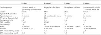
In all, four patients – cardiac diagnoses: hypoplastic left heart syndrome in two, tricuspid atresia in one, and complex cyanotic heart disease with a functionally univentricular heart in one – with plastic bronchitis at a median age of 31 months (range 21.5–45 months) with a median weight of 12.7 kg (range 9.5–14.8 kg) presented at our clinic (Table 1).
Haemodynamic parameters of all patients, materials used for stent implantation, diameters of the LPA measured during catheterisation before and after stent implantation and the mean follow-up time of each patient.

AV=atrioventricular; BiB=balloon-in-balloon (pfm medical ag; Powerflex™ balloon Cordis International, Waterloo, Belgium); CP=Cheatham-platinum (CP StentTM NuMED Inc., Best, The Netherlands; IntraStent™ Max™ LD Stent ev3, Paris, France; VACS II Paediatric Balloon Catheter OSYPKA AG, Rheinfelden-Herten, Germany); CVP=central venous pressure; dTp=transpulmonary pressure gradient; LPA=left pulmonary artery; MGA=malposition of the great arteries; PS=pulmonary stenosis; Qs=flow in systemic circuit; TCPC=total cavopulmonary connection
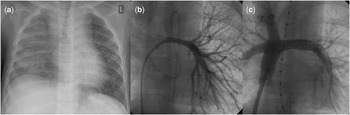
A complete separation of the pulmonary and systemic circulation by an extracardiac non-fenestrated total cavopulmonary connection had been performed at median 20.5 months (18–21 months) preceding the hospital admission. On admission, three patients had no signs of a pulmonary infection. There was one patient who was treated with antibiotics. All patients underwent a complete cardiological examination – clinical, ECG, Holter, echocardiogram, chest x-ray – in combination with cardiac catheterisation. The x-rays show in all patients a right-sided atelectasis as shown in Figure 1. All haemodynamic data are depicted in Table 1.
( a ) X-ray and LPA (left pulmonary artery) angiogram of Patient no. 3 before ( b ) and after ( c ) stent implantation. Notice the almost doubled diameter. This x-ray is made before catheter intervention. Notice also that atelectasis is located on the right side.

The median central venous pressure was 14–15 mmHg (range 12–17 mmHg). All four patients had a significant stenosis of the left pulmonary artery, which was treated by stent implantation. The diameter of the left pulmonary artery was augmented from a median value of 4.8 mm (range 3.9–6 mm) to a median value of 8.7 mm (range 7.8–10.4 mm; Table 1).
All patients were treated with type 5 phosphodiesterase inhibitors (Sildenafil) before catheter intervention. In addition, a therapy with the endothelin receptor antagonist Bosentan was initiated. Inhalative treatment consisted of tissue plasminogen activator, dornase alfa, Salbutamol, and NaCl 5.85%. In three cases, tissue plasminogen activator was applied until symptoms improved. There was one child with recurrent expectorations of casts who received it over a 5-month period without improvement. All children had a history of chylothorax during their peri-operative periods and were thus placed on a diet including medium chain triglycerides. Of these children, three are free of symptoms with a median follow-up time of 13.5 months (range 8–25 months). Patient no. 3 is still expectorating casts.
Discussion
Plastic bronchitis occurs in several non-cardiac disorders, but manifestation after Fontan staged palliation is associated with a poorer outcome.Reference Fredenburg, Johnson and Cohen 1 , Reference Madsen, Shah and Rubin 2 This series of cases shows significant improvement of plastic bronchitis in three of four patients after successful stenting of the left pulmonary artery in combination with pulmonary vasodilator and inhalative therapy.
Considering the increasing number of patients receiving total cavopumulmonary connection, the incidence of plastic bronchitis is also expected to increase. However, the reported patient series are small, and often only case reports are available. The pathophysiology is not completely understood. Some suggest that plastic bronchitis is linked to poor haemodynamics. A multifactorial aetiology is discussed and most probable.Reference Madsen, Shah and Rubin 2 , Reference Seear, Hui, Magee, Bohn and Cutz 3 , Reference LaRue, Gossett, Stewart, Backer, Mavroudis and Jacobs 4 It is assumed that high central venous pressures lead to an abnormal response of the bronchial epithelium resulting in excess mucus production.Reference Seear, Hui, Magee, Bohn and Cutz 3 The fact that three of our four patients showed sustained improvement of symptoms after stenting of the left pulmonary artery led to the assumption that improving haemodynamics plays an important role in the treatment of plastic bronchitis. Even though the patients presented with left pulmonary artery stenosis, radiological findings indicated impaired ventilation of the right lung. Central venous pressures were not in a range to explain disruption of the alveolar-epithelial barrier. A possible explanation would be that cast formation was directly caused by the stenosis as a result of elevated pulmonary blood flow and shear stress of the counter side. To prove this hypothesis, further investigation and repeated cardiac catheterisation after stenting of the left pulmonary artery would have been helpful, which was not done so far in our patients. Another possible explanation would be that the stenosis impaired systemic venous drainage and thus increased flow in the thoracic duct. The combination of impaired haemodynamics, caused by the stenosis, and elevated central venous pressure may have induced chylo-alveolar fistulas, as discribed by Parikh et al.Reference Parikh, Witte and Samson 5 Owing to the fact that all patients were on a specific pulmonary vasodilator therapy in conjunction with the catheter intervention, it is not clear which therapy caused the improvement of symptoms.
Symptomatic therapy of plastic bronchitis consists of mucolytic therapy either by adding water to the airway (NaCl 5.85%), depolymerising the mucin network (acethylcysteine),Reference Brogan, Finn and Pyskaty 6 or by solving abnormal polymers in the mucus including deoxyribonucleic acid (dornase alfa).Reference Manna, Shaw, Tibby and Durward 7 In some cases, fibrinolytic agents like inhaled urokinase or tissue plasminogen activator had been used with unknown clinical efficacy. Heath et al reported on inhaled tissue plasminogen activator that reduced the cast weight but not in a dose-dependent manner. The authors concluded that a fibrinolytic treatment is more effective than a mucolytic one.Reference Heath, Ling and Racz 8 However, optimal treatment for this condition remains unclear.
Reducing the aetiology of this complex complication only to mechanics seems simplistic.Reference Madsen, Shah and Rubin 2 The fact that not all of the patients were free of symptoms after intervention indicates that impaired haemodynamics plays an important role, but the aetiology of plastic bronchitis is most likely multifactorial. However, any effort should be undertaken to optimise haemodynamics in these patients to improve their prognosis. Alternatively, fenestration of the Fontan tunnel, Fontan take-down, and cardiac transplantation has been suggested in the literature.Reference Fredenburg, Johnson and Cohen 1
In conclusion, we report on four patients with Fontan physiology with severe plastic bronchitis and stenosis of the left pulmonary artery. In three of them, symptoms improved substantially after successful stenting.
Acknowledgement
None.