Introduction
Outside of a specialist otolaryngology clinic, examination of the ear is generally limited to otoscopy with a conventional monocular otoscope. As the speculum channel is used for the light source attachment, there is no accessibility for instrumentation of the ear. As such, it is difficult to perform any therapeutic intervention safely, and therefore the otoscope is restricted to a mainly diagnostic role. Furthermore, if the external auditory canal is impacted with cerumen, debris or secretions, then further examination is limited.
Current practice for further examination or intervention requires the use of a clinical microscope. The microscope facilitates the employment of therapeutic interventions,Reference Hickey, Buckley and O'Connor1 but its use is associated with a significant learning curve. When performed by inexperienced practitioners, it can be uncomfortable for patients and pose a risk of complications. Additionally, clinical microscopes are an expensive investment which require an adequately sized room for their use and ongoing maintenance as well as not being easily portable.
Otoendoscopes provide an alternative approach to ear examination and procedures in clinic, but this again is associated with a learning curve even for experienced ENT surgeons and requires costly equipment and maintenance.Reference Pothier, Hall and Gillett2 A lower cost portable alternative for otological assessment and treatment is the use of surgical loupes with an ear speculum. However, it takes time for clinicians to adjust to using surgical loupes, and the focal distance and field of view can be challenging in the restricted working area of the ear canal. Loupes have been used in otological practice,Reference McClelland, Marzouk, Coyle and Tatla3 but have not been widely described or adopted into routine use.
Smartphone technology has been embraced in this digital health era, and numerous medical devices utilise this equipment, including in otolaryngology.Reference Wong and Fung4 This technology can be used to facilitate telemedicine systems which are especially vital in the current coronavirus disease 2019 (Covid-19) pandemic.Reference Wosik, Fudim, Cameron, Gellad, Cho and Phinney5 There are several smartphone-enabled video otoscopes now available to both clinicians and the general public.
The majority of the current devices allow users to examine the external auditory meatus and tympanic membrane but do not permit procedures to be performed. The Tympa Health device (London, UK) uses a smartphone to visualise the tympanic membrane through a conventional speculum with the separation of the light delivery channel to allow instruments to be passed through the speculum. Thus, it is possible to perform tasks like microsuction and foreign body removal under direct vision. However, the learning curve and usability of such devices is not yet known, especially in users who may predominantly work in non-hospital settings.
Therefore, the aim of our study was to compare and assess self-reported confidence of novices in using smartphone-enabled video otoscopes, a microscope and surgical loupes for skill acquisition in ear examination and basic otological procedures.
Materials and methods
This study involved quality improvement of standard medical care using CE-marked devices. All data obtained was anonymised and unidentifiable. It was granted exemption from ethical board review by the St George's University of London research ethics office.
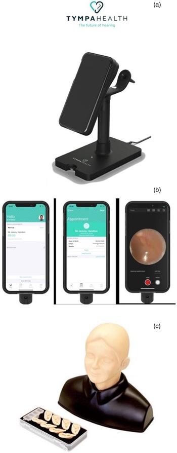
For examination and procedures with a smartphone-enabled otoscope, we used two smartphone-enabled video otoscopes (Tympa Health). This device includes a casing that houses a smartphone, an arm that attaches to an ear speculum and a handle (Figure 1a). The camera and torch from the smartphone align to allow visualisation through the speculum. A user interface application on the smartphone allows optimisation of digital images obtained and storage if required (Figure 1b). The gap between the smartphone and speculum provides space to allow the user to introduce instruments through the speculum. For examination and procedures via otomicroscopy, two portable clinical operating microscopes (Labomed Prima, Fremont, USA) with adjustable interpupillary distances were used. Surgitel EVC250M Oakley flip down surgical loupes (Oakley, Ann Arbor, USA) with ×2.5 magnification with adjustable interpupillary distances were used in combination with a headlight for surgical loupes guided otoscopy.
(a) Tympa Health smartphone-enabled otoscope on a stand. (b) Application on iPhone that enables optimisation and storage of images. Names shown are fictitious, and any resemblance to actual persons, living or dead, is purely coincidental. (c) Kyoto Kagaku Ear Examination Simulator.

Twenty-nine final-year medical students participated in the study. Prior to teaching, all students undertook a pre-study questionnaire about their knowledge, confidence and experience of techniques for otoscopy and aural microsuction as well as questions regarding smartphone use. Questions regarding confidence were scored using a Likert-like scale between 1 to 5, where 1 is very unconfident and 5 is very confident (see Figure 1 in the supplementary material, available on The Journal of Laryngology & Otology website).
Participants underwent standardised lecture-based teaching on ear anatomy and ear examination delivered by a senior registrar. Participants then underwent small group hands-on teaching delivered by senior ENT registrars on otological examination and procedural techniques using a clinical microscope, surgical loupes and a smartphone-enabled video otoscope. Teaching for each modality lasted for 30 minutes.
During the small group teaching with each modality, participants were asked to complete ear examination on volunteer patients with no pathology and on a Kyoto Kagaku Ear Examination Simulator model (Kyoto Kagaku, Kyoto, Japan; Figure 1c). The Kyoto Kagaku Ear Examination Simulator model has interchangeable attachments of different silicone model ears (pinna, ear canal, tympanic membrane), each with a different pathology. Participants were asked if they were able to identify simple pathology such as tympanic membrane perforation and differentiate between a normal and abnormal tympanic membrane. Furthermore, participants performed tasks such as foreign body removal and suction clearance of wax from the model ear using a Zoellner suction tube and instruments such as Jobson Horne probe and crocodile forceps. Task completion was observed by the teaching faculty.
Participants were asked to rate their confidence using each modality using a Likert-like questionnaire and to state their preference of modality for performing examination, comfort when using, performing microsuction and for education. Questions regarding confidence were scored using a Likert-like scale between 1 to 5, where 1 was very unconfident, 2 was unconfident, 3 was neutral, 4 was confident and 5 was very confident (see Figure 2 in the supplementary material, available on The Journal of Laryngology & Otology website).
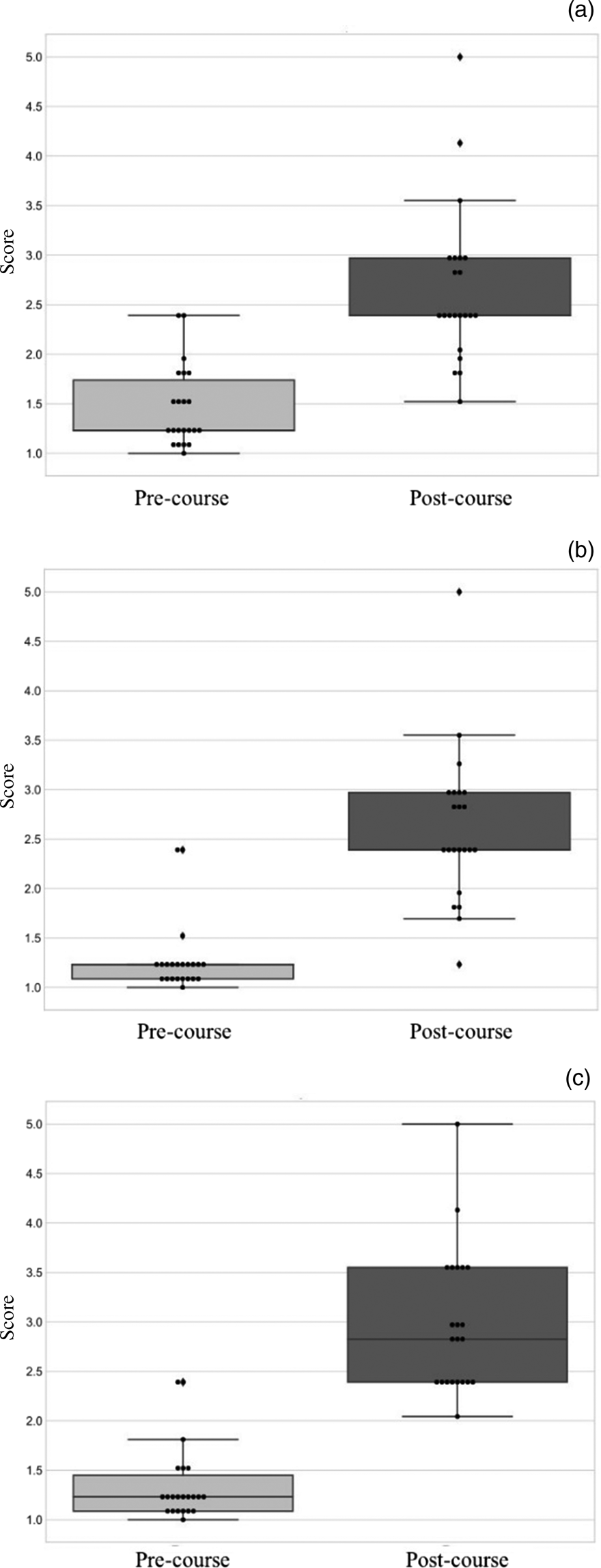
Confidence level pre- and post-teaching (course) for each modality for ear examination and procedures using (a) microscope, (b) loupes and (c) smartphone.

Results
Participants had limited experience in ear examination. The pre-teaching questionnaire identified the participants’ knowledge regarding ear examination and ear procedures. In regard to ear examination, 29 participants stated it could be performed with an otoscope, 10 participants stated it could be performed with a microscope and one participant stated it could be performed with a smartphone-enabled video otoscope. Twenty-seven participants had used an otoscope, one had used a microscope, none had used surgical loupes and one had used a smartphone for a medical examination previously. Participants used their smartphones between 0.25 and 15 hours a day (mean: 5.05 hours). A summary of the experience reported by the cohort can be seen in Table 1.
Methods for wax removal and general ear examination known by the cohort prior to the course

Participants were able to report (mention) more than one method. They also reported their experience with each method.
Participant's confidence in ear examination increased with training and was highest for smartphone-enabled otoscopy. Mean confidence scores for ear examination pre-teaching with use of a microscope, surgical loupes and a smartphone-enabled video otoscope were 1.52, 1.13 and 1.24, respectively (1 = least, 5 = most). After teaching, all modalities demonstrated a significant increase in confidence in ear examination (Wilcoxon two-tailed score, p < 0.0001) and mean scores with use of a microscope, surgical loupes and a smartphone-enabled video otoscope were 3.66, 3.69 and 4.26, respectively. Confidence in using the smartphone-enabled video otoscope post-teaching was highest (one-way analysis of variance, p = 0.015; Table 2 and Figure 2).
Confidence scores pre- and post-teaching, using the three modalities for ear examination and procedures

For scores, 1 = least and 5 = most
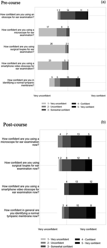
Table 3 shows how the confidence reported by the participants shifted after having gone through the training. Prior to the course, the overall cohort reported being less confident with all methods explored (microscope, surgical loupes and smartphone-enabled video otoscopes). After having received the instruction, the participants reported an increase in their confidence. Figure 3 shows this information in context to the questions asked of the participants.
Confidence reported pre- and post-teaching (course) in a scale from very unconfident (1) to very confident (5).

Confidence level reported pre- and post-teaching (course) using a scale

For scores 1 = very unconfident and 5 = very confident
Participants were able to differentiate a normal versus abnormal tympanic membrane post-training and preferred smartphone-enabled otoscopy. One of the main areas of interest and importance in ear examination in non-hospital care settings is the detection of normal versus abnormal tympanic membranes. The first step in this is obtaining a clear and confident view of the tympanic membrane and recognising anatomy. Learners were asked which technique they preferred. For teaching anatomy, over 60 per cent of the participants preferred smartphone-enabled video otoscopes, 27.58 per cent preferred the microscope and 10.34 per cent preferred the loupes. For the recognition of tympanic membrane abnormality, 79.31 per cent of participants reported preferring the smartphone-enabled video otoscope for the successful differentiation of normal tympanic membranes, followed by the microscope in second place (10.34 per cent) and finally the loupes (10.34 per cent; Table 4)
Preferred modality for ear examination and procedures

Participants preferred smartphone otoscopy for learning and performing microsuction. The ability to perform basic therapeutic interventions was assessed on all three devices. The faculty observed participants performing microsuction on models with simulated wax. Smartphone-enabled video otoscope was the preferred method for learning (65.51 per cent) and performing (51.72 per cent) microsuction (Table 4).
Being able to carry out medical procedures in a comfortable manner is important not only for the patient, but also for the medical practitioner. We asked our participants to rate what method they found most comfortable to use for diagnosis and interventions. The smartphone-enabled video otoscope was the most comfortable method with 68.98 per cent of the participants preferring it. This was followed by 24.13 per cent preferring the microscope and 6.89 per cent preferring loupes (Table 4).
Discussion
Microscope otoscopy has long been considered the ‘gold standard’ for otological examination. The benefits of higher magnification, superior depth perception with a binocular view and the ability to perform procedures with instruments through a speculum with one or even two hands have ensured that it is the workhorse examination technique of the otolaryngologist in clinic and in the operating room. Although the three-dimensional view and set up is superior, it is only used in limited settings. The use of a microscope in clinic for ear examination is usually only feasible in a hospital specialist setting owing to the cost, maintenance and logistical requirements necessary. Therefore, non-specialists commonly do not have access, training or familiarity with micro-otoscopy, and their practice is limited to use of the otoscope in a diagnostic capacity alone. Here we show that novice practitioners can learn basic diagnostic and therapeutic skills in ear examination using a variety of tools and are most comfortable with smartphone-enabled video otoscopes.
The introduction of smartphone-enabled video otoscopes has improved the quality of diagnostic views that are possible from a mobile otoscopy device. Cellscope® is one such device that has been used in the USA. This device includes a case which fits onto an iPhone® with an attachable otoscope head piece connecting an ear speculum which corresponds on the case to the rear-facing camera of the iPhone. In a single centre prospective study by Moshtaghi et al., this device was compared with microscope otoscopy in an otology clinic.Reference Moshtaghi, Sahyouni, Haidar, Huang, Moshtaghi and Ghavami6 A single smartphone-enabled video otoscope acquired image with a short patient history resulted in the correct diagnosis in 96 per cent of normal tympanic membranes and 100 per cent of microscope-confirmed abnormal tympanic membranes. This has been supported in our study with the use of smartphone-enabled video otoscopes being the preferred method for identifying normal and abnormal tympanic membranes by participants, even over the microscope. Chan et al. demonstrated that smartphone-enabled video otoscopes were preferred by a group of paediatricians over conventional otoscopes for diagnosing acute otitis media.Reference Chan, Silverstein, Bryan, McCracken, Little and Shane7 This shows that the smartphone-enabled video otoscope, in terms of diagnostic reliability, has a level of parity with microscope otoscopy and superiority over monocular otoscopy.
Smartphone otoscopes have also been utilised to improve access to otology assessment and treatment in settings that would usually have limited provision of ENT services. Cupris® TYM otoscope is a smartphone-enabled video otoscope that has been used in rural locations in low- and middle-income countries.Reference Mandavia, Lapa, Smith and Bhutta8 The device has been used to capture images and videos from ear examinations that were then reviewed by another ENT surgeon in a remote location. In these settings, the device was shown to be a valid tool for the diagnosis of ear disease. However, ear examination was limited using the smartphone-enabled video otoscopes in paediatric populations because of wax impaction, as was seen in over a third of otoscopies in a study conducted in Greenland.Reference Demant, Jensen, Bhutta, Laier, Lous and Homøe9
There has been a rapid increase in the number of publications on the use of the endoscope in ear surgery,Reference Kapadiya and Tarabichi10 and the endoscope has been utilised for ear procedures in the out-patient setting.Reference Pothier, Hall and Gillett2 Smartphone adaptors have been developed over the last decade to enable the attachment of an endoscope and to utilise the smartphone camera to take endoscopic images.Reference Stew, Dharmawardana, Kao, Elias and Ooi11 In the UK, endoscope-I® has been used to provide high-quality photo documentation using a smartphone camera and visual access of the tympanic membrane facilitated by an otoendoscope.Reference Mistry, Coulson and George12
Smartphone enabled video otoscopes and smartphone attachments that facilitate otoendoscope attachments currently offer diagnostic images that supersede conventional otoscopy making them a viable alternative in terms of mobile otoscopy. However, most current devices do not enable the user to perform procedures as there is little or no space to pass instruments through a speculum or around an endoscope. Therefore, if wax impaction obscures the view of the tympanic membrane, then dewaxing will be required using conventional methods such as microsuction using a microscope. The Tympa Health smartphone enabled video otoscope facilitates the user to perform therapeutic procedures owing to the distance between the speculum and the smartphone camera that allows handheld instruments to pass through the speculum under direct vision.
Our study demonstrated that participants were confident using a smartphone enabled video otoscope for ear examination and procedures after receiving brief training and that they preferred these devices over conventional methods for performing microsuction. As smartphones are used routinely in everyday life, our participant group used them on average for 5 hours a day (although 1 participant reporting use of 15 hours daily may have been a misinterpretation of the question), devices that utilise smartphones likely feel familiar and are easily adoptable especially in surgical specialties.Reference Mobasheri, Johnston, Syed, King and Darzi13
Exposure to ENT in the UK undergraduate curriculum is variable with some medical schools not providing a dedicated ENT rotation and the majority of final year medical students not feeling prepared to practise ENT.Reference Ferguson, Bacila and Swamy14,Reference Powell, Cooles, Carrie and Paleri15 This has been a longstanding problem resulting in UK medical students lacking confidence in performing otoscopy.Reference Fisher and Pfleiderer16 This issue is not limited to the UK with recent evidence being published that US medical students were not satisfied with their exposure to otoscopy training.Reference Niermeyer, Philips, Essig and Moberly17
Smartphone-enabled video otoscopes provide a valuable teaching tool for ENT education especially for novices and non-specialists.Reference Hakimi, Lalehzarian, Lalehzarian, Azhdam, Nedjat-Haiem and Boodaie18,Reference Schuster-Bruce, Davies, Conchie, Shamil and Waddell19 The preference of novices in our study for using the smartphone-enabled video otoscopes for learning ear anatomy and pathology supports this. One of the difficulties in teaching ear anatomy through conventional otoscopy and microscopy is the difficulty in sharing the view of the tympanic membrane with the trainer or learners without using a teaching arm or a camera connection on the otoscope head. Smartphone-enabled video otoscopes can overcome this obstacle by providing a magnified high-resolution image on a screen that can be viewed by both the trainer and trainee. Instructors are therefore able to demonstrate anatomy and identify landmarks in real time. Furthermore, trainers can provide feedback on otoscopy technique and on the performance of otological procedures that can be undertaken through the speculum. Trainers can directly oversee and guide a trainee through a procedure such as microsuction or foreign body removal. Furthermore, it provides the potential for more complex procedures to be performed under supervision that can also be recorded and played back to further facilitate learning.
• Aural microsuction is usually undertaken in a secondary care setting through micro-otoscopy
• Smartphone-enabled video otoscopes have been used by clinicians for ear examination but not external ear procedures
• Our study shows novices were most confident in using smartphone-enabled video otoscopes over loupes and microscopes
• Smartphone-enabled video otoscopes are easily used by novices and could be used by non-specialists after minimal training
• This technology would enable procedures previously restricted to delivery in hospital ENT departments to be used in other clinical settings
The Covid-19 pandemic has changed medical practice for the foreseeable future.Reference Fontanarosa and Bauchner20 The role of telemedicine has expanded rapidly as access to hospital care has become limited in order to protect patients and clinicians.Reference Hollander and Carr21 The feasibility of a telemedicine otology referral service has been explored in the UK, and a study by Cottrell et al. found that use of the endoscope-I system by general practitioners (family medicine) to refer patients to ENT specialists was well received and felt by some users to improve communication between non-specialist clinicians and ENT specialists.Reference Cottrell, George, Coulson and Chambers22 In the Covid-19 era, smartphone-enabled video otoscopes have been used by patients to provide otolaryngologists with otoscopic images which have been used to diagnose and guide treatment.Reference Meng, Dai, Hang and Wang23 Use of devices such as the smartphone-enabled video otoscopes by general practitioners would enable effective triaging and management of patients with otological symptoms with real time feedback from ENT specialists. Implementation of these patient pathways would prevent unnecessary visits to hospitals for specialist referrals, improving patient satisfaction and importantly, in the Covid-19 era, patient safety.
Limitations
Our study was focused on senior medical students, who had limited prior ENT experience. The procedures were performed mainly on simulated ear examination models. In order to investigate the adoptability of smartphone-enabled video otoscopes further, studies including both participants who have less training in clinical medicine and clinicians with more experience such as ENT trainees and consultants are required.
Formal assessment of diagnostic and procedural skill acquisition was limited to observation by faculty to minimise pressure placed on the students. However subsequent work and assessment will be needed to evaluate task-based performance of these skills. Furthermore, extending the study to include procedures (i.e. microsuction and manual removal of wax) performed using smartphone-enabled video otoscopes in patients and qualitative assessment of patient experience would help determine the validity of this technology in clinical use. Furthermore, statistical analysis of data was limited because of the relatively small sample size.
Conclusion
Smartphone-enabled video otoscopes provide a combination of high-quality diagnostic application, ease of use by novices for ear procedures and a shared digital view enabling direct supervision and recording of procedures. Our findings show that this technology can be used by novices in ENT, and therefore could be used by non-specialists after minimal training. This would enable procedures that were previously restricted to delivery in hospital ENT departments to be provided in other clinical settings such as family medicine practices, small emergency medicine departments and community pharmacy centres. With the current rate of ENT referrals rising in the UK,24 this might help ease the burden on hospital ENT out-patients by providing a means for routine ear procedures to be performed by qualified staff in alternative locations outside the hospital. The necessity for these new models of care that embrace technological advances and deliver care outside of the hospital is especially crucial in the current Covid-19 pandemic.
Supplementary material
The supplementary material for this article can be found at https://doi.org/10.1017/S0022215121004102.
Competing interests
Annakan Navaratnam, Anushri Khetani, Deepak Chandrasekharan, Rakesh Mistry and Joseph Manjaly have no conflict of interest. Jesus Rogel-Salazar is an employee of Tympa Health Technologies Ltd. Taran Tatla, Arvind Singh and Krishan Ramdoo are patent holders and shareholders in Tympa Health Technologies Ltd.