Implications
The natural form of vitamin E is preferentially accumulated by sows and piglets. In the present study, we show that micellized d-α-tocopherol added to sows in water at threefold lower doses than the synthetic form (dl-α-tocopherol) in feed allows to reach similar α-tocopherol concentration and a predominant –R stereoisomer distribution in piglets. The present paper contributes to better understanding the oral natural vitamin E needs of the sow and piglets during gestation and lactation period.
Introduction
Vitamin E is an important nutrient for health and survival of newborn pigs. It constitutes an efficient agent in tissues to control oxidative stress by capturing free radicals and other reactive substances (Halliwell, Reference Halliwell1994) and it may increase the immune response (Babinszky et al., Reference Babinszky, Langhout, Verstegen, den Hartog, Joling and Nieuwland1991) by its effects on cell protection. Maternal feeding constitutes the main transmission vehicle of this nutrient that provides the newborn piglet with the first defences against oxidative damage (Debier, Reference Debier2007). However, serum vitamin E suffers an important decline after weaning (Chung et al., Reference Chung, Mahan and Lepine1992) because of lower feed intake and increased stress that result in increased disease susceptibility.
It has been demonstrated that sows and piglets (Lauridsen et al., Reference Lauridsen, Engel, Jensen, Craig and Traber2002) preferentially accumulated the natural form of vitamin E (RRR-α-tocopherol) over the synthetic form (all-rac-α-tocopherol). In addition, micellized natural vitamin E has been found to be more effective at elevating plasma α-tocopherol (Pumfrey et al., Reference Pumfrey, Deyonge, Slusark, Garber, Roeder, Sanchez and Schelling1993; Amazan et al., Reference Amazan, Rey, Fernandez and López-Bote2012).
We hypothesized that oral micellized natural vitamin E given at threefold lower doses than the synthetic form in feed might improve serum α-tocopherol concentration of sows, the α-tocopherol stereoisomer transfer, and oxidative status and immune response in piglets after weaning.
The objectives of the present research were first to study how the vitamin E supplementation source (natural form orally v. the synthetic form in feed) and the dose (1/2 v. 1/3), with respect to the synthetic form administrated to sows and piglets, affect the concentration of serum α-tocopherol, α-tocopherol stereoisomers accumulation, antioxidant capacity and immune response of weaned piglets; and second, to study which strategy based on feeding sows or piglets and the equivalent dose of vitamin E in drinking water is required to limit undesirable effects of oxidative stress in piglets after weaning.
Material and methods
All the experimental procedures used in this study were in compliance with the Spanish guidelines for the care and use of animals in research (BOE, 2005).
Animals and experimental diets
Experiment 1
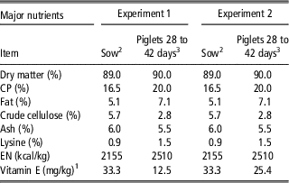
Thirty-six sows (CEP porcino, Aguilafuente, Segovia, Spain) were randomly distributed into three groups of each. One group was fed a standard diet (Table 1) supplemented with 30 mg/kg of synthetic α-tocopheryl acetate and the other two groups were supplemented with natural α-tocopherol at different doses (1/2 and 1/3 respect to the synthetic form) in drinking water. Pregnant sows were housed in groups and their individual feeding (2.3 kg/day) was electronically controlled during the last week of pregnancy (107 days of gestation). During the 28 days of lactation, sows were housed individually and were given 5.1 kg/day of feed. The transition from 2.3 to 5.1 kg/day was gradual within the 1st week of lactation being slightly restricted, and thus they consumed all the feed. Average weights of sows were 250 (±8.98) kg at 107 day of gestation and 210 (±8.85) kg at 25 day of lactation. As water was provided ad libitum, natural vitamin E was included in the first 5 l to assure its complete intake. The natural vitamin E added to the drinking water for sows was an emulsion of 70 mg/g of RRR-α-tocopherol (H2E-Vitapherole E 7% WS FG, VitaeCaps, S.A., Talavera de la Reina, Toledo, Spain). Hence, sows supplemented with the synthetic form of vitamin E (30-SINT) received an average amount of 75 mg/day during the last week of gestation and 150 mg/day during the lactation period and those supplemented with the natural form (1/2-NAT and 1/3-NAT) received the correspondence 1/2 or 1/3 lower amount when compared with the synthetic dose according to the feeding period.
Chemical composition and ingredients of the experimental diets

1 Groups that received natural vitamin E in water were not supplemented with vitamin E in feed (all-rac-α-tocopherol). Analysed concentration in this feed was 9.18 μg of α-tocopherol/g of feed (sows) and 9.75 μg of α-tocopherol/g of feed (piglets).
2 Ingredients (%): Barley: 36.3; corn: 12; wheat: 5; soya meal: 15; wheat bran: 20; pork lard: 2.8; beetroot pulp: 5; molasses: 1; calcium carbonate: 0.99; bicalcium phosphate: 0.82; salt: 0.5; l-lysine: 0.16; methionine: 0.01; choline chloride: 0.03; premix: 0,3 (per kg/diet: vitamin A: 9000 IU; vitamin D3: 1750 IU; vitamin E (all rac α-tocopheryl acetate): 30 mg; vitamin B1: 1 mg; vitamin B2: 3.5 mg; vitamin B12: 0.02 mg; vitamin B6: 0.50 mg; nicotinic acid: 30 mg; biotin: 0.05 mg; pantothenic acid: 10 mg; vitamin K3: 0.5 mg; choline chloride: 200 mg; Fe (ferrous carbonate): 50 mg; Cu (pentahydrate sulphate): 15 mg; Co (heptahydrate sulphate): 0.45 mg; Zn (oxide): 90 mg; Mn (monohydrate sulphate): 35 mg; I (potasium iodure): 0.5 mg; Se (sodium selenite): 0.0675 mg; 6-fitase EC 3,1,3,26: 750 FTU; BHT and ethoxyquin: 0.1 mg.
3 Ingredients (%): Barley: 22.9; corn: 23.5; wheat 10; soya meal 44: 8; extruded soya: 10; fish meal: 7.56; acid serum: 12.48; pork lard: 2.55; l-lysine 50%: 0.77; l-threonine: 0.17; dl-methionine 99%: 0.22; calcium carbonate: 0.76; bicalcium phosphate: 0.67; salt: 0.15; premix: 0.2% (per kg/diet: vitamin A: 6000 IU; vitamin D3: 12 000 IU; vitamin E: 12.5 mg; vitamin B1: 0.5 mg; vitamin B2: 2.0 mg; vitamin B12: 0.012 mg; vitamin B6: 1.0 mg; nicotinic acid: 12 mg; pantothenic acid: 6 mg; choline chloride: 100 mg; Fe (monohidrate sulphate and ferrous carbonate): 70 mg; Cu (pentahydrate sulphate): 20 mg; Co (heptahydrate sulphate): 0.42 mg; Zn (oxide): 75 mg; Mn (monohydrate sulphate): 35 mg; I (potasium iodure): 0.4 mg; Se (sodium selenite): 0.112 mg; 6-phitase EC 3,1,3,26: 500 FTU; BHT and ethoxyquin: 0.3 mg).
After weaning (average number of weaned piglets per sow: 7.1±0.8), those piglets from each sow with weights closer to the mean weaning weight were selected and randomly distributed to further groups, one of which was fed with synthetic α-tocopheryl acetate (15 mg/kg, average intake: 3.33 mg/day) and the other with natural α-tocopherol in drinking water (the same product described for sows) at different doses (1/2 and 1/3 respect to the synthetic form; average intakes: 1.7 and 1.1 mg/day, respectively). This experimental design resulted in seven piglet dietary treatments: (1) piglets from sows fed synthetic α-tocopheryl acetate that were fed with the same tocopherol form in their feed (30-SINT-15-SINT); (2) piglets from sows fed synthetic α-tocopheryl acetate in feed that were provided the natural form in drinking water at 1/2 dose (30-SINT-1/2-NAT) when compared with the synthetic form intake; (3) piglets from sows fed synthetic α-tocopheryl acetate in feed that were provided the natural form in drinking water at 1/3 dose (30-SINT-1/3-NAT) when compared with the synthetic form intake; (4) piglets from sows supplemented with 1/2 dose of natural α-tocopherol in drinking water that were provided with 15 mg/kg synthetic α-tocopheryl acetate in feed (1/2-NAT-15-SINT); (5) piglets from sows fed 1/2 dose of natural α-tocopherol in drinking water that were supplemented with 1/2 dose of natural α-tocopherol in drinking water (1/2-NAT-1/2-NAT); (6) piglets from sows supplemented with 1/3 dose of natural α-tocopherol in drinking water that were provided with 15 mg/kg synthetic α-tocopheryl acetate in feed (1/3-NAT-15-SINT); and (7) piglets from sows fed 1/3 dose of natural α-tocopherol in drinking water that were supplemented with 1/3 dose of natural α-tocopherol in drinking water (1/3-NAT-1/3-NAT). As water was provided to the piglets ad libitum during the post-weaning period, vitamin E was administered into the first 0.7 l to assure the complete intake. Dietary treatments were administered for 14 days after weaning.
Experiment 2
This experiment was carried out in the same conditions described in experiment 1. The experiment started at day 30 of gestation (–84-day prepartum) and finished 42 days after farrowing. Three dietary treatments were established according to the sow vitamin E supplementation: (1) sows supplemented with α-tocopheryl acetate in feed (150 mg/day; 30-SINT); (2) sows supplemented with 150 mg/day of natural vitamin E in the drinking water (1/1-NAT); and (3) sows supplemented with 50 mg/day of natural vitamin E in the water (1/3-NAT). After weaning, piglets fed the same feed (3.33 mg dl-α-tocopheryl acetate/day) until 42 days of age. As described in experiment 1, natural vitamin E for sows was an emulsion of 70 mg/g of RRR-α-tocopherol (H2E-Vitapherole E 7% WS FG, VitaeCaps, S.A., Talavera de la Reina, Toledo, Spain).
Samples collection
Blood samples were taken at the sow tail vein on different days prepartum and postpartum (experiment 1: day –7 prepartum and after 2, 14 and 28 days postpartum; experiment 2: days –14 and –84 prepartum and 7 and 27 postpartum) by puncture into vacuum tubes. Milk (5 ml) and colostrum samples were collected from a representative number of sows (n=10 per treatment) by hand-milking. In piglets, blood samples were collected from the jugular vein (n=7 per treatment in experiment 1 and n=12 in experiment 2). All blood samples were immediately placed on ice after collection. The serum was then separated by centrifugation at 600×g for 10 min at 4°C and the supernatant was kept in a freezer at –80°C until analysis. Analyses were carried out within the next 2 months.
Laboratory analysis
Tocopherol quantification in serum, colostrum and milk
The α-tocopherol concentration in serum from sows and piglets was quantified as described by Rey et al. (Reference Rey, Daza, López-Carrasco and López-Bote2006) by direct extraction. Thus, serum samples were mixed with 0.054 M dibasic sodium phosphate buffer adjusted to pH 7.0 with HCl and absolute ethanol. After mixing, tocopherol was extracted with hexane by centrifugation. The upper layer was evaporated to dryness and dissolved in ethanol before analysis. Extraction in colostrum and milk samples was carried out by saponification in the presence of KCl (1.15%) and KOH (50%; Butriss and Diplock, Reference Butriss and Diplock1984). Tocopherols were analysed by reverse phase HPLC (HP 1100, equipped with a diode array detector; Agilent Technologies, Waldbronn, Germany) as described (Rey et al., Reference Rey, Daza, López-Carrasco and López-Bote2006). Identification and quantification were carried out by means of a standard curve (R 2=0.999) built using the pure compound (Sigma, Alcobendas, Madrid, Spain). All samples were analysed in duplicate.
Tocopherol quantification in the tissues
α-Tocopherols in subcutaneous fat was analysed using the procedure described by Rey et al. (Reference Rey, Daza, López-Carrasco and López-Bote2006). In this case, samples were saponified in the presence of pyrogalol (3% in ethanol), KCl (1.15%) and KOH (50%) and afterwards mixed with hexane.
To extract α-tocopherol in Longissimus dorsi muscle and liver, the direct extraction procedure and HPLC analysis was the same as described before for serum samples.
α-Tocopherol stereoisomers in the muscle
Distribution of the stereoisomers of α-tocopherol in the Longissimus dorsi muscle was determined by the method described by Lauridsen and Jensen (Reference Lauridsen and Jensen2005). Briefly, the heptane extract containing 1 to 2 μg of α-tocopherol in 9 ml was evaporated to dryness under a nitrogen stream. Then the α-tocopherol extract was derivatized to its methyl ester. Chromatographic separation was achieved on a Chiralcel OD-H column 25 (0.46 cm; 5-μm particle size, cellulose tris [3,5-dimethylphenylcarbamate]; Daicel Chemical Industries, Ltd, Tokyo, Japan). This method allows the separation of the eight stereoisomers of α-tocopherol into five peaks. Peak 1 contains all four 2S forms (2SR/SR/S); Peak 2 contains the 2RSS-α-tocopherol; Peak 3 contains 2RRS-α-tocopherol; Peak 4 contains 2RRR-α-tocopherol (=natural α-tocopherol); and Peak 5 contains 2RSR-α-tocopherol.
Oxidative status of serum samples
The ferric-reducing antioxidant power (FRAP) was measured using the procedure described by Benzie and Strain (Reference Benzie and Strain1999). The FRAP reagent was prepared fresh by mixing 10 vol of acetate buffer (300 mM) with one volume of 10 mmol TPZ solution (2,4,6-tripyridyl-s-triazine in 40 mM HCl) and 1 vol of 20 mM aqueous ferric chloride. A 100-μl aliquot of the sample extract was mixed with 3 ml of the working FRAP solution. The absorbance of the samples was recorded after 0 and 4 min at 593 nm. Results were expressed as μM.
The thiobarbituric acid-reactive substances (TBARS) were measured in serum according to a modification of the spectrophotometic method of Buege and Aust (Reference Buege and Aust1978). Perchloric acid was added to serum samples to precipitate proteins. The protein-free supernatant was collected, iron-induced with 1 mM FeSO4 and mixed in a proportion 1 : 2 with thiobarbituric acid (TBA) reagent (0.026 M TBA, 0.92 M trichloroacetic acid in 250 ml of water with 62.5 mL HCl 1 M). Then samples were centrifugated at 600×g for 10 min and absorbance was read at 532 nm using a UV-Vis spectrophotometer (ScanGo, ThermoFisher Scientific, Alcobendas, Spain). TBARS concentrations were calculated using 1.56×10⁵ M−1×cm−1 as the molar absorption coefficient. Results were expressed as μmoles MDA (malondialdehyde)/l serum.
Determination of IgA, IgM and IgG antibody levels in piglet serum
Immunoglobulins A, G and M were determined in serum piglet using a pig ELISA quantification kit (Bethyl Laboratories, Inc., USA). Briefly, for IgM determination, purified antibody (porcine IgM) was diluted in coating buffer (0.05 M carbonate-bicarbonate at pH 9.6) and incubated at room temperature for 60 min in a 96-well plate. The plate was then washed five times with washing solution (50 mM tris, 0.14 M NaCl, 0.05% Tween 20, Ph 8.0) to remove the non-coated antibody. Blocking solution (50 mM Tris, 0.14 M NaCl, 1% BSA, pH 8.0) was added to each well and the plate incubated at room temperature for 30 min. The blocking solution was removed and the plate washed. Sample was then added and the plate incubated for 1 h, after which time the diluted HRP-conjugated pig detection antibody was added. Finally, tetramethylbenzidine (TMB) substrate solution was added and incubated for 15 min. After incubation with the substrate, the reaction was quenched and the absorbance measured at 450 nm. Similar protocols were used for the IgA and IgG quantifications. A standard curve was prepared for each ELISA batch, and the final values were expressed in mg/mL.
Statistical analysis
The experimental unit for analysis of all data was the individual sow or piglet. Data were analysed following a completely randomized design using the GLM procedure contained in SAS (version 8; SAS Institute Inc., Cary, NC, USA). A repeated measurement test was carried out to study the effects of time and treatment on serum status and interaction. A comparative analysis between means was performed using the following orthogonal contrasts (experiment 1): (1) synthetic vitamin E in feed v. natural in drinking water; (2) sow supplementation dose (1/2 v. 1/3 of natural vitamin E in water); (3) sow supplementation source (synthetic vitamin E in feed v. natural in drinking water); and (4) piglet supplementation dose (1/2 v. 1/3); (5) Piglets supplementation source (synthetic in feed v. synthetic in feed and natural in drinking water). In experiment 2, the following comparisons were made: (1) synthetic vitamin E in feed v. natural in drinking water; (2) synthetic vitamin E in feed v. 1/3 natural in drinking water; (3) synthetic vitamin E in feed v. 1/1 natural in drinking water; and (4) 1/3 natural vitamin E v. 1/1 natural vitamin E in drinking water.
Data are presented as the mean of each group and s.e.m. together with the significance levels (P-value). Differences between means were considered statistically significant at P<0.05.
Results and discussion
Transfer of α-tocopherol to piglets
Piglets suffer a marked decline in α-tocopherol concentration after weaning (Chung et al., Reference Chung, Mahan and Lepine1992; Mahan et al., Reference Mahan, Kim and Stuart2000; Lauridsen et al., Reference Lauridsen, Engel, Jensen, Craig and Traber2002). As micellized natural vitamin E (d-α-tocopherol) was expected to be more efficiently accumulated than the synthetic form (Pumfrey et al., Reference Pumfrey, Deyonge, Slusark, Garber, Roeder, Sanchez and Schelling1993; Pagan et al., Reference Pagan, Kane and Nash2005) and that water consumption would be more consistent than feed intake during the post-weaning period (Wilburn et al., Reference Wilburn, Mahan, Hill, Shipp and Yang2008), we designed various strategies to study the effectiveness of supplementing piglets and/or sows with vitamin E. Supplementation ratios to piglets and/or sows were 1 : 1, 1 : 2 and 1 : 3 (natural vitamin E in water: synthetic in feed) trying to find a real equivalence between both forms of vitamin and administration vehicles that minimize piglet serum α-tocopherol decline after weaning. These ratios were based on previous results (Wilburn et al., 2008) that reported a water to feed ratio⩽2 : 1 when using the acetylated form (synthetic v. natural). Doses were close to or under the range of minimum daily vitamin E requirement for pregnant sows (44 IU/kg) and piglets (11 IU/kg) recommended in the NRC guidelines (NRC, 1998), and therefore higher differences among groups would be expected.
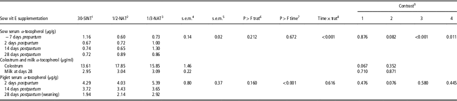
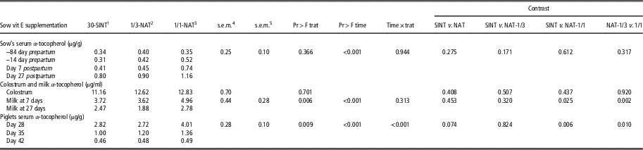
Serum, colostrum and milk α-tocopherol concentrations from sows and piglets until weaning (day 28) are presented in Table 2. The α-tocopherol concentrations in serum, colostrum and milk for the group supplemented with the synthetic form were similar to the values reported by other authors using similar feed inclusion level (Mahan et al., Reference Mahan, Kim and Stuart2000). Oral supplementation of sows with natural vitamin E as a micellized form (d-α-tocopherol) at the lowest dose (1/3-NAT) resulted in a similar concentration of α-tocopherol in serum to those supplemented with the synthetic form in the feed (30-SINT) at days 2, 14 and 28 postpartum. In addition, the sows that received natural vitamin E in water tended to have higher values in colostrum than those supplemented with the synthetic form (P=0.067), although in milk samples at day 28 postpartum differences were not significant (experiment 1). This result would partly indicate that the micellized natural alcohol form in water was more efficiently accumulated than the synthetic form. Results from experiment 2 support this observation (Table 3) when using a 1 : 1 ratio of natural in water to synthetic in feed supplementation regime. Moreover, as expected, sow serum α-tocopherol concentration increased with supplementation time (P<0.001; Table 2) with it being interesting to note that the increase was higher when sows were supplemented with the natural vitamin E (contrast 3: interaction time×source, P<0.001), mainly with 1/3-NAT (contrast 4: interaction time×dose: P=0.011). Other authors have previously demonstrated that natural vitamin E was a superior source compared with synthetic (Chung et al., Reference Chung, Mahan and Lepine1992; Mahan et al., Reference Mahan, Kim and Stuart2000; Yang et al., Reference Yang, Mahan, Hill, Shipp, Radke and Cecava2009) and that the supplementation was more effective in increasing serum α-tocopherol when administered in the water supply than when it was added to the diet (Wilburn et al., Reference Wilburn, Mahan, Hill, Shipp and Yang2008). Moreover, micellized natural vitamin E is better absorbed in plasma because micelles that transport vitamin E solubilized in their core (Pumfrey et al., Reference Pumfrey, Deyonge, Slusark, Garber, Roeder, Sanchez and Schelling1993; Pagan et al., Reference Pagan, Kane and Nash2005). However, to the best of our knowledge, there is no information in the literature in which a lower dose resulted in higher absorption and serum accumulation. In the unique study carried out with supplementation in drinking water, Wilburn et al. (Reference Wilburn, Mahan, Hill, Shipp and Yang2008) used higher doses (50, 100, 150 ppm) of α-tocopheryl acetate, the accumulation of which depends on the presence of an esterase enzyme present in the digestive secretions. Our results might indicate that under minimal doses, absorption of lower amounts of the micellized alcohol form administered orally (1/3- NAT v. 1/2-NAT) would be more efficiently accumulated. This observation was corroborated in experiment 2 in which a similar trend was observed, although differences were not statistically significant (P=0.171; Table 3). Moreover, it was found that the higher the vitamin E concentration in sow serum, colostrum and milk, the higher the vitamin E levels in piglet serum postpartum (P=0.076; Table 2). At day 39 of age (Table 4, experiment 1), neither piglet supplementation source nor dose significantly affected α-tocopherol accumulation in the serum, muscle, subcutaneous fat or liver. It was found that only piglets supplemented with the natural micellized alcohol form in water tended to show higher accumulation of α-tocopherol in subcutaneous fat than those supplemented with the synthetic form (30-SINT-15-SINT; P=0.082), despite the lower doses administered in water to piglets or their mothers. Moreover, it is interesting to note that the sow vitamin E source might have some influence on α-tocopherol accumulation in piglet fat (P=0.159) at day 39. There is no information in the literature regarding the effects of sow vitamin E supplementation in drinking water on piglet tissue α-tocopherol levels. According to Lauridsen et al. (Reference Lauridsen, Engel, Jensen, Craig and Traber2002), piglet fat and the brain were the only tissues in which α-tocopherol concentrations increased from 7 to 21 days of lactation when mothers were supplemented. Other investigators have found a tendency for greater vitamin E concentration in piglets of sucking sows receiving high levels of dl-α-tocopheryl acetate (Mahan, Reference Mahan1991; Hiridoglou et al., Reference Hiridoglou, Farnworth and Butler1993; Pinelli-Saavedra et al., Reference Pinelli-Saavedra, Calderón de la Barca, Hernández, Valenzuela and Scaife2008) or d-α-tocopheryl acetate (Mahan et al., Reference Mahan, Kim and Stuart2000) because of the high absorption from sow’s milk.
α-Tocopherol concentration in sows and piglets serum from −7 to 28 days postpartum and composition of colostrum and milk as affected by sow vitamin E supplementation (experiment 1)

1 30-SINT: sows supplemented with α-tocopheryl acetate in feed (150 mg/day).
2 1/2-NAT: sows supplemented with 75 mg/day of natural vitamin E (d-α-tocopherol) in water.
3 1/3-NAT: sow supplemented with 50 mg/day of natural vitamin E (d-α-tocopherol) in water.
4 s.e.m. for treatment and interactions.
5 s.e.m. for time and interactions; piglets n=7; sows n=10.
6 P-value for treatment effect.
7 P-value for time effect.
8 P-value for treatment×time interaction.
9 Contrast: (1): Sow’s vit E source effect; (2): Sow’s vit E dose effect; (3) Time×source; (4) Time×dose.
α-Tocopherol accumulation in sows serum, colostrum and milk, and in piglet serum from 28 to 42 days as affected by sow vitamin E supplementation (experiment 2)

1 30-SINT: sows supplemented with α-tocopheryl acetate in feed (150 mg/day).
2 1/3-NAT: sows supplemented with 50 mg/day of natural vitamin E (d-α-tocopherol) in water.
3 1/1-NAT: sow supplemented with 150 mg/day of natural vitamin E (d-α-tocopherol) in water.
4 s.e.m. for treatment and interactions.
5 s.e.m. for time and interactions; piglets n=12; sows n=10.
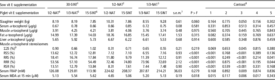
α-Tocopherol, α-tocopherol stereoisomers, FRAP and MDA concentrations in piglet tissues at slaughter (39 days) as affected by sow or piglet vitamin E supplementation (experiment 1)

1 30-SINT: sows supplemented with α-tocopheryl acetate in feed (150 mg/day).
2 1/2-NAT: sows supplemented with 75 mg/day of natural vitamin E (d-α-tocopherol) in water.
3 1/3-NAT: sow supplemented with 50 mg/day of natural vitamin E (d-α-tocopherol) in water.
4 1/2-NAT: piglets supplemented with 1.7 mg/day of natural vitamin E (d-α-tocopherol) in water.
5 1/3-NAT: piglets supplemented with 1.1 mg/day of natural vitamin E (d-α-tocopherol) in water.
6 15-SINT: piglets supplemented with α-tocopheryl acetate in feed (3.33 mg/day).
7 piglets n=7.
8 Contrast: (1): SINT v. others; (2): Sow’s vit E dose effect; (3): Sow’s vit E source effect; (4) Piglet vit E dose effect; (5): Piglet vit E source effect.
9 2S: represents the sum of SSS-, SSR-, SRS- and SRR-α-tocopherol.
The predominant importance of sow over piglet supplementation was observed when α-tocopherol stereoisomers in the muscle at 39 day of age were quantified (Table 4). Those piglets from sows supplemented with the natural source of vitamin E (1/2-NAT-1/2-NAT; 1/2-NAT-15-SINT; 1/3-NAT-1/3-NAT; 1/3-NAT-15-SINT) had higher RRR (P<0.001) and lower RRS (P<0.001), RSS (P<0.001) and RSR (P<0.001) α-tocopherol stereoisomers proportions at day 39 of age than those from sows supplemented with the synthetic form (30-SINT-1/2-NAT; 30-SINT-1/3-NAT; 30-SINT-15-SINT). Lauridsen et al. (Reference Lauridsen, Engel, Jensen, Craig and Traber2002) previously reported that lactating sows and suckling piglets preferentially incorporate RRR- over all-rac-α-tocopherol into milk, plasma and tissues. Hoppe and Krennrich (Reference Hoppe and Krennrich2000) and Stone et al. (Reference Stone, LeClair, Ponde, Baggs and Reis2003) found similar results in humans. This is because of the higher affinity of the α-tocopherol transfer protein for the single stereoisomer (-RRR) of natural vitamin E, against the equimolar amounts of the other eight isomers presented in the synthetic form (Hosomi et al., Reference Hosomi, Arita, Sato, Kiyose, Ueda, Igarashi, Arai and Inoue1997). Furthermore, it is of interest to highlight that the α-tocopherol stereoisomer profile reflects the vitamin E source provided to sows to a higher extent than that supplied to piglets after weaning. Hence, only RRR and RRS α-tocopherol stereoisomers in the piglet muscle were statistically affected (P=0.04) by the type of vitamin E supplemented to piglets. Meanwhile, more marked effects (P<0.001) were observed in all the α-tocopherol stereoisomers as affected by the sow’s vitamin E source supplementation. These results are of emphasized interest and would indicate that the vitamin E source given to sow is a determinant factor to reach higher –R stereoisomer accumulation in the piglet.
This sow-source supplementation effect was confirmed in experiment 2 in which all groups of piglets received the same feed after weaning. Hence, piglets from sows supplemented with the natural micellized alcohol form at low dose in water (1/3-NAT) did not show different α-tocopherol accumulation in serum than those from sows supplemented with the synthetic form in feed. Moreover, piglets from sows supplemented with a similar concentration to the synthetic (1/1-NAT) showed higher α-tocopherol accumulation in serum than the 30-SINT (P=0.006) and 1/3-NAT (P=0.010) groups 1 and 2 week after weaning. These results would corroborate that the equivalent dose of the natural form in water to the synthetic form in feed is 1 : 3. Hence, our results would agree with those reported by Wilburn et al. (2008) as water to feed ratio⩽2 : 1.
Piglet serum α-tocopherol concentration decreased with time (P<0.001), especially from weaning to day 35 post-weaning (Tables 2 and 3). This has been widely reported in the literature and is attributed to increased stress (Lauridsen et al., Reference Lauridsen, Engel, Jensen, Craig and Traber2002; Moreira and Mahan, Reference Moreira and Mahan2002; Wilburn et al., Reference Wilburn, Mahan, Hill, Shipp and Yang2008). However, it is interesting to note that the magnitude of the decrease was greatest in groups with higher serum concentrations of α-tocopherol at weaning (P<0.001, interaction time×treatment; Table 3). Hence, results from experiment 2 show that at weaning serum α-tocopherol concentration in piglets from sows supplemented with natural vitamin E in drinking water at a similar dose than the synthetic form (1/1-NAT v. 30-SINT) were 1.42 times higher than those from sows fed the synthetic vitamin E. One week after weaning the concentration was 1.35 times higher and at day 14 post-weaning (42 days of age) it was very similar. In experiment 1 (Table 4), in which not only sows but also piglets were supplemented after weaning with both sources in either feed or drinking water at low doses, a similar 1.04-fold increase was found at day 39, thus indicating that the amount of vitamin E provided to weaning piglets was insufficient to prevent the drop in serum α-tocopherol. In a recent study, Amazan et al. (Reference Amazan, Rey, Fernandez and López-Bote2012) reported a lower vitamin E decrease 5 days post-weaning in piglets born from supplemented sows (52.5 mg/day) that were provided with natural vitamin E in drinking water (5 mg/day), in addition to the synthetic form in the feed.
Piglet performance
In experiment 1, natural vitamin E supplementation to sows significantly increased piglet weight (BW) at weaning (P=0.036). Hence, those piglets born from sows supplemented with the natural form had higher BW than those from sows supplemented with the synthetic form (8.1 v. 7.5 kg), but differences in BW at 42 days of age were not statistically significant (9.6 v. 10.2 kg). Daily feed intake (DFI; 0.23 kg±0.01) was not statistically different between treatments. In experiment 2, BW at 28 days (6.67 kg±0.34), BW at 42 days (8.80 kg±0.26) and DFI (0.24 kg±0.01) were not statistically affected. There is a lack of information in the literature concerning the effect of natural vitamin E supplementation in drinking water on these parameters. Wilburn et al. (2008) found no effect of adding natural vitamin E to the diet or drinking water on average daily gain for any post-weaning period, but to the best of our knowledge no more information has been reported on pigs.
Oxidative status in piglet serum
To evaluate oxidative stress of piglets after weaning depending on sow and piglet vitamin E source and dose supplementation, MDA and the total antioxidant capacity measured as FRAP were quantified (Table 4). The MDA concentrations were similar to those found by Shakardi-Nagy et al. (Reference Shakardi-Nagy, Meng-Chuan, Guan-Yeu, Kirwan, Chueh Chao, Tschanz and Brenna2003) for piglets after 28 days of age that were supplemented with different vitamin E concentrations. In addition, FRAP values were within the values expected in the literature for piglets of these ages (Lauridsen and Jensen, 2005). In experiment 1, it is interesting to note that low doses of oral natural vitamin E supplementation to sows and/or piglets did not increase the oxidative stress of piglets at 39 days of age when compared with the use of the synthetic form in feed. Some investigators have previously reported the effectiveness of dietary vitamin E supplementation on controlling MDA (Hacisevki et al., Reference Hacisevki, Torun and Cengel2012) and FRAP values (Benzie and Strain, Reference Benzie and Strain1999; Hamilton et al., Reference Hamilton, William, Benzie, Mulholland and Strain2000). However, there is not much information in which these parameters are evaluated according to source or supplementation dose in water of piglets or their mothers. Moreover, FRAP and MDA values increased (P=0.009) and decreased (P=0.006), respectively, in piglet serum at 39 days by the vitamin E source provided to sows. This result may indicate that supplementation to sows with the natural form of vitamin E in water could protect other compounds or antioxidants presented in piglet serum and increase the serum ferric-reducing antioxidant power. Hamilton et al. (Reference Hamilton, William, Benzie, Mulholland and Strain2000) reported some interactions between vitamin E and other antioxidants that resulted in increased FRAP values. Amazan et al. (Reference Amazan, Rey, Fernandez and López-Bote2012) reported that FRAP of serum was affected by natural vitamin E supplementation of piglets in accordance with the differences observed in serum vitamin E concentration. Other authors (Van Zoeren-Grobben et al., Reference Van Zoeren-Grobben, Lindeman, Houdkamp, Brand, Schrijver and Berger1994) reported that breast-fed children possessed more efficient antioxidant barrier in the blood and experienced less oxidative stress than formula-fed children because natural milk provided available antioxidants. Our results might indicate the importance of RRR-α-tocopherol stereoisomer, which is preferentially incorporated by piglets born from sows supplemented with the natural form, to control the oxidative stress. Brigelius-Flohé and Traber (Reference Brigelius-Flohé and Traber1999) reported highly different biological activities between stereoisomers, explained by differences in absorption based on the rat resorption gestation test. However, other compounds affected by the vitamin E supplementation form may participate to control the oxidative status of newborn piglets.
Piglet immune response
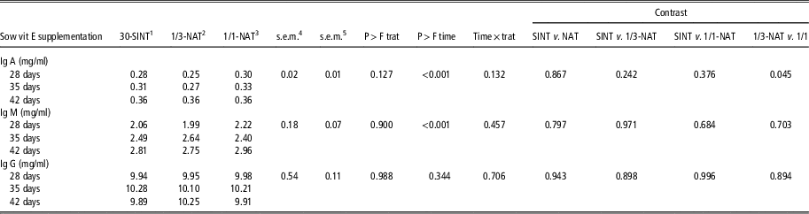
The IgM, IgA and IgG levels in the serum of piglets from the different dietary treatments are presented in Table 5. Immunoglobulin levels in piglet serum at day 39 were not affected by natural vitamin E supplementation at low doses in drinking water of piglets or sows when compared with the synthetic form in the feed. There is a lack of information in the literature on immunoglobulin concentrations as affected by vitamin E supplementation in drinking water. Nemec et al. (Reference Nemec, Butler, Hidiroglou, Farnworth and Nielsen1994) reported no effect on the plasma of sows and their piglets when diets were supplemented with dl-α-tocopheryl acetate at 44 IU/kg during gestation and 220 IU/kg during lactation. Similarly, Bonnette et al. (Reference Bonnette, Kornegay, Lindemann and Hammerberg1990) using four dietary levels of vitamin E (from 11 to 550 IU/kg feed) found no effects on the humoral and cell-mediated immunity in 4-week weaning piglets. Amazan et al. (Reference Amazan, Rey, Fernandez and López-Bote2012) also reported that immunoglobulin levels in piglet serum were not affected by natural vitamin E supplementation in drinking water. In contrast, other authors (Babinszky et al., Reference Babinszky, Langhout, Verstegen, den Hartog, Joling and Nieuwland1991) observed that vitamin E-enriched diets (136 IU dl-α-tocopheryl acetate/kg v. 13 IU/kg) fed to sows increased the immune response of their progeny. In a similar way, vitamin E provided to piglets through the diet (Fragou et al., Reference Fragou, Fegeros, Xylouri, Baldi and Politis2004) or parenteral (Hidiroglou et al., Reference Hidiroglou, Batra, Farnworth and Markham1995) improved immune status. In a recent study, Bondo and Jensen (Reference Bondo and Jensen2011) reported that oral administration of RRR-α-tocopherol to pregnant mares enhanced immunoglobulin status in foals. In the present study, IgA tended to be higher (P=0.145) at day 39 in piglets supplemented with natural vitamin E when compared with those supplemented with the synthetic vitamin (30-SINT-15-SINT), although differences were not statistically significant. In the second experiment (Table 6), IgA of piglet serum was also affected by the dose of natural vitamin E supplementation. Hence, piglets from sows supplemented with 1/3-NAT had lower (P=0.045) IgA at days 28, 35 and 42 of age when compared with those from sows supplemented 1/1-NAT. These results would confirm that, although threefold lower doses of natural vitamin E in drinking water of sows when compared with the synthetic supplementation produce similar piglets’ serum vitamin E concentration and oxidative status, the general status of the animal is not good enough. It is interesting to note that results of the present study were obtained when using doses below the minimal NRC recommendations and that some authors (Peplowski et al., Reference Peplowski, Mahan, Murray, Moxon, Cantor and Ekstrom1981) have suggested that to optimize the immune response in pigs, levels of vitamin E of 20 times higher are needed.
Igs concentration in piglets serum at 39 days of age as affected by sow or piglet vitamin E supplementation (experiment 1)

1 30-SINT: sows supplemented with α-tocopheryl acetate in feed (150 mg/day).
2 1/2-NAT: sows supplemented with 75 mg/day of natural vitamin E (d-α-tocopherol) in water.
3 1/3-NAT: sow supplemented with 50 mg/day of natural vitamin E (d-α-tocopherol) in water.
4 1/2-NAT: piglets supplemented with 1.7 mg/day of natural vitamin E (d-α-tocopherol) in water.
5 1/3-NAT: piglets supplemented with 1.1 mg/day of natural vitamin E (d-α-tocopherol) in water.
6 15-SINT: piglets supplemented with α-tocopheryl acetate in feed (3.33 mg/day).
7 piglets n=7.
8 Contrast: (1): SINT v. others; (2): Sow’s vit E dose effect; (3): Sow’s vit E source effect; (4) Piglet vit E dose effect; (5): Piglet vit E source effect.
Immunoglobulins concentration in piglets serum at weaning and 1 and 2 weeks thereafter as affected by sow’s vitamin E supplementation (experiment 2)

1 30-SINT: sows supplemented with α-tocopheryl acetate in feed (150 mg/day).
2 1/3-NAT: sows supplemented with 50 mg/day of natural vitamin E (d-α-tocopherol) in water.
3 1/1-NAT: sows supplemented with 150 mg/day of natural vitamin E (d-α-tocopherol) in water.
4 s.e.m. for treatment and interactions.
5 s.e.m. for time and interactions; piglets n=12.
On the other hand, in experiment 2 (Table 6), IgM and IgA increased after weaning with piglet age (P<0.001), whereas IgG remained unchanged. A similar evolution in immunoglobulin concentrations of young pigs was reported by Bourne and Curtis (Reference Bourne and Curtis1973), who described a decrease in IgA and IgM concentrations during the first 2 weeks of age and a subsequent increase. In addition, Nemec et al. (Reference Nemec, Butler, Hidiroglou, Farnworth and Nielsen1994) reported a drop in the IgM of piglets from sows without vitamin E supplementation when compared with those supplemented with 88 IU/kg, whereas a drop in IgA was observed in all groups.
In conclusion, oral supplementation of sows with low doses of micellized d-α-tocopherol is an interesting feeding strategy, when compared with the use of the synthetic form in feed, to reach similar α-tocopherol concentration in piglets. A predominant importance of sow over piglet vitamin E supplementation is also observed on stereoisomer distribution in piglets. The equivalence ratio in sows of natural micellized d-α-tocopherol in water:synthetic α-tocopherol in feed has been established as 1 : 3; however, a higher ratio (1 : 2) or doses of micellized α-tocopherol in water are needed to optimize the immune response of piglets.
Acknowledgements
This research was supported by project CDTI IDI-20090944. The authors would like to thank Prebia Feed Extracts (Talavera de la Reina, Toledo, Spain) for providing the natural vitamin E and PigChamp Pro Europa (Segovia, Spain) for the farm installations supply. D. Amazan was recipient of a FPI grant.