
Introduction
Rapid growth in the utilization of ionizing radiation in modern life has created a growing demand for new hybrid electronic materials that can readily detect such radiation. Traditionally, radiation detection is achieved with inorganic semiconducting materials such as silicon, cadmium zinc telluride, or mercury iodide, which directly convert ionizing radiation into an electrical signal.[Reference Owens and Peacock1] However, these rigid materials suffer from severe limitations with processing into large-area pixelated detector matrices, an inability to conform to various regions of the human body, and also require correction factors to measure the radiation dose delivered to biological species because the device materials do not have a water-equivalent density.[Reference Basirico, Ciavatti, Cramer, Cosseddu, Bonfiglio and Fraboni2] A radiation detector that could remedy these issues would have wide-ranging applications, including radiation protection, dosimetry measurements for radiotherapy, X-ray imaging in diagnostic radiology, or personal health monitoring in a range of high-risk industries.[Reference Fraboni, Ciavatti, Basiricò and Fraleoni-Morgera3] Demand from such applications has created intense interest in printed electronics, a field that focuses on the manufacture of electronic circuits and devices on mechanically flexible substrates at high speeds across large areas using cheap roll-to-roll (R2R) processing techniques.[Reference Hwang, Jung, Heo, Scholes, Watkins, Subbiah, Jones, Kim and Vak4–Reference Bergqvist, Österberg, Melianas, Ever Aguirre, Tang, Cai, Ma, Kemerink, Gedefaw, Andersson and Inganäs7] However, realization of the potential for printed electronics in such applications requires two critical parallel developments. Firstly, innovative manufacturing techniques that are faster, cheaper, and more eco-friendly than conventional methods must be developed.[Reference Seyler, Haid, Kwon, Jones, Bäuerle, Holmes and Wong8,Reference Al-Mudhaffer, Griffith, Feron, Nicolaidis, Cooling, Zhou, Holdsworth, Belcher and Dastoor9] Simultaneously, new electronic materials must be developed that are compatible with these solution-based manufacturing techniques and allow manipulation of the nanoscale architecture of multilayer thin films to create highly functional electronic devices.[Reference Lipomi and Bao10,Reference Griffith, Cooling, Elkington, Muller, Belcher and Dastoor11]
Organic electronic (OE) materials that exhibit charge selectivity or semiconducting behavior have attracted particular interest as ideal candidates for printed electronics applications. The suitability of OE materials for this purpose arises from the ability to easily modify their chemical, physical, and electronic properties and control their film-forming mechanisms through conventional wet chemistry. Examples of electronic devices prepared from these innovative materials include thin-film transistors,[Reference Elkington, Wasson, Belcher, Dastoor and Zhou12] solar cells,[Reference Brabec, Zerza, Cerullo, De Silvestri, Luzzati, Hummelen and Sariciftci13] sensors,[Reference Rogers, Someya and Huang14] large-area OLED displays,[Reference Reineke, Lindner, Schwartz, Seidler, Walzer, Lüssem and Leo15] and photodetectors.[Reference Griffith, Willis, Kumar, Holdsworth, Bezuidenhout, Zhou, Belcher and Dastoor16] Precise control of nanoscale morphology in multilayer OE stacks is required to achieve device functionality, a process typically achieved with carefully calibrated laboratory fabrication and processing techniques. However, controlling OE material nanoscale structure and morphology across large areas during printing fabrication remains a significant unsolved challenge due to the crude thermodynamic levers available.[Reference Pastorelli, Schmidt, Hösel, Søndergaard, Jørgensen and Krebs17] One potential avenue to circumvent this challenge is to create discrete nanoparticles where the desired structure is imprinted through chemically directed assembly using surfactants.[Reference Holmes, Marks, Cave, Feron, Barr, Fahy, Sharma, Pan, Kilcoyne, Zhou, Lewis, Andersson, van Stam, Walker, Moons, Belcher and Dastoor18,Reference Sankaran, Glaser, Gärtner, Rödlmeier, Sudau, Hernandez-Sosa and Colsmann19] Not only does this approach disperse the OE nanoparticles into greener solvents such as water and alcohols but also ensures that the thermodynamic control of film morphology is decoupled from the printing process. This nanoengineering approach enables the dual benefits of exquisite nanoscale film structure and low-cost, large-area printing of electronic devices to be simultaneously realized.[Reference Ameri, Al-Mudhaffer, Almyahi, Fardell, Marks, Al-Ahmad, Fahy, Andersen, Elkington, Feron, Dastoor and Griffith20]
Direct radiation sensors have been previously fabricated using OE materials. The most promising results have been observed with single-crystal semiconductors such as TIPS pentacene and rubrene,[Reference Lai, Cosseddu, Basiricò, Ciavatti, Fraboni and Bonfiglio21] although solution-based polymers such as poly(triarylamine) have also been demonstrated.[Reference Intaniwet, Mills, Shkunov, Thiem, Keddie and Sellin22] These devices exhibited radiation sensitivity in the range of 100 nC/Gy/mm2 to 1 µC/Gy/mm2, with operating voltages as low as 1 V for single-crystal molecules and up to 100 V for the polymer semiconductors. Indirect ionizing radiation sensors, where a secondary scintillator material absorbs radiation and has a subsequent luminescence absorbed by an organic photodetector, have also been fabricated from OE materials. In these studies, inorganic scintillators such as terbium or europium-doped gadolinium oxide (GOS:Tb, GOS:Eu) have been incorporated into P3HT:PC61BM solar cells and demonstrated impressive sensitivities on the order of μC/Gy/mm2.[Reference Büchele, Richter, Tedde, Matt, Ankah, Fischer, Biele, Metzger, Lilliu, Bikondoa, Macdonald, Brabec, Kraus, Lemmer and Schmidt23,Reference Agostinelli, Campoy-Quiles, Blakesley, Speller, Bradley and Nelson24] Attempts to replace the scintillator with a printable organic molecule have been reported, although the detection sensitivity was reduced by orders of magnitude.[Reference Oliveira, Correia, Sowade, Etxebarria, Rodriguez, Mitra, Baumann and Lanceros-Mendez25]
In this work, we report the development of a fully printable radiation-sensing device prepared from a mixed nanoparticle system composed of a radiation scintillator [1-phenyl-3-mesityl-2-pyrazoline (PMP)] and a semiconducting polymer [violanthrone-79 (VA79)]. Combining the materials inside a nanoparticle allows the absorbed radiation to be efficiently transmitted from PMP to VA79 through Förster resonance energy transfer (FRET). A combination of steady-state and time-resolved absorbance and photoluminescence measurements are used to demonstrate successful FRET in both the solid-state films and blended nanoparticle material systems. These materials are then used to create a simple radiation sensor, which successfully demonstrates the detection of gamma radiation from a 137Cs source.
Investigating the potential for energy transfer
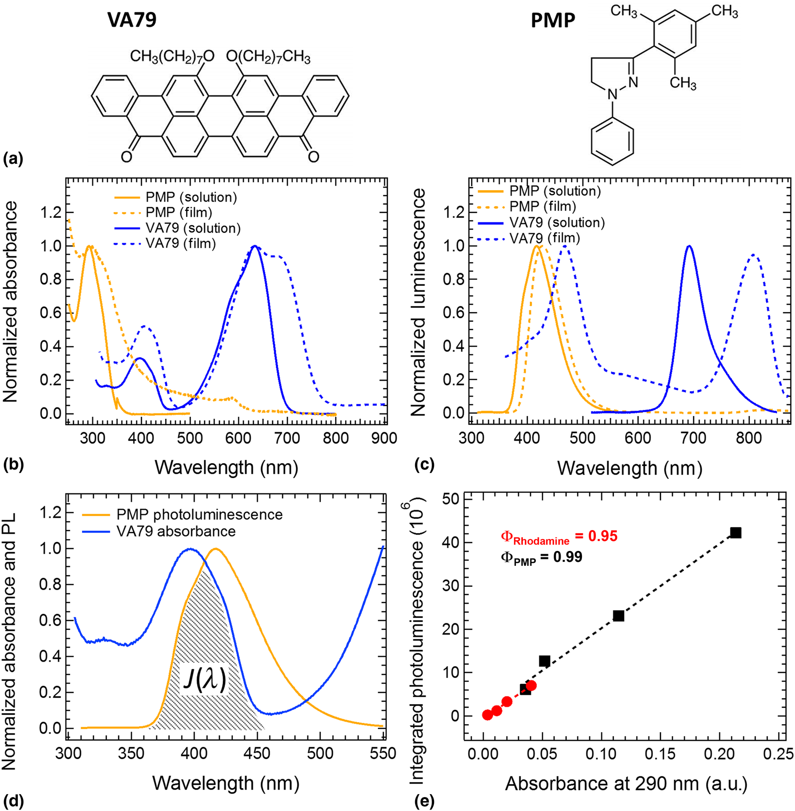
Figure 1 shows the chemical structures of the PMP scintillator and the VA79 semiconducting polymer along with their solution and solid-state UV–visible and photoluminescence spectra. Details of the experimental measurement procedures are included in Supplementary Material. The PMP scintillator has an absorption peak at 292 nm with an extinction coefficient of 18,400/M/cm in chloroform solution [Fig. 1(b)]. This peak represents the π–π* transition that is photoexcited by ionizing radiation to induce scintillation. The photoluminescence peak occurs at 417 nm in solution, representing a substantial Stokes shift of 125 nm [Fig. 1(c)]. This large shift allows the luminescence to be detected in the visible spectrum without the use of a third wavelength-shifting component that is often required in organic scintillator detectors.[Reference Fraboni, Ciavatti, Basiricò and Fraleoni-Morgera3] The solid-film absorbance and photoluminescence show minor broadening in comparison to the solution spectra, which is attributed to increased intermolecular dipole interactions. The VA79 semiconductor shows a weaker blue absorption band at 396 nm with an extinction coefficient of 8900/M/cm and a stronger red absorption peak at 633 nm with an extinction coefficient of 26,900/M/cm in chloroform solution (Supplementary Fig. S1). A single photoluminescence peak occurs at 692 nm in solution, indicating that the higher energy blue absorbance band does not exhibit radiative emission in solution. However, in the solid state, substantial differences are observed in the VA79 photophysics. The blue absorption peak shows a higher oscillator strength with respect to the red peak, and the red absorbance peak shows a double structure and substantial broadening that could represent vibronic structure or molecular aggregation. The photoluminescence now exhibits radiative emission from both the blue and red absorbance bands, with Stokes shifts of 63 and 118 nm, respectively. This distinct behavior is suggestive of more complex solid-state ordering in VA79 that can act as an easily identifiable marker for VA79 material inside nanoparticulate structures.
(a) Chemical structures of the radiation scintillator [1-phenyl-3-mesityl-2-pyrazoline (PMP)] and the semiconducting polymer [violanthrone-79 (VA79)]. (b) Solution (solid lines) and solid-state (dashed lines) UV–visible absorbance spectra for PMP and VA79. (c) Solution (solid lines) and solid-state (dashed lines) photoluminescence spectra for PMP and VA79. The excitation wavelengths employed were 315 and 400 nm for PMP and VA79, respectively. (d) Normalized spectra comparing the photoluminescence output of PMP with the absorbance band of VA79. The spectral overlap, J(λ), is shown as the shaded region. (e) The integrated area under the photoluminescence curves plotted as a function of the absorbance at the excitation wavelength of 290 nm for rhodamine 6G (red circles) and PMP (black squares).

Figure 1(d) compares the spectral overlap of the PMP emission with the VA79 blue absorption band. Substantial overlap is observed, which is indicative of ideal conditions for the photodetection of the radiation-induced scintillation from PMP with the semiconducting polymer. This process could occur through either radiative emission from PMP and re-absorption by VA79, or by direct transfer of the PMP excited state to the VA79 molecule through a coupling of the two molecular dipoles. In general, this second process, referred to as FRET, would be preferred for maximizing radiation sensitivity as it restricts the loss of energy from non-radiative decay of the PMP excited state. The efficiency of energy transfer,  $\emptyset _{_{{\rm FRET}}}$, is inversely proportional to the sixth power of the distance, r, between the PMP and VA79 transition dipoles:[Reference Clegg26]
 $$\emptyset _{{\rm FRET}}\; = \; \; \displaystyle{1 \over {1 + \; {\lpar {r/R_0} \rpar }^6}}$$
where R 0 is the Förster radius associated with the energy transfer between two materials and represents the molecular separation at which the energy transfer yield will be 0.5. R 0 can be computed analytically from the photoluminescence yield of the donor material,  $\emptyset _{_{{\rm PMP}}}$, the extinction coefficient of the acceptor, ε VA79, the spectroscopic overlap of the two materials, J(λ), the orientation of their transition dipoles, κ 2, and the refractive index of the solvent, η:[Reference Lakowicz27]
 $$R_0^6 \; = \; 8.8\; \times 10^{-25}\; \displaystyle{{\kappa ^2\emptyset_{{\rm PMP}}J\lpar \lambda \rpar } \over {\eta ^4}}$$
The only unknown parameter in Eq. (2) is the photoluminescence yield of PMP, which was subsequently determined by comparison with the photoluminescence output of standard dye rhodamine 6G according to:[Reference Brouwer28]
 $$\emptyset _{{\rm PMP}} = \displaystyle{{F_{{\rm R6G\;}} \; f_{{\rm PMP}}\; \eta _{{\rm R6G}}^2} \over {F_{{\rm PMP}}\; f_{{\rm R6G}}\eta _{{\rm PMP}}^2}} \emptyset _{{\rm R6G}}$$
where  $\emptyset _{_{{\rm R6G}}}$ is the photoluminescence yield of the rhodamine 6G standard (0.95),[Reference Brouwer28] the standard F R6G and F PMP are the integrated areas under the emission spectra of each material, f R6G and f PMP are the absorption at the excitation wavelength, and
 $\eta _{_{{\rm R6G}}}$ and
 $\eta _{_{{\rm PMP}}}$ are the refractive indices of the solvents, chloroform and ethanol, respectively. Figure 1(e) shows the obtained data (extracted from the absorbance and photoluminescence spectra in Supplementary Figs. S2 and S3), which result in a measured photoluminescence yield of 0.99 ± 0.05 for PMP according to Eq. (3). Inserting the spectral overlap between PMP and VA79 determined from Fig. 1(d) into Eq. (2) with the experimental value of
 $\emptyset _{_{{\rm PMP}}}=0.99$ reveals a Förster radius of R 0 = 3.7 nm for this material system. This value is comparable to common bioluminescent molecules used to probe protein structures through FRET processes[Reference Dacres, Wang, Dumancic and Trowell29] and is sufficiently large to expect a high degree of direct energy transfer in solid-state blends where the material nanostructure can be intimately mixed on this size scale.
Nanoparticle synthesis and energy transfer analysis
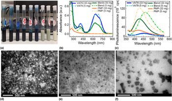
After verifying the potential for energy transfer between PMP and VA79, binary PMP:VA79 nanoparticles were prepared from a 1:1 weight ratio of starting materials with varying concentrations of the SDS surfactant using the miniemulsion technique as outlined previously (with specific details for this work reported in Supplementary Material).[Reference Holmes, Marks, Cave, Feron, Barr, Fahy, Sharma, Pan, Kilcoyne, Zhou, Lewis, Andersson, van Stam, Walker, Moons, Belcher and Dastoor18]Figure 2(a) shows images of the nanoparticle suspensions of pure VA79, the blended VA79/PMP mixture and pure PMP. All suspensions visually appeared to be composed of uniformly suspended material with little evidence of phase separation of materials. UV–visible absorbance and photoluminescence spectra of the nanoparticle suspensions were obtained on samples prepared by diluting the stock suspensions shown in Fig. 2(a) by a factor of 1000 such that the peak absorbance was below 0.5 in order to minimize nonlinear optical processes. The absorbance spectra of the VA79 nanoparticles show an onset at 780 nm [Fig. 2(b)], consistent with the solid-state behavior observed in Fig. 1(b). Furthermore, the 5 mg sample shows distinct evidence of peak splitting in the main absorption band located at ~650 nm distinct evidence of solid-state absorption behavior. The PMP suspensions show an absorbance peak at 320 nm, again in agreement with peaks observed for the solid films in Fig. 1(b). The blended VA79/PMP NP suspensions show an absorbance onset at 780 nm and some fine structure to the absorbance peak centered at 650 nm, consistent with the presence of solid-state VA79. They also show substantially enhanced absorbance at 318 nm, indicating the presence of PMP in the suspensions and implying the nanoparticles contain both component materials inside the particle structure. Curiously, the photoluminescence of the 33 mg particles appears at 415 nm, in the same position as the PMP sample, while the photoluminescence for the blended samples prepared with 5 mg of SDS appears at 462 nm, in agreement with the position of the pure VA79 emission [Fig. 2(c)]. This result could indicate a different chemical composition for the particles prepared with different SDS content, or could perhaps be a result of the SDS tuning the particle size and influencing energy transfer interactions between materials in the blend. The presence of nanoparticles in the suspensions was confirmed through transmission electron microscopy (TEM) imaging, with sample preparation and instrument settings described in Supplementary Material. Nanoparticles were detected in all the samples prepared with 5 mg of SDS [Figs. 2(d)–2(f)]; however, the 33 mg samples were covered with excess SDS and became unstable under the electron beam leading to very poor quality images. The particle sizes determined from a digital analysis of the TEM images with ImageJ were determined to be 27 ± 5 nm for pure VA79, 26 ± 7 nm for the blended VA79/PMP mixture, and 108 ± 20 nm for the pure PMP materials. Scanning electron microscopic imaging and dynamic light scattering (DLS) measurements verified these particle sizes and the presence of a thick SDS coating for the 33 mg SDS samples (Supplementary Fig. S6). DLS measurements also revealed that the samples prepared with 5 mg of SDS were on average 50–75% larger than those prepared with 33 mg of SDS (Supplementary Fig. S7). These results show clear evidence that a blended nanoparticle can be prepared from the radiation scintillator PMP and the semiconducting polymer VA79, with the nanoparticle size easily tuned by the SDS surfactant content. Solid films cast onto ITO-glass electrodes for the purposes of radiation sensor devices showed uniform thin films with an even distribution of nanoparticles throughout the films. The nanoparticle sizes were consistent with the particle sizes determined in aqueous suspension (Supplementary Fig. S8) and indicate a uniform connection of individual nanoparticles into a continuous solid film without aggregation or phase separation.
(a) Photographs of the nanoparticle suspensions prepared from pure VA79 (left three samples), blended VA79 and PMP (center three samples), and pure PMP (right three samples). (b) UV–visible absorbance spectra and (c) photoluminescence spectra for nanoparticle suspensions prepared from VA79 (blue), the VA79/PMP blend (green), and PMP (orange) with either 33 mg (solid line) or 5 mg (dashed line) of SDS surfactant. TEM images of nanoparticles prepared with 5 mg of SDS surfactant for (d) VA79, (e) the VA79/PMP blend, and (f) PMP.

In order to investigate whether energy transfer was indeed successfully induced in these samples, three distinct sample sets were prepared. The first sample set was composed of mixed chloroform solutions, where the PMP content was systematically varied from 0 to 50% by weight while keeping the total solid concentration at 20 mg/mL for all samples. In order to obtain appropriate concentrations for absorbance and photoluminescence data collection, these samples were diluted by a factor of 2000 prior to measurement. The second sample set involved solid film spin cast from the mixed 20 mg/mL chloroform solutions at 5000 RPM, creating samples with a thickness of 70 ± 10 nm. The third sample set consisted of the pure VA79, blended VA79/PMP, and pure PMP nanoparticles prepared with 5 mg of SDS.
Photoluminescence spectra were collected for the three sets of samples. These spectra were corrected as described in Supplementary Material to account for the differing amounts of VA79 in each mixture in a sample set and then further corrected for the fractional absorbance of both VA79 and PMP at the excitation wavelength of 315 nm. The corrected photoluminescence spectra then represent the emission output from VA79 (or PMP) after ensuring an identical number of photons were absorbed by the VA79 (or PMP) component of each sample, and that there is also an identical nominal amount of VA79 (or PMP) molecules present for emission in all samples.
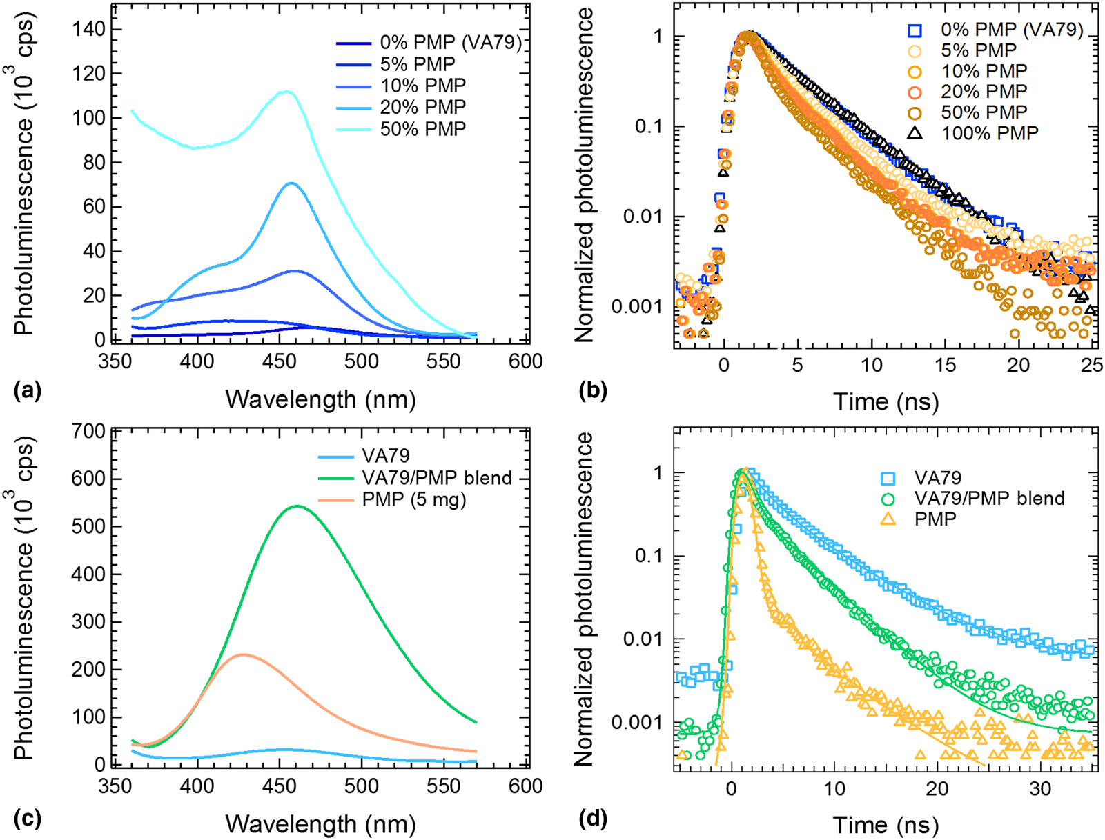
The corrected photoluminescence spectra for the chloroform solutions showed emission peaks that were identical within the noise of the measurement for all mixture ratios (Supplementary Fig. S4), indicating that none of the photons absorbed by the PMP molecules at 315 nm are transferred to VA79 in solution. This result is consistent with the expected separation between molecules in the 10 µM solutions being of the order of hundreds of nanometers, while the required distance for energy transfer is of the order of 10 nm as calculated earlier. In contrast, the solid films showed substantial growth of the VA79 emission peak at 462 nm with the addition of increasing PMP content [Fig. 3(a)]. At the maximum loading of 50%, the photoluminescence peak was increased by a factor of 16.2 compared to the pure VA79 film. The PMP peak at 410 nm was also correspondingly quenched, although observation of this required the spectra to be renormalized to correct for the linear absorptance of PMP at 315 nm rather than VA79 (Supplementary Fig. S5). This enhanced photoluminescence in the mixed solid films implies a strong energy transfer from PMP to VA79 in the mixed films. This energy transfer conclusion was confirmed by analyzing the photoluminescence lifetime of the PMP molecules in the mixed films. Samples were excited with sub-ns pulses from a 340 nm pulsed LED and monitored at an emission wavelength of 410 nm where only the PMP emits [Fig. 1(c)]. The VA79 lifetime was obtained for comparison by monitoring the emission at 770 nm. Figure 3(b) shows that the lifetime of the PMP is systematically quenched from its natural value as increasing amounts of PMP are added. The lifetimes were fitted with a double exponential function, with a time constant of 3.1 ns carrying the dominant (95%) weighting for PMP and a longer time constant of 5.2 ns (5% weighting) required to obtain a satisfactory fit. This dominant component decreased systematically with an increasing VA79 ratio, from a value of 3.1 ns for pure PMP films down to 1.2 ns for the 50% VA79:PMP film (Supplementary Table S1). The longer component showed similar behavior, decreasing from 5.2 ns down to 1.9 ns for the 50% mixed film. This behavior supports the observations from the steady-state photoluminescence of the PMP excited state being quenched due to energy transfer to VA79 in these mixed systems.[Reference Gopich and Szabo30]
(a) Steady-state and (b) time-resolved photoluminescence for solid films prepared with varying weight ratios of VA79 and PMP. (c) Steady-state and (d) time-resolved photoluminescence for nanoparticle suspensions prepared with 5 mg of SDS and VA79, PMP, and a VA79/PMP blend. Steady-state spectra were acquired by exciting at 315 nm, while the time-resolved spectra were acquired by exciting at 340 nm and measuring emission at 410 nm.

The nanoparticle suspensions exhibited a similar result, with the corrected steady-state VA79 photoluminescence peak showing an increase in magnitude by a factor of 18.9 in the VA79/PMP-blended nanoparticles compared to the pure VA79 sample, indicative of strong intermolecular energy transfer within the blended nanoparticles [Fig. 3(c)]. The time-resolved photoluminescence again required a double exponential fit to adequately account for the experimental data; however, the dominant (94% weighting) time constant for PMP became much faster (0.4 ns), while the slower component remained at 5.0 ns. The blended samples showed a quenching of the slower component to 3.7 ns; however, the fast component was not quenched [Fig. 3(d) and Supplementary Table S2]. We postulate that the faster component may be due to oxygen quenching introduced into the water-based nanoparticle suspensions, although this requires further investigation in future studies.
Radiation sensitivity of the blended mixtures
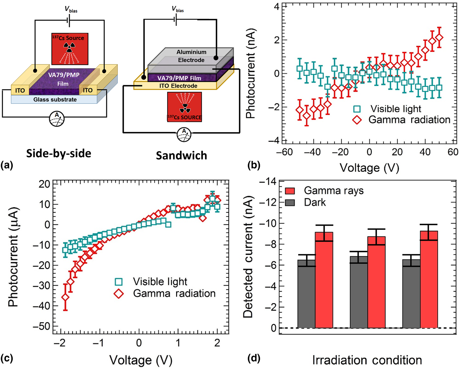
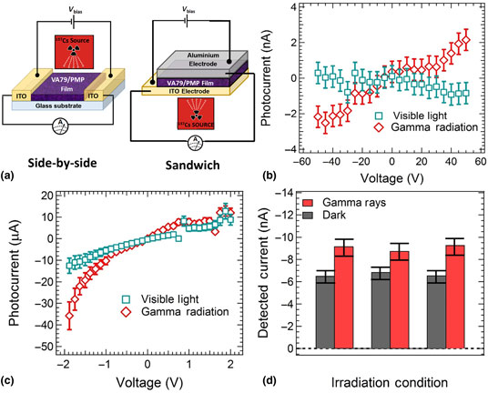
After confirming energy transfer from the PMP scintillator to the VA79 semiconductor, simple photoresistive radiation sensors were prepared from both the pure VA79 and the blended samples. Electronic devices were fabricated in two different configurations to investigate the influence of sensor geometry on device sensitivity. A side-by side electrode configuration was prepared from a spin-coated 300-nm layer of the organic film deposited across a 20-µm isolated channel between two opposing 5 mm2 ITO electrodes. A sandwich configuration was also prepared by spinning a 300-nm organic film onto an ITO electrode and then thermally evaporating an aluminum contact on top of this film to define an active area of 5 mm2 [Fig. 4(a)]. Four identical devices were prepared for both sensor geometries, with error statistics calculated as the standard error around the average current output across the four devices. This standard error is shown as the magnitude of the error bars on the data in [Figs. 4(b)–4(d)]. The side-by-side electrode configuration exhibited a relatively low current response due to the 20-µm transit distance for charges through the semiconductor. Accordingly, a large bias voltage ranging from −50 to +50 V was applied across these ITO contacts in order to induce a current response from these devices. For the pure VA79 sample, illumination of the semiconductor with a Xenon lamp caused a statistically significant increase in the current above the relatively large dark background when the bias voltages were increased to values exceeding ±25 V (Supplementary Fig. S8). This observation was attributed to a photoinduced change in conductivity of VA79 as has been previously observed for other organic semiconductors.[Reference Elkington, Wasson, Belcher, Dastoor and Zhou12] However, irradiation of these pure VA79 samples with a 137Cs gamma ray source at a dose rate of 0.5 mGy/s created a negligible change in the photocurrent that could not be detected outside the noise level of the dark current (Supplementary Fig. S8b).
(a) The two different device structures employed to create a radiation sensor from the mixed VA79/PMP material system. Photocurrent plotted as a function of bias voltage for sensors fabricated with a 50% mixture of VA79 and PMP in the (b) side-by-side device configuration and (c) sandwich configuration. Data is shown for sensors under illumination from a Xenon lamp (green squares) and under irradiation from a 5 µCi 137Cs source at a dose rate of 0.5 mGy/s. (d) Repeated cycling from dark (black) to gamma-ray exposure (red) for the same side-by-side 50% VA79/PMP devices at a bias voltage of −45 V.

In contrast, upon the addition of the PMP scintillator to create a blended VA79/PMP organic layer, the side-by-side sensors exhibited a minor decrease in the output current under illumination from the Xenon lamp that was only statistically significant at very high bias voltages (exceeding ±45 V). However, a detectable increase in the photocurrent was observed under irradiation from the gamma source at a dose rate of 0.5 mGy/s for bias voltages exceeding ±25 V [Fig. 4(b) and Supplementary Fig. S9]. The response was observed to be linear with respect to bias voltage, which is consistent with the symmetric electrode workfunctions creating an Ohmic device response. This radiation sensitivity result is attributed to the PMP absorbing the gamma radiation and transferring the excited state to the VA79 in order to influence the semiconductor conductivity and suggests that the energy transfer observed between these two materials can create a successful sensor for ionizing gamma radiation. However, it is noted that the side-by-side sensor provides relatively small increases in the photocurrent 2.2 ± 0.7 nA above a proportionally larger background dark current of 9.7 ± 0.5 nA for the bias voltage of −45 V where maximum sensitivity was observed. This represents a sensitivity of approximately 130 nC/Gy/mm2, although there is significant uncertainty in this value given the single-dose rate monitored. To improve this output response ratio, new devices were fabricated with a sandwich geometry that reduces the transit length of charges from 20 to 0.3 µm, while simultaneously expanding the cross-sectional contact area from 0.0015 mm2 in the side-by-side device to 5 mm2 in the sandwich geometry. The sandwich-type sensors exhibited much larger raw output currents and could also be operated at bias voltages an order of magnitude lower than the side-by-side sensors (±2 V), indicative of similar electric field strength being achieved across the thinner transit path in the sandwich devices at lower external bias voltage. The current response exhibited diode-like behavior, with a limited current that plateaued in forward bias, consistent with the asymmetric ITO and aluminium contacts creating a Schottky junction for this sensor configuration. The photocurrent signal varied from −3.5 ± 0.9 µA at a bias voltages of −0.3 V to −40 ± 9 µA at a bias voltage of −2.0 V under exposure from the gamma radiation source. This signal was readily distinguishable from the background dark current values of −0.3 ± 0.2 and −4 ± 1 µA, respectively, and represents a device sensitivity of 300–400 nC/Gy/mm2 estimated for the single-dose rate of 0.5 mGy/s. The sandwich devices also showed a current response under illumination from the Xenon lamp; however, the signal from gamma radiation exposure was higher and clearly distinguished from the visible light output at bias voltages exceeding −1 V [Fig. 4(c) and Supplementary Fig. S10]. Finally, the reproducibility of gamma sensitivity response was investigated by repeatedly switching the side-by-side device under a bias voltage of −45 V from dark conditions to exposure under the gamma radiation source for 2 s at a time. The device showed a reproducible current output of 9.0 ± 0.4 nA under gamma radiation exposure, which relaxed back to a reproducible dark current output of 6.6 ± 0.3 nA, indicating a stable and consistent photocurrent response of 2.4 ± 0.7 nA that could be resolved outside the noise of the background dark current after multiple exposure events.
This work has shown that the combination of organic scintillator PMP and organic semiconductor VA79 can induce the transfer of absorbed energy in the PMP molecule to the VA79 molecule with high efficiency. Blended nanoparticles were created from the material combination in order to reduce the intermolecular distance, creating a highly efficient energy transfer pathway. Creation of simple photoresistive radiation-sensing films from these materials showed a successful detection of gamma radiation which was not present in the control semiconducting sample without the PMP scintillating component. These results highlight a pathway toward low-cost flexible-printed radiation sensors that are water equivalent due to the organic active materials.
Supplementary material
The supplementary material for this article can be found at https://doi.org/10.1557/mrc.2019.132.
Acknowledgements
Financial support for this work through the University of Newcastle Strategic Investment Award (10.23285) is gratefully acknowledged. The authors thank Dr. Yun Lin and the University of Newcastle Electron Microscopy and X-ray Unit for technical assistance with electron microscopy measurements. This work was performed in part at the Materials node (Newcastle) of the Australian National Fabrication Facility (ANFF), which is a company established under the National Collaborative Research Infrastructure Strategy to provide nano- and microfabrication facilities for Australia's researchers.