Introduction
Pneumomediastinum is defined as the presence of free air within the mediastinum.Reference Newcomb and Clarke1 Pneumomediastinum can be spontaneous or secondary to trauma, viscous perforation (ruptured abdominal organ) or infection with gas-producing organisms.Reference Macia, Moya, Ramos, Morera, Escobar and Saumench2 Patients with spontaneous pneumomediastinum can present with a myriad of symptoms, including chest pain, dyspnoea, neck pain and odynophagia.Reference Macia, Moya, Ramos, Morera, Escobar and Saumench2 Otolaryngologists are likely to become involved in the care of patients with spontaneous pneumomediastinum when such patients present with head and neck symptoms; hence, an awareness of this condition is important.
This case report describes an atypical presentation of spontaneous pneumomediastinum, and subsequent management within an otolaryngology department.
Case report
A 22-year-old man presented to the emergency department of a large teaching hospital complaining of pain in his neck and left anterior chest. The pain was worse on deep inspiration, when recumbent and on swallowing. He had not vomited, had no dyspnoea and was four hours post-prandial. His past medical history was significant for asthma.
On examination, the patient was afebrile with a respiratory rate of 16 breaths per minute, a pulse of 60 bpm, blood oxygen saturations of 99 per cent on air and a blood pressure of 108/70 mmHg. He was tender along the costochondral junctions of the sixth to the 10th ribs on the left, and his heart sounds were normal.
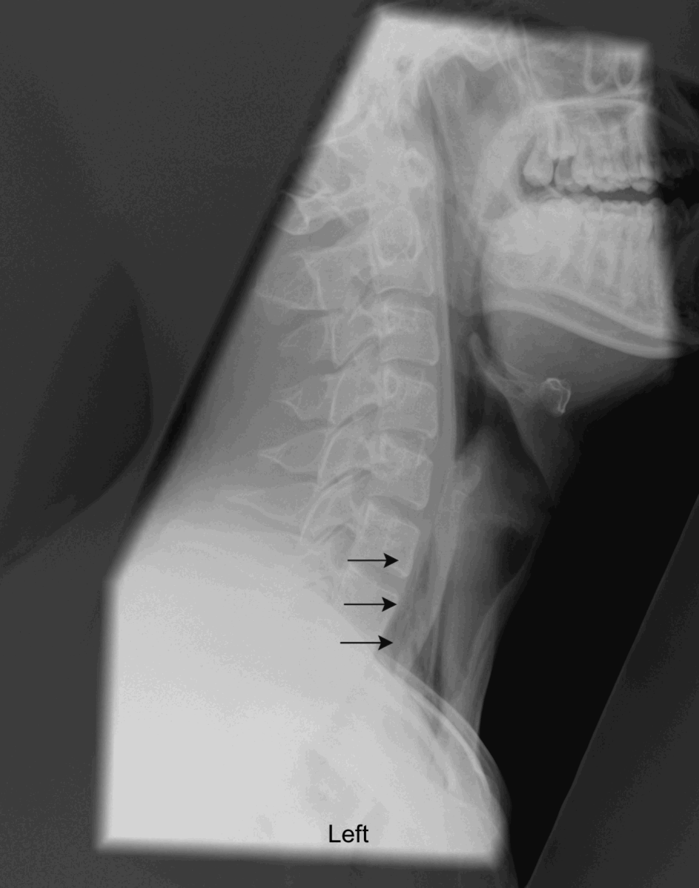
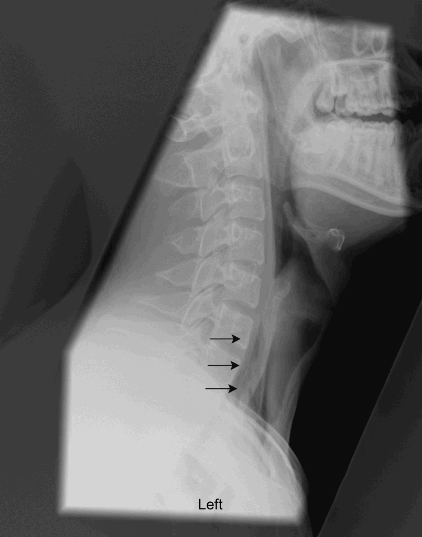
A lateral soft tissue neck radiograph (Figure 1) reviewed in the emergency department did not show any foreign body, and the patient was discharged. This radiograph was later reported as showing surgical emphysema of the neck, and so the patient was recalled to the emergency department, 15 hours after initial discharge.
Lateral X-ray of the neck for soft tissue detail, showing surgical emphysema (arrows). There is no evidence of a radiopaque foreign body.
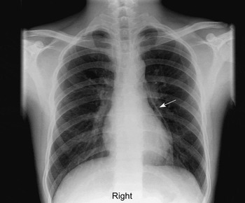
Subsequently, the patient was admitted to the otolaryngology department. No abnormality was seen on flexible nasendoscopy. A chest radiograph (Figure 2) confirmed pneumomediastinum. The patient was placed on a nil by mouth regime and commenced on intravenous antibiotics, following advice from the microbiology department.
Chest X-ray performed the day after initial presentation, showing pneumomediastinum (arrow).

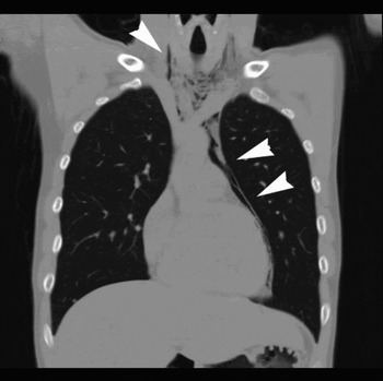
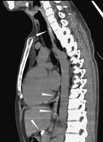
In order to exclude oesophageal perforation, further investigations were required. A contrast computed tomography (CT) scan was selected in preference to a contrast swallow test, and demonstrated a normal oesophagus with surgical emphysema and pneumomediastinum (Figures 3 and 4).
Coronal computed tomography (CT) of the neck, chest and abdomen with oral and intravenous contrast, confirming the X-ray findings. There is no leak of contrast. Such a CT is useful to confirm ‘silent’ pneumomediastinum and possible leak of contrast material.

Sagittal computed tomography scan showing oral contrast in the upper and lower oesophagus and in the stomach (arrowheads).

The patient was commenced on a soft diet, and was discharged three nights after admission. At the time of writing, he was well with no recurrence of the pneumomediastinum.
Discussion
Spontaneous pneumomediastinum is uncommon, ranging in incidence from one in 800 to one in 30 000 patients, and usually has a benign, self-limiting course.Reference Newcomb and Clarke1, Reference McMahon3, Reference Munsell4 It is associated with young, thin men, and links with a possible underlying elastic tissue disorder have been postulated.Reference Macia, Moya, Ramos, Morera, Escobar and Saumench2, Reference Jougon, Ballester, Delcambre, Mac Bride, Dromer and Velly5 Other associations include smoking (seen in 33 per cent of cases) and asthma (in 22 per cent).Reference Newcomb and Clarke1, Reference Macia, Moya, Ramos, Morera, Escobar and Saumench2
Macklin and Macklin used an animal model to elucidate the pathogenesis of spontaneous pneumomediastinum.Reference Macklin and Macklin6 After lung insufflation and alveolar rupture, hot gelatine was used to help show the pathway of air from alveolus to mediastinum. A pressure gradient from the alveoli to the mediastinum of 40 mmHg encouraged alveoli rupture. Air then tracked into the mediastinum. Spontaneous pneumomediastinum has thus been linked with circumstances that would induce raised alveolar pressure via the Valsalva manoeuvre. The use of illicit drugs, such as cocaine, has also been associated with spontaneous pneumomediastinum, although the postulated mechanism for this is breath-holding rather than secondary to any pharmacological effect.Reference Barbera Mir, Vallejo Galvete, Velo Plaza, Aviles Ingles, Ocon Alonso and Lahoz Navarro7
The most common presenting symptoms of spontaneous pneumomediastinum are chest pain and dyspnoea.Reference Macia, Moya, Ramos, Morera, Escobar and Saumench2 Symptoms such as neck pain are seen in 72 per cent of cases, and therefore presentation to otolaryngologists is common.Reference Munsell4 Vomiting is an associated symptom in 9.8 per cent of cases, and hence Mackler's triad (i.e. vomiting, subcutaneous emphysema and low chest pain), which is classically associated with Boerhaave syndrome, may be present, compounding the diagnostic difficulties.Reference Macia, Moya, Ramos, Morera, Escobar and Saumench2, Reference Mackler8, Reference Forshaw, Khan, Strauss, Botha and Mason9 Other important differential diagnoses include cardiac, pulmonary or musculoskeletal conditions. Clinical signs associated with spontaneous pneumomediastinum have a variable incidence. Hamman's sign, described as praecordial crunching or crackles synchronous with the heart sounds on auscultation, is only encountered in approximately one-third of patients.Reference Hamman10
Investigations which expedite diagnosis are important. Chest radiographs can be negative in up to 30 per cent of cases of spontaneous pneumomediastinum.Reference Kaneki, Kubo, Kawashima, Koizumi, Sekiguchi and Sone11 There is also a significant time lag between the emergence of clinical and radiological signs.Reference Panacek, Singer, Sherman, Prescott and Rutherford12 When the diagnosis is in doubt due to a confounding history and clinical signs, other investigations may be used. A CT scan with orally administered, water soluble contrast may be of value in such circumstances.
• The incidence of spontaneous pneumomediastinum ranges from one in 800 to one in 30 000
• Risk factors for spontaneous pneumomediastinum include asthma, male sex and slim build
• Clinical symptoms and presentation are variable, with Hamman's sign being present in only one-third of cases
• Chest radiographs can be negative in 30 per cent of cases, with a time lag between clinical and radiological signs
• A computed tomography scan with orally administered, water soluble contrast can aid diagnosis
Spontaneous pneumomediastinum is typically managed conservatively with bed rest, analgesia, oxygen, antibiotics and bronchodilators.Reference Macia, Moya, Ramos, Morera, Escobar and Saumench2 Associated complications include pneumothorax, which may require chest drain insertion.Reference Newcomb and Clarke1 This is in contrast with the clinical picture of oesophageal rupture, which has a mortality rate of 8 to 60 per cent.Reference Wilde and Mullany13
Conclusion
Although spontaneous pneumomediastinum is uncommon, awareness amongst otolaryngologists is important due to its variable presentation and clinical course. Important and potentially life-threatening differential diagnoses must be excluded using appropriate investigations. As in the case presented, a CT scan with orally administered, water soluble contrast can be beneficial when the diagnosis, and hence the management plan, is in doubt.