INTRODUCTION
The Sarcoptes mite was regarded as one of the main infectious causes of mortality in the cheetah (Acinonyx jubatus) population in the Masai Mara ecosystem (Kenya) (Mwanzia et al. Reference Mwanzia, Kock, Wambua, Kock and Jarret1995; Ngoru and Mulama, Reference Ngoru and Mulama2002). This threatened population is estimated at only 61 animals (Gros, Reference Gros1998), sharing habitat with other wild and domestic animals that are known to be affected by the Sarcoptes mite (Siegmund et al. Reference Siegmund, Fraser, Archibald, Blood, Handerson, Howell, Kitchell, Siegmund and Fraser1973; Mugera et al. Reference Mugera, Bwangamoi, Wandera, Mugera, Bwangamoi and Wandera1979; Blood and Radostitis, Reference Blood, Radostitis, Blood and Radostitis1989; Ngoru and Mulama, Reference Ngoru and Mulama2002; Pence and Ueckermann, Reference Pence and Ueckermann2002; Kahn et al. Reference Kahn, Line, Allen, Anderson, Jeffcoh, Quesenberry and Radostitis2005). The Masai Mara ecosystem is inhabited by pastoral communities and the interaction between wildlife and livestock creates a rich platform for disease transmission in this shrinking ecosystem (Holmes, Reference Holmes1996). The illegal incursion of livestock into protected areas occurs mostly during the dry season, which increases the chance of wildlife/livestock interaction. Major outbreaks of disease in this interface system have been associated with droughts, when limited water and food supplies cause increased interaction between livestock and wildlife (Kock et al. Reference Kock, Wambua, Mwanzia, Wamwayi, Ndungu, Barett, Kock and Rossiter1999).
A number of mite species that affect domestic animals and wildlife (Pence and Ueckermann, Reference Pence and Ueckermann2002; Kahn et al. Reference Kahn, Line, Allen, Anderson, Jeffcoh, Quesenberry and Radostitis2005; Alasaad et al. Reference Alasaad, Walton, Rossi, Bornstein, Abu-Madi, Soriguer, Fitzgerald, Zhu, Zimmermann, Uade Ugbomoiko, Pei and Heukelbach2011c) are of zoonotic importance (Bornstein et al. Reference Bornstein, Morner, Samuel, Samuel, Pybus and Kocan2001; Fischer et al. Reference Fischer, Holt, Harumal, Currie, Walton and Kemp2003; Kahn et al. Reference Kahn, Line, Allen, Anderson, Jeffcoh, Quesenberry and Radostitis2005; Navarro-Gonzalez et al. Reference Navarro-Gonzalez, Serrano, Casas-Díaz, Velarde, Marco, Rossi and Lavín2009; Alasaad et al. Reference Alasaad, Ndeereh, Rossi, Bornstein, Permunian, Soriguer and Gakuya2012). The Sarcoptes mite is a contagious skin disease (Zumla and Croft, Reference Zumla and Croft1992) and is spread by direct and indirect contact with other diseased animals or objects that have been in contact with affected animals (Siegmund et al. Reference Siegmund, Fraser, Archibald, Blood, Handerson, Howell, Kitchell, Siegmund and Fraser1973). The introduction of a single case of sarcoptic mange into overcrowded living conditions can lead to an epidemic (Obasanjo et al. Reference Obasanjo, Wu, Conlon, Karanfil, Pryor and Moler2001) causing potentially devastating mortality in both wild and domestic animals (Bornstein et al. Reference Bornstein, Morner, Samuel, Samuel, Pybus and Kocan2001).
Sarcoptic mange has been reported in a number of different wildlife species in the Masai Mara ecosystem (Mwanzia et al. Reference Mwanzia, Kock, Wambua, Kock and Jarret1995; Ngoru and Mulama, Reference Ngoru and Mulama2002), although a general study on the various aspects of Sarcoptes mite epidemiology and affected cheetah treatment has yet to be performed. Any such epidemiological and control study would have to confront the difficulties inherent to a complex wild/domestic animal system. All previous observational studies of mange-like skin diseases in cheetahs are from the Koiyaki and Lemek community conservancies in Masai Mara (Mwanzia et al. Reference Mwanzia, Kock, Wambua, Kock and Jarret1995).
The aim of our study was to contribute to knowledge of the epidemiology of the sarcoptic mange in the threatened cheetah population from the wildlife/livestock ecosystem Masai Mara, and to report on the treatment strategies undertaken thus far.
MATERIALS AND METHODS
Study area
The study was carried out in the Masai Mara ecosystem (Kenya), which comprises the Masai Mara National Reserve (NR), the Mara Conservancy and the surrounding community ranches of Koiyaki, Siana, Lemek, Olkinyei and Ol Choro Orogwa. The Masai Mara N.R. and Mara Conservancy form the protected area of the Masai Mara ecosystem (10°13′ and 10°45′ S, and 34°045′ and 35°25 E), which covers approximately 1,850 km2 and is located at the northern tip of the Serengeti National Park (Tanzania).
In the study area, all members of the ‘Big Five’ animal group (lion, leopard, African elephant, African buffalo, and Black Rhinoceros) are present. The wildebeest are the dominant herbivores, and their numbers are estimated in millions. Around July, these animals migrate north from the Serengeti plains in search of fresh pasture, and return to the south around October. The migrants are followed by predators, most notably lions and hyenas. Other herbivores are present including Thomson's and Grant's gazelles, impalas, topi, elands, duikers, Coke's hartebeests, zebras and Masai giraffes. A pastoral community whose livelihood is dependent on livestock lives in the surrounding area and, as a result, interaction between livestock and wildlife is frequent.
Animal data
Observational data of mange-like skin disease was collected over a period of 2 years (November 2007–November 2009). The disease was recorded based on clinical observation, if at least 3 of the following lesions were observed in an animal: pruritus, alopecia, crust formation, skin roughening and poor body condition. The study area was divided into 8 blocks (Fig. 1). Our study was a combination of opportunistic and systematic (cross-sectional) sampling. Affected lions, wildebeests and dogs were opportunistically reported by KWS veterinarians and Masai Pastoralists. For cheetahs, Thomson's gazelles and all domestic animals we carried out monitoring 4 times per year (October, January, April and July). Monitoring was carried out by 2 veterinarians and 1 technician. A distance of about 200 km per 2 days was covered per each block in each time (October, January, April and July). Domestic animals were randomly selected at both household and individual levels. The total number of herds of studied cattle, goats and sheep were 15, 20 and 32, respectively. Wild animals were observed using binoculars and the naked eyes. Since the Masai Mara area is quite secure for wildlife with high visitation, most wild animals are not shy and can be approached closely. The animals observed were cheetah (Acinonyx jubatus), Thomson's gazelle (Eudorcas thompsonii), sheep (Ovis aries), cattle (Bos indicus), goat (Capra hircus), dog (Canis lupus familiaris), wildebeest (Connochaetes taurinus) and lion (Panthera leo). GPS co-ordinates of all individual animals and/or herds observed to have clinical signs of mange, as well as of healthy herds and animals, were recorded. The size of herds varied from 50 up to 300 animals. The seasons were divided into the dry season (January–March and July–September) and wet season (April–June and October–December). The studied wild and domestic species were monitored for Sarcoptes-like lesions in all blocks except block 8 (Ol Choro Orogwa), due to its inaccessibility.
Map showing the study blocks in the Masai Mara ecosystem (Kenya) together with the distribution of affected animals of all species.

A stratified random sampling method was used to identify the sampling units. Domestic animal stratification was based on the study blocks (community ranches) that were closest to the protected areas or to where cheetahs are known to occur.
Within the strata, mangy animals were randomly sampled. Cheetah sex and age classes (adults ⩾1 year and cubs <1year) were determined.
Capture and treatment of mange-infected animals
Animals were determined to be affected through observation of clinical signs. Any animal that showed a mange-like skin condition was a candidate for treatment. Wild herbivores were captured by chemical immobilization through darting using etorphine hydrochloride (M99® 9·8 mg/ml, Novartis South Africa Pty Ltd, Isando, South Africa) combined with Xylazine hydrochloride (Ilium Xylazil-100 100 mg/ml, Troy Laboratories Pty Ltd, Smithfield, Australia) while wild carnivores were captured using ketamine hydrochloride (100 mg/ml Agraket®, Agrar Holland Bv) combined with Xylazine hydrochloride. Mangy domestic animals were treated since it was easy to restrain them. The immobilized wild animals were treated in the field with 0·2 mg/kg bwt of injectable 1% Ivermectin (Kalamectin 1% w/v, Kela NV, St Lenaartseweg, Belgium), administered subcutaneously.
After sampling and treatment, the herbivores were revived with diprenorphine hydrochloride (M5050® 12 mg/ml, Novartis South Africa Pty Ltd, Isando, South Africa) and Atipamezole hydrochloride (Antisedan® 5 mg/ml, Pfizer laboratories Pty Ltd, Sandton, South Africa) immediately, while the carnivores were revived using Atipamezole hydrochloride after 30 to 45 min of immobilization to allow Ketamine hydrochloride to wear off. Domestic animals were re-treated after 1 month, if necessary. Treated wild animals were ear-tagged to follow their progress.
Ethics
The Committee of the Department of Veterinary and Capture Services of the Kenya Wildlife Service (KWS) approved the study including animal capturing and treatment protocols. KWS guidelines on Wildlife Veterinary Practice-2006 were followed. All KWS veterinaries were guided by the Veterinary Surgeons Act Cap 366 Laws of Kenya that regulates veterinary practices in Kenya.
Parasite collection and identification
Affected areas of skin from representative samples randomly selected (namely: 8 cheetahs, 10 Thomson's gazelle, 9 dogs, 51 sheep, 5 wildebeest, 2 cattle, 1 goat and 2 lions) were scraped with a scalpel until they bled to obtain hairs and crusts for parasitological examination. The scrapings were placed in universal bottles containing 70% ethanol and transported to the laboratory. A portion was removed from the alcohol and clarified in KOH to recover parasites for microscopy (Alasaad et al. Reference Alasaad, Rossi, Soriguer, Rambozzi, Soglia, Pérez and Zhu2009a). All mites were identified as S. scabiei or Psoroptes ovis on the basis of known morphological criteria (Fain, Reference Fain1968; Margaret and Rusell, Reference Margaret, Rusell, Margaret and Rusell1978; Sanders et al. Reference Sanders, Froggatt, Wall and Smith2000).
Data analyses
R Package V.2.11.1. software was used. A (log +1) transformation had been applied to all variables prior to analysis. General Linear Models (GLMs) with binomial, quassibinomial distribution and logit link were tested following the proposal of Vicente et al. (Reference Vicent, Höfle, Garrido, Fernández-de-Mera, Juste, Barral and Gortazar2006). After transformation, Linear Model with R (lm function) was applied.
RESULTS
The clinical observation together with the examination of the skin samples revealed that cheetah, Thomson's gazelle, wildebeest, lion, dog, goat and cattle all suffered from infestation by Sarcoptes scabiei, while sheep were affected by P. ovis.
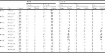
The estimated total number of mangy and healthy animals in the studied area is shown in Table 1. The prevalence of mange was highest in cheetahs 12·8% (n = 47), followed by dogs 4·7 (n = 279), while the lowest was 0·09% in cattle (n = 2311) and goats (n = 1174). The prevalence of affected Thomson's gazelle and sheep was 0·81% (n = 10 788) and 0·76% (n = 6699), respectively. Two herds of cattle (total 15 herds), 1 herd of goats (total 20 herds) and 13 of sheep (total 32 herds) were affected with mange mite giving a herd prevalence of 13·3%, 5% and 40·6% respectively. Only 2 lions and 5 wildebeest were found to be affected with sarcoptic mange. The prevalence of sarcoptic mange was 12·5% in female cheetahs and 9·6% in male cheetahs. The prevalence in adult cheetahs was 11·8% as opposed to cub cheetahs which was 15·3%. In both cases the differences were not statistically supported (P > 0·5). During the study period 2 cheetahs, a cub and an adult female died due to complications of mange infestation, while we did not identify any carcass of mangy Thomson's gazelle, but we cannot discard the possibility that affected carcasses were removed by scavengers.
Number of affected (mangy) and healthy animals in the Masai Mara ecosystem, stratified by block, year and season (dry and wet)

—, Not estimated.
Affected animals were observed in 6 of the 7 blocks studied (Fig. 1). No affected animals were observed in block 6. The greatest prevalence of affected animals was in blocks 1 to 5. Affected animals of different species were observed in the different blocks, namely cheetahs in blocks 1, 3, and 5; Thomson's gazelles in blocks 2, 3, 5 and 7; sheep in blocks 4 and 5; dogs in blocks 4 and 5; wildebeest in block 1. The affected lions were observed in block 2, while cattle and goat were observed in block 5. In terms of seasonality, affected animals were observed in blocks 1, 2, 3 and 5 during the dry season and in blocks 3 and 4 during the wet season.
Although we observed 5 affected wildebeest in 2 herds of about 100 animals which gave a prevalence of 2·0%, the sightings were from a single block at a single point in time, hence we did not carry out statistical analysis. That was the same case with the 2 lions that were observed as the study progressed. The 2 wild species were not included in the cross-sectional study.
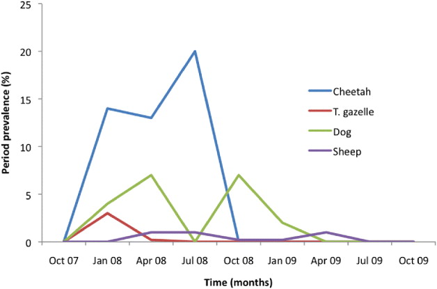
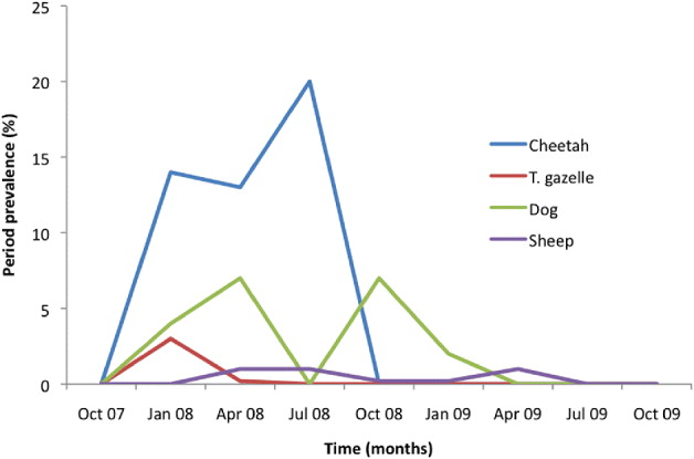
The temporal prevalence during the study period in all species is shown in Fig. 2. At the beginning prevalence increased steadily in cheetahs, Thomson's gazelles and dogs, with the peak infestation point in the first 2 being reached in January 2008. The peak infestation in dogs occurred later in April 2008. There was another peak in July 2008 in cheetahs and in dogs in October 2008, while the prevalence in sheep had a different pattern. No infestation was observed in cheetahs after October 2008 or in dogs after April 2009. Prevalence in Thomson's gazelles dropped steadily in April 2008 and was low up to April 2009.
The prevalence of Sarcoptes infestation in the study species from Masai Mara in the period 2007–2009.
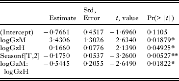
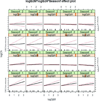
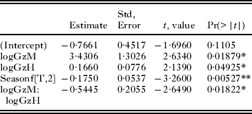
For all studied species (with the exception of cheetah) there was no statistical association with the geographical blocks or seasons (dry or wet). The prevalence of the Sarcoptes mite in cheetah was not associated to the geographical blocks, animal sex or the presence of affected domestic animals. Cheetah infection with S. scabiei was associated with the season (dry more than wet season; F = 10·1822, P = 0·007) and the interaction between the total number of healthy gazelles and the prevalence of infected gazelles (F = 7·4489, P = 0·018). The high prevalence of mangy gazelles had a negative effect on cheetah; nonetheless this negative effect gradually decreased when the number of healthy gazelles increased. The best model was season effect with an interaction between mangy and healthy gazelle (logMangyCheetah ∼ logMangyGazelle: logHealthyGazelle + Season) (For more details see Table 2 and Fig. 3).
GLM Model coefficients analysing the relationship between mangy cheetahs and seasons (wet and dry), and the total number of mangy and healthy gazelles (GzM, mangy gazelle; GzH, healthy gazelle; Seasonf [T.2], wet season. Pr(>|t|), significance level. Residual standard error: 0·117. Multiple R-squared = 0·5473. Adjusted R-squared = 0·4266. F-statistic = 4·534. P-value = 0·01337.)
Trellis of effects plots of the interaction logGzM (mangy gazelle), logGzH (healthy gazelle) and LogCh (mangy cheetah). All variables had been (log(x + 1) transformed. Horizontal axis: Mangy gazelles (GzM). Vertical axis: Mangy Cheetah (Ch). Each partial graph represents different values of healthy gazelles (GzH). Increasing values from left bottom corner to top right.

The treated animals recovered completely within approximately 1 month. Re-treatment (1 month after the first dose) was only necessary in a few cases in domestic animals, while none of the wild animals was re-treated during the study period. As the study progressed the numbers of observed affected animals declined due to the continuous treatment of all sampled animals, and by July 2009 no positive cases in any of the species studied were found, and none of the dead animals were reported to be caused by Sarcoptes mite (Fig. 2).
DISCUSSION
In this study S. scabiei was isolated in all animals that were positive for mange except sheep, where P. ovis was isolated. These observations agree with those reported by other authors who have described sarcoptic mange as the commonest in wildlife (Pence and Ueckermann, Reference Pence and Ueckermann2002), and psoroptic mange likewise as the commonest in sheep (Mugera et al. Reference Mugera, Bwangamoi, Wandera, Mugera, Bwangamoi and Wandera1979; Blood and Radostitis, Reference Blood, Radostitis, Blood and Radostitis1989; Kusiluka and Kambarase, Reference Kusiluka and Kambarase1996). Although affected sheep were consistently observed in the ecosystem throughout the study period, only Psoroptes mites were ever observed. This confirms the idea that there is no mange transmission between sheep and the other studied wild and domestic animals.
Our prevalence study of the mange-like skin disease revealed that cheetahs had the highest prevalence (12·8%) of all the studied animals.
Prevalence in female cheetahs was higher than in male cheetahs, and also higher in cubs than in adults. Although these prevalences are not significantly different, the greater prevalence in females and cubs does suggest that these two groups of cheetahs are more vulnerable since they live together and the probability of transmission is higher. No clear age structuring of scabietic wild animals was reported in many cases (Rossi et al. Reference Rossi, Fraquelli, Vesco, Permunian, Sommavilla, Carmignola, Da Pozzo and Meneguz2007). However, some reports indicate higher prevalences in young animals, and a decrease with advancement of age (Tikaram and Ruprah, Reference Tikaram and Ruprah1968; Munang'andu et al. Reference Munang'andu, Siamudaala, Matandiko, Munyeme, Chembensofu and Mwase2010; Alasaad et al. Reference Alasaad, Ndeereh, Rossi, Bornstein, Permunian, Soriguer and Gakuya2012).
Our study also revealed low prevalence in Thomson's gazelle, which, nevertheless, is still significant since it is to our knowledge the first report of mange in free-ranging Thomson's gazelles in Kenya. The sample size (n = 10 788) was very large and since this gazelle is a favourite prey item of the cheetah (Ngoru and Mulama, Reference Ngoru and Mulama2002; Hayward et al. Reference Hayward, Hofmeyer, O'Brien and Kerley2006), there is also a high probability that transmission occurs between these two species. Our study further shows that, although dogs had the second highest prevalence, affected dogs were observed exclusively in market places, which suggests that there transmission to cheetahs and other wild animals is unlikely in the study area.
No association was found between Sarcoptes infection in cheetah and sympatric domestic animals, and this suggests limited, if any, interaction between wild and domestic animals in the transmission and maintenance of this pathogen. This is in contrast with other disease models, in which the expansion of humans and their livestock into wildlife areas had resulted in increased risk of transmission (Bengis, Reference Bengis, Kock and Fisher2002; Jones et al. Reference Jones, Patel, Levy, Storeygard, Balk, Gittleman and Daszak2008). However, in this study affected cheetahs were found close to affected Thomson's gazelles, suggesting that there could be transmission of mites from Thomson's gazelles to cheetahs during feeding. Noteworthy, this gazelle is the favourite prey item of the cheetah (Ngoru and Mulama, Reference Ngoru and Mulama2002; Hayward et al. Reference Hayward, Hofmeyer, O'Brien and Kerley2006; Gakuya et al. Reference Gakuya, Rossi, Ombui, Maingi, Muchemi, Ogara, Soriguer and Alasaad2011). The high prevalence of mangy Thomson's gazelles had a negative effect on cheetahs, nonetheless this negative effect was contrasted when a high number of healthy gazelles was present. The specificity of S. scabiei by its host species has been the subject of an ongoing debate (Warburton, Reference Warburton1920; Burgess, Reference Burgess1994). This debate has been advanced with the advent of PCR technology and the molecular marker systems in the genetic era. Studies on the coding sequences in combination with microsatellite markers provided support for a genetic differentiation of S. scabiei (Walton et al. Reference Walton, Choy, Bonson, Valle, McBroom, Taplin, Arlian, Mathews, Currie and Kemp1999, Reference Walton, Dougall, Pizzutto, Holt, Taplin, Arlian, Morgan, Currie and Kemp2004; Alasaad et al. Reference Alasaad, Soglia, Sarasa, Soriguer, Pérez, Granados, Rasero, Zhu and Rossi2008, Reference Alasaad, Soglia, Spalenza, Maione, Soriguer, Pérez, Rasero, Ryser Degiorgis, Nimmervoll, Zhu and Rossi2009b, Reference Alasaad, Schuster, Gakuya, Theneyan, Jowers, Maione, Molinar-Min, Soriguer and Rossi2011b). In Europe, it was shown that the Sarcoptes mite is host-taxon derived in its effect on wildlife (Rasero et al. Reference Rasero, Rossi, Soglia, Maione, Sacchi, Rambozzi, Sartore, Soriguer, Spalenza and Alasaad2010), and that host-parasite specificity is temporally stable in the short term (Alasaad et al. Reference Alasaad, Oleaga, Casais, Rossi, Molinar-Min, Soriguer and Gortazar2011a). Nevertheless, the studied wild animals in Europe lacked any clear predator-prey interaction and putative inter-specific transmission models such as the cheetah and the Thomson's gazelle in this study could not be explored.
Our study also revealed greater numbers of affected cheetah in the dry as opposed to the wet season. A winter rise in the number of Sarcoptes outbreak episodes has been reported worldwide, especially in Europe, in several wild and domestic hosts (Bornstein Reference Bornstein, Morner, Samuel, Samuel, Pybus and Kocan2001; Rossi et al. Reference Rossi, Fraquelli, Vesco, Permunian, Sommavilla, Carmignola, Da Pozzo and Meneguz2007). This seasonal variation is thought to be related to the fertility of Sarcoptes mites, where the maximum egg production by adult female mites occurs in the autumn, and is low to null during January to July (Sokolova et al. Reference Sokolova, Radchenko and Lange1989). However, the seasonality of Sarcoptes cases in Africa is more related to the dry season, suggesting that stress factors such as the scarcity of water and grazing pastures during the dry season may lead to greater individual susceptibility to mite infection (Malan et al. Reference Malan, Horak, de Vos and van Wyk1997; Munang'andu et al. Reference Munang'andu, Siamudaala, Matandiko, Munyeme, Chembensofu and Mwase2010; Alasaad et al. Reference Alasaad, Ndeereh, Rossi, Bornstein, Permunian, Soriguer and Gakuya2012).
We noted an association between prevalence and the temporal effect in all the study animals. Mange prevalence was higher in 2007/2008 than in 2008/2009 and by July 2009 patent cases were hard to find. All wild animals treated were ear-tagged and we didn't recapture any that was affected by Sarcoptes that had been ear-tagged. However, it was difficult to estimate the recovery time of the wild animals due to environmental difficulties, and to the fact that our study was based on transect visits with trimester intervals, while animals may completely recover from sarcoptic mange infection in less than 3 months. For instance, Munang'andu et al. (Reference Munang'andu, Siamudaala, Matandiko, Munyeme, Chembensofu and Mwase2010) reported a recovery prevalence of 82% in mangy African buffaloes after a single dose of ivermectin, and Goldust et al. (Reference Goldust, Rezaee and Hemayat2012) reported a cure rate of 85·9% at a 2-week interval with a single dose of ivermectin in humans. It would be tempting to assume that treatment of clinically affected individuals belonging to a range of sympatric hosts contributed to control of sarcoptic mange at the population level but, with all evidence, the trial was not designed to test it. As a consequence, the alternative hypothesis that incidence of mange spontaneously decreased during the study period, as registered elsewhere (Pence and Uecke, Reference Pence and Ueckermann2002), cannot be ruled out. For further studies, radio-equipment of treated wildlife would be desirable to improve the quality of individual follow-up and adequately monitor the health of in-contact conspecifics.
In conclusion, our study shows that there is no evidence of an association between Sarcoptes infection in wild and domestic animals. Cheetah infection with Sarcoptes was associated with climatic stressors and the presence of mangy Thomson's gazelles ‘the favourite prey’; however, this negative effect was contrasted when a high number of healthy gazelles was present. Cheetahs may preferentially select mangy preys, since the affected preys could have a reduced flight response compared with healthy individuals. More investigation is needed to demonstrate the efficacy of the therapeutic treatment of mangy individuals as a strategy for effective and sustainable conservation of threatened cheetahs within Sarcoptes outbreak areas.
ACKNOWLEDGEMENTS
We thank the staff of the Department of Veterinary and Capture Services, Kenya Wildlife Service for assisting in data collection.
FINANCIAL SUPPORT
The research was supported by Kenya Wildlife Service, Proyecto de Excelencia RNM 06400 (Junta de Andalucía, Spain) and Juan de la Cierva Grant (Ministerio Innovación y ciencia, Spain).