Introduction
Congenital long QT syndrome is a hereditary arrhythmia disorder that features a prolonged QT interval on the electrocardiogram and can cause syncope or sudden death by triggering a characteristic polymorphic ventricular tachycardia known as torsade de pointes. To date, approximately 18 causative genes have been identified. Of them, KCNQ1 (type 1), KCNH2 (type 2), and SCN5A (type 3) are known as the primary causative genes, accounting for approximately 40%, 40%, and 10% of all cases, respectively. Reference Takase, Ikeda and Shimizu1
SCN5A (NM_000335.5) encodes the cardiac sodium channel. Gain-of-function mutations cause congenital long QT syndrome type 3, whereas loss-of-function mutations are known to cause Brugada syndrome, sick sinus syndrome, and progressive cardiac conduction disorders. Furthermore, variants exhibiting characteristics of both gain-of-function and loss-of-function mutations exist, such as p.Glu1783Lys (historically reported as p.Glu1784Lys). They are collectively termed SCN5A-related overlap syndromes. Reference Imai, Kusano and Aiba2
This report describes a family in which overlapping SCN5A-related phenotypes were suspected based on family history despite the absence of obvious abnormalities on the initial electrocardiogram at presentation, and the diagnosis was made following Holter monitoring. Informed consent was obtained from the affected family members for the reporting of this case.
Case
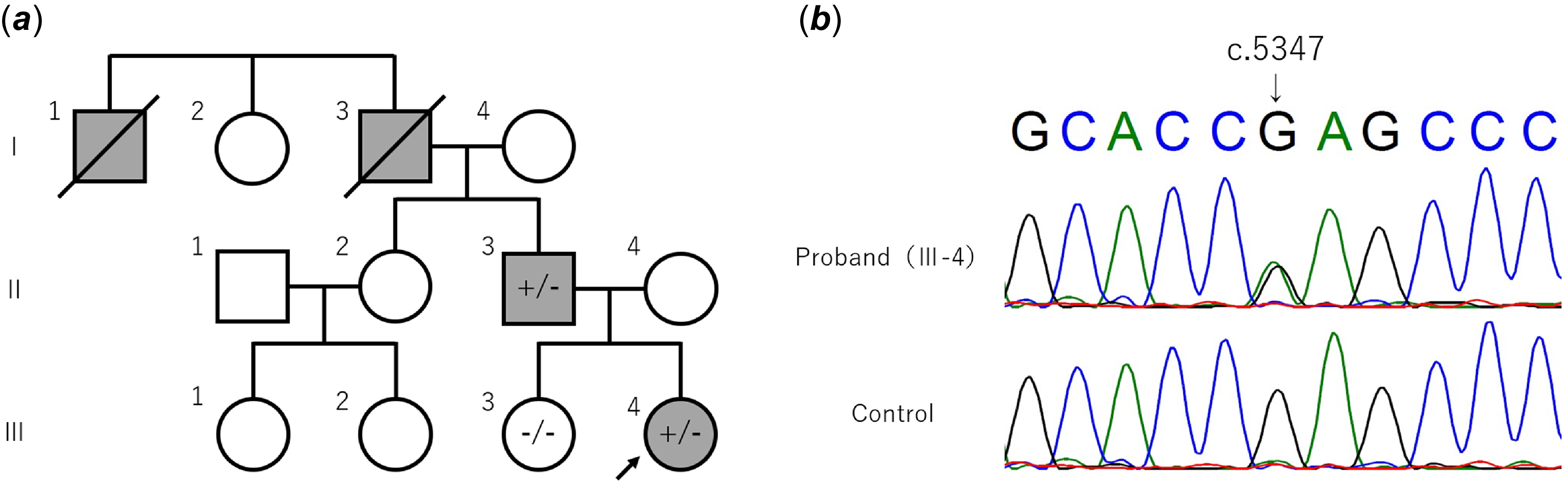
The proband (III-4) is a 10-year-old girl with no history of illness. Her school electrocardiogram screening at age 6 years showed no abnormalities, but screening at age 9 years indicated possible QT prolongation, leading to her presentation at our hospital. Her 12-lead electrocardiogram at presentation showed sinus rhythm at a heart rate of 91 beats per minute with no notable abnormalities (Figure 1a). Her QTc(F) was 423 ms at rest and 410 ms after triple-master stress testing, suggesting that long QT syndrome was unlikely. Her family history was negative for long QT syndrome, but her father (II-3) had been diagnosed with Brugada syndrome (Supplemental fig. 1) and had an implantable cardioverter-defibrillator (Figure 2a). In addition, there was a history of sudden death (I-1) and pacemaker implantation (I-3) in her paternal family line. Considering the possibility of overlapping SCN5A-related phenotypes, a Holter ECG was performed. Minimum, average, and maximum heart rates were 50, 77, and 150 beats per minute, respectively, with a total of 105,825 beats. No significant arrhythmia was detected. Though no abnormalities were observed on resting or exercise electrocardiograms, the QTc(F) during sleep was markedly prolonged at 550 ms (Figure 1b). On the other hand, a high right precordial lead electrocardiogram (V1 and V2 in the second intercostal space) did not reveal a diagnostic Brugada pattern, and signal-averaged ECG was negative for late potentials (filtered QRS duration, 99 ms; RMS40, 22.8 μV; LAS40, 37 ms). Based on these electrocardiogram findings and the family history, overlapping SCN5A-related phenotypes were strongly suspected. Following appropriate genetic counselling and obtaining written consent, genetic testing of both the patient and her father was performed at the National Cerebral and Cardiovascular Center (Osaka, Japan). Next-generation sequencing examined KCNQ1, KCNH2, SCN5A, KCNJ2, CACNA1C, CASQ2, RYR2, CALM1, CALM2, and CALM3. In both individuals, the SCN5A c.5347G>A (rs137854601), p.Glu1783Lys variant was confirmed in a heterozygous state. The proband subsequently began taking mexiletine, and a tendency towards QTc(F) shortening during sleep was observed. The older sister (III-3) of the proband showed no abnormalities on a 12-lead electrocardiogram or Holter monitor, with no SCN5A variants on genetic testing.
The proband’s electrocardiograms. (a) Initial 12-lead electrocardiogram. Sinus rhythm with a heart rate of 91 beats per minute. QTc(F) is 423 ms, with no ST changes suggestive of Brugada syndrome. (b) Waveform during sleep on the Holter electrocardiogram. QTc(F) is 550 ms.

Figure 1. Long description
Panel A: A 12-lead electrocardiogram graph. The horizontal axis represents time in seconds, and the vertical axis represents electrical activity in millivolts. The graph shows multiple leads labeled from V1 to V6 and aVR to aVF. The heart rate is 91 beats per minute, and the QTc(F) interval is 423 milliseconds. No ST changes suggestive of Brugada syndrome are visible. Panel B: A waveform graph from a Holter electrocardiogram during sleep. The horizontal axis represents time in seconds, and the vertical axis represents electrical activity in millivolts. The QTc(F) interval is 550 milliseconds.
Genetic information of this family. (a) Pedigree chart. The arrow indicates the proband. “+” indicates SCN5A c.5347G>A-positive, and “-” indicates SCN5A c.5347G>A-negative. (b) Sanger sequencing of SCN5A of the proband and healthy control sample with no genetic relationship.

Sanger sequencing was performed to confirm the variant. Polymerase chain reaction was performed using PrimeSTAR GXL DNA polymerase (Takara, Shiga, Japan) and the following primers: forward primer, TCTTTGCCCTCATGATGTCCC; reverse primer, ATCTGGATCTTCAGGGCGTC. A Big Dye Terminator 3.1 Cycle Sequencing Kit (Thermo Fisher Scientific, Waltham, MA, USA) with ABI PRISM 3130xL (Applied Biosystems, Foster City, CA, USA) was used to read the sequencing (Figure 2b). All reagents and equipment were used according to the manufacturers’ instructions.
Discussion
A family case, in which overlapping SCN5A-related phenotypes were suspected based on family history and QT prolongation during sleep on a Holter ECG, despite a normal ECG at presentation, that was confirmed by genetic testing, was reported.
In SCN5A-related inherited arrhythmia, different ECG patterns may be present in family members with the same variant, as in the present family, or a single patient may show both QT prolongation and Brugada phenotype. Reference Abu Dogoshh, Konstantino and Haim3 The p.Glu1783Lys in SCN5A identified in the present case was first reported as a pathogenic variant in 1999 Reference Wei, Wang and Alings4 and is now recognised as a frequent causative variant for overlap syndrome. Reference Makita, Behr and Shimizu5
In congenital long QT syndromes, cases like the present one where resting 12-lead ECGs do not show QT prolongation are not uncommon. Reports indicate that this occurs in 36% of type 1, 19% of type 2, and 10% of type 3 cases, and evaluation typically occurs during the recovery phase after exercise stress testing. Reference Priori, Schwartz and Napolitano6 However, in type 3, which manifests during sleep, exercise-induced variability is minimal, so significant findings may not be confirmed on outpatient electrocardiograms.
School-based electrocardiogram screening for children is most widely implemented in Japan, and in recent years, other countries have also begun introducing it. Reference Zimatore, Gallotta and Campanella7 Long QT syndrome is one condition that can be diagnosed early through this screening. The Japanese school cardiac screening guidelines do not mandate Holter monitoring for suspected QT prolongation. Reference Sumitomo, Baba and Doi8 However, this case highlights the utility of Holter monitoring. At he very least, for cases with a family history of Brugada syndrome, active consideration should be given to performing Holter monitoring. In such patients, genetic testing should also be considered.
Supplementary material
The supplementary material for this article can be found at https://doi.org/10.1017/S1047951126124263.
Acknowledgements
The authors would like to express their gratitude to Dr Yasuya Inden (Department of Cardiology, Nagoya University Hospital) for providing the data for the patient’s father. The authors thank FORTE Science Communications (https://www.forte-science.co.jp/) for English language editing.
Financial support
This work was supported by the Japan Society for the Promotion of Science KAKENHI Grant No. 25K10708.
Competing interests
The authors declare no conflict of interest.
Ethical standard
The authors assert that all work reported complies with the ethical standards of the Declaration of Helsinki and institutional and national research committees.