It has been recognised for many years that antisense oligonucleotides – short single-stranded nucleic acid sequences that inhibit target gene expression via sequence-specific, Watson–Crick base-pair hybridisation to sense mRNA – can be employed to selectively reduce the expression of disease-related proteins in vivo, and consequently to improve disease phenotypes. A large number of biotechnology companies have emerged in that time with a view to translating the promise of ‘antisense’ in cell culture and animal models to successful pharmaceutical products. Target proteins as varied as the low-density lipoprotein (LDL)–cholesterol component apolipoprotein B100 (Refs Reference Kastelein1, Reference Lemonidis2, Reference Wang3), the skeletal muscle protein dystrophin (Refs Reference Aartsma-Rus4, Reference Alter5, Reference Arechavala6), hepatitis C viral proteins (Ref. Reference McHutchison7) and intracellular signalling molecules (often proto-oncogenes such as MYC; Refs Reference Devi8, Reference Iversen9, Reference Leonetti10) have all been shown to be ‘knocked down’ in animal models using antisense oligonucleotides and tested in clinical trials. Since the approval in 1998 of the first antisense drug, fomivirsen (Vitravene®; Refs Reference Crooke11, Reference Galderisi, Cascino and Giordano12), which targets a cytomegalovirus mRNA, there has been a notable dearth of subsequent approvals for therapeutics of this type, due to a variety of factors preventing promising antisense candidates from reaching the clinic. Recently, however, there have been some positive clinical trial data from Isis Pharmaceuticals (Refs Reference Kastelein1, Reference Kastelein13), and several high-impact publications demonstrating excellent outcomes of antisense therapy in animal models of human disease (Refs Reference Alter5, Reference Yin, Lu and Wood14, Reference Madsen15, Reference Henke16). Thus, it is timely to review the pharmaceutical science underlying the in vivo effects of antisense oligonucleotides. We focus here on the nature of the biological barriers to in vivo knockdown and strategies that have already overcome each barrier or may do so in the future, and then analyse the efficacy and potency of antisense therapeutics after intravenous administration.
Natural deoxyribonucleotides are not used as antisense oligonucleotides in vivo because of stability and delivery problems, and several chemical modifications have been introduced over the past decade to improve the efficiency of antisense therapeutics. The pharmaceutical chemistry of antisense oligonucleotides is reviewed elsewhere (Refs Reference Urban and Noe17, Reference Kaur, Babu and Maiti18, Reference Faria and Ulrich19, Reference Prakash and Bhat20, Reference Crooke21). The modifications considered in this article include: (1) first-generation phosphorothioate (PS) DNA antisense; (2) second-generation 2'-O-alkyl (particularly 2'-O-methoxyethyl; MOE) ‘gapmer’ chimaeric DNA–RNA antisense (Ref. Reference Crooke21); (3) phosphorodiamidate morpholino (PMO) antisense (Ref. Reference Iversen22); (4) peptide nucleic acid (PNA) (Ref. Reference Nielsen23); and (5) locked nucleic acid (LNA) antisense (Refs Reference Kaur, Babu and Maiti18, Reference Jepsen, Sorensen and Wengel24). These modifications and others have achieved the goal of producing nuclease-resistant antisense agents that retain high efficacy in vitro. 2'-O-modification and LNA chemistries do not support RNase H activity, and oligonucleotides containing these chemistries therefore require regions free of 2'-modification to recruit this high-efficiency silencing mechanism. Monia and colleagues demonstrated that a region containing five or more 2'-deoxy nucleotides was required for RNase H activity (Ref. Reference Monia25). A major innovation allowing the benefits of MOE, 2'-O-methyl (OMe) and LNA chemistries to be realised without the loss of efficacy has thus been the use of gapmer strategies (Refs Reference Monia25, Reference Fluiter26, Reference Grunweller27, Reference Monia28) in which central regions of the oligonucleotide consisting of nucleotides whose chemistry supports RNAse H activity – that is, unmodified phosphodiester (PO) internucleoside linkages (PO or PS) – are flanked by 2′-modified regions (eg. MOE–PS–MOE). PMO oligonucleotides differ from the other modifications described above in that these agents are designed to work via steric inhibition of translation rather than degradation of mRNA transcripts.
This review focuses mainly on the barriers to efficacy of second- and third-generation antisense oligonucleotides in vivo, with a view to informing the use of antisense as both a research tool in animal models and a therapeutic platform in humans. Much of the research described herein is from animal studies that have revealed insights into antisense mechanisms; where informative human data are available, these have also been included.
General pharmacokinetic properties of antisense oligonucleotides
Antisense oligonucleotides exhibit predictable pharmacokinetic properties given their size and charge. They can exist as free molecules within the plasma or exhibit strong plasma protein binding depending on sequence and modification. Antisense oligonucleotides almost universally exhibit biphasic plasma concentration–time profiles, usually with a rapid initial decrease in plasma concentration within the first hour after intravenous injection in mice (Ref. Reference Yu29), rats (Ref. Reference McMahon30) and humans (Ref. Reference Yu29) (t 1/2α of around 3–30 min), even for nuclease-stable antisense (Refs Reference Yu31, Reference Raoof32), and a slower second phase of clearance over the next 24 h (t 1/2β) (Refs Reference McMahon30, Reference Zhang33). In humans, a similar pharmacokinetic profile is observed for PS oligonucleotides (Refs Reference Zhang33, Reference Sewell34, Reference Marcucci35). A cross-species comparison for ISIS 301012 found only small differences between the pharmacokinetic properties of the drug in mice, rats, monkeys and humans (Ref. Reference Yu29). For nuclease-stable oligonucleotides, the biphasic nature of the fall in plasma concentration represents a combination of renal excretion, tissue distribution and tissue clearance, considered in the following sections.
Tissue distribution
The kidney and liver are the predominant destinations for antisense oligonucleotides. More than 80% of the antisense oligonucleotide detected in tissues after intravenous administration is found in these two organs, and these observations are valid for both charged (Ref. Reference Yu31) and uncharged (Ref. Reference McMahon30) antisense oligonucleotides, and also after oral (Refs Reference Raoof32, Reference Cossum36) or intradermal (Ref. Reference Cossum36) delivery. When the spleen is included in the analysis, the proportion of dose within these tissues rises above 90% in many cases.
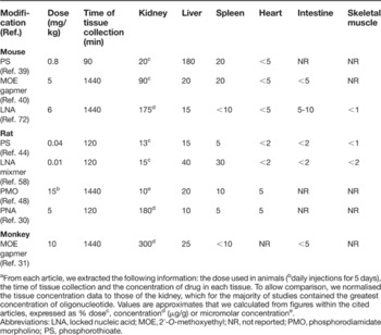
Table 1 shows representative examples of tissue distribution for oligonucleotides of varying chemistries. The major determinants of tissue distribution remain unclear to some extent, but blood flow, specific cellular uptake (e.g. reticuloendothelial function) and preference for organs with fenestrated vascular endothelia all appear to be relevant factors, as considered further in the next section. The liver and kidney receive large proportions of cardiac output, and these organs accumulate significant proportions of an administered oligonucleotide dose, with the kidney predominant for all chemical modifications other than PS (Table 1). However, oligonucleotide distribution is not always directly proportional to the blood flow to that organ. Collectively, skeletal muscle receives >20% of cardiac output in mice (Ref. Reference Barbee37) and rats (Ref. Reference Delp38), yet retains <5% of oligonucleotide across a variety of modification types and experimental protocols (Table 1), and the heart and gastrointestinal tract also show discordance between the proportion of cardiac output received and the proportion of oligonucleotide they contain (Table 1). Thus, there is preferential distribution of oligonucleotide to liver, kidney and spleen (and bone marrow) at the expense of other tissues.
Tissue distribution of the major classes of modified antisense oligonucleotides after systemic administrationa

a From each article, we extracted the following information: the dose used in animals (bdaily injections for 5 days), the time of tissue collection and the concentration of drug in each tissue. To allow comparison, we normalised the tissue concentration data to those of the kidney, which for the majority of studies contained the greatest concentration of oligonucleotide. Values are approximates that we calculated from figures within the cited articles, expressed as % dosec, concentrationd (μg/g) or micromolar concentratione.
Abbreviations: LNA, locked nucleic acid; MOE, 2′-O-methoxyethyl; NR, not reported; PMO, phosphorodiamidate morpholino; PS, phosphorothioate.
Tissue clearance of oligonucleotide
The clearance of oligonucleotide from the liver, kidneys and spleen is slow, and due in large part to nuclease degradation. The tissue half-life of most nuclease-resistant oligonucleotides is extremely long, with reported values generally expressed in days rather than hours (Refs Reference Geary39, Reference Geary40). The rate of clearance, and resultant duration of action of the oligonucleotide, is dependent on the modification, with MOE oligonucleotides demonstrating tissue half-lifes greater than 10 days across a number of tissues and across species (Ref. Reference Geary40).
Barriers to efficacy of antisense therapeutics
Overview
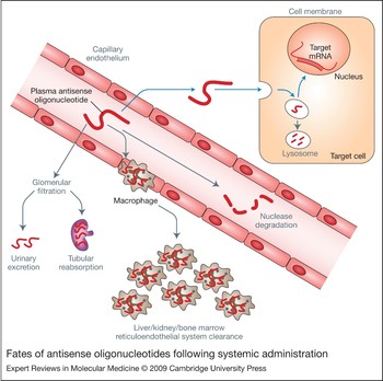
In order to gain access to complementary mRNA species within tissues after systemic administration, antisense oligonucleotides must avoid nuclease degradation, reticuloendothelial system uptake and renal excretion, traverse the microvascular endothelium, cross target cell membranes and escape the endosome–lysosome system (Fig. 1). The combination of these barriers has contributed to the requirement for larger doses to be used in animal experiments and clinical trials, and thus greater class-related and drug-specific toxicity. The failure of antisense oligonucleotides in clinical trial has resulted from deficiencies of both efficacy and safety (Ref. Reference Rayburn and Zhang41). In many cases it has been difficult to measure target knockdown as an indicator of efficacy. For those cases where clinical efficacy has been lacking, it is often difficult to assign this to a failure of stability, delivery to the target cell type, uptake, endosomal escape, mRNA cleavage, or choice of the therapeutic target. For example, the development of GEM91 by Hybridon was halted when dose-limiting thrombocytopaenia occurred in Phase II trial, but in any case the efficacy seen in Phase I trial (as indicated by levels of viraemia) was not replicated in the Phase II trial according to a press release (Ref. 42) – greater understanding of the fate and efficacy during the development of GEM91 may have prevented the failure or indicated that development should be halted at an earlier point. Where target knockdown can conveniently be correlated with disease outcomes, such as for ISIS301012 (Refs Reference Kastelein1, Reference Kastelein13), the close evaluation of efficacy during each stage of development allows for more informed drug development.
Fates of antisense oligonucleotides following systemic administration. (a) Successful gene silencing after systemic administration requires the movement of the antisense oligonucleotide from the blood into the interstitium, then into the target cell (nucleus) to reach the target mRNA (blue arrows). (b) However, there are several barriers and mechanisms to prevent successful knockdown (grey arrows and grey labels). Antisense oligonucleotides may be degraded in the blood, or removed by renal excretion or uptake by tissue macrophages. Antisense oligonucleotides may not reach their target cells because they are unable to escape across the vascular wall. Once in the interstitium, antisense oligonucleotides may be prevented from reaching their intracellular target (mRNA) by an inability to cross the cell membrane or escape the endosome–lysosome pathway.

Barrier 1: nuclease degradation
Studies using radiolabelled phosphodiester antisense oligonucleotides have shown that these agents are likely to have little utility in vivo because of rapid nuclease degradation (Refs Reference Roivainen43, Reference Lendvai44, Reference Kuhnast45), with <15% of intact drug detectable in rat plasma 20 min after dosing (Ref. Reference Lendvai44). The major modified antisense chemistries have overcome this problem to a large extent. PS oligonucleotides (Ref. Reference Roivainen43), MOE–PS–MOE gapmers, LNA oligonucleotides (Ref. Reference Fluiter46) and LNA–DNA–LNA gapmers (Ref. Reference Fluiter26) are stable in serum for extended periods, whereas OMe–PS–OMe gapmers show moderate serum stability in vivo (Ref. Reference Agrawal47). A study using a gel-shift assay to detect degradation of PNA in rat tissue and urine samples after intravenous administration found no detectable degradation products (Ref. Reference McMahon30). PMO oligomers show good serum stability in rats after intravenous injection, as does the PMO portion but not the cell-penetrating peptide portion of PMO–cell-penetrating peptide conjugates (Ref. Reference Amantana48). The extent of modification is an important factor here, with fully modified PS being more stable than partially modified PO/PS sequences (Ref. Reference Agrawal47).
Barrier 2: clearance from the blood by the kidney and reticuloendothelial system
Clearance via the kidney
The degree of renal excretion of antisense oligonucleotides varies widely depending on the chemical modification, dose and species. Unmodified oligonucleotides are rapidly metabolised and degradation products renally excreted (Ref. Reference Fluiter26). PS oligonucleotides often display far lower rates of renal clearance; some PS sequences are able to avoid glomerular filtration by binding to plasma proteins such as albumin (Ref. Reference Sawai49). The degree of renal excretion has been reduced to a low level using PS antisense oligonucleotides selected for high plasma-protein-binding affinity; two antisense molecules that exhibited >90% plasma protein binding showed slow plasma clearance in monkeys, while an antisense with <80% plasma protein binding exhibited rapid plasma clearance (Ref. Reference Yu31). This study also demonstrated that, in mice, the proportion of PS oligonucleotide excreted in the urine increased at doses >5 mg/kg, suggesting saturation of albumin binding. Furthermore, MOE gapmers with increasing proportion of phosphodiester internucleoside linkages show increasingly greater renal excretion than those containing fully phosphorothioated central regions (Ref. Reference Geary50). It is pertinent to emphasise that not all PS oligonucleotides share the property of low renal clearance: an early study in humans found that a low dose (0.1 mg/kg) resulted in ∼50% urinary excretion within 24 h in human immunodeficiency virus (HIV) patients (Ref. Reference Zhang33), possibly as a result of a sequence that exhibits low affinity for albumin. Nonetheless, PS oligonucleotides can be developed to exhibit low urinary excretion in humans: <10% of the administered dose of ISIS 104838 (Ref. Reference Sewell34) and ISIS 2302 was found in urine 24 h after intravenous dosing, and the low level of urinary excretion is retained in the MOE gapmer ISIS 301012 in humans (Ref. Reference Yu29). Methodological differences may account for some of the variability in reported plasma levels and urinary excretion; for example isotope choice and placement can affect the quantitation of urinary excretion rates for radiolabelled drug (Ref. Reference Geary39).
Non-PS oligonucleotides generally exhibit greater urinary excretion than PS oligonucleotides. Urinary excretion of OMe–PS–OMe gapmer oligonucleotides has been shown to be extensive (30–50%) over 72 h (Ref. Reference Agrawal47), although this study employed a high dose, which may have increased the level of urinary excretion as discussed above. PNA oligomers appear to undergo rapid and extensive urinary clearance: in rats, >90% of a 5 mg/kg intravenous dose was recovered as parent drug in the urine (Ref. Reference McMahon30), although this characteristic may not hold for PNAs containing positively charged residues (Ref. Reference Sazani51). A comparison of LNA and PS oligonucleotide urinary excretion (Ref. Reference Fluiter46) revealed far greater renal excretion of the LNA than the PS 30 min after intravenous injection in nude mice. Although the albumin-binding affinities for these modifications are often untested, it is possible to speculate that a lower affinity for albumin was responsible for the rapid urinary excretion of LNA oligonucleotides. For chemistries with low albumin-binding affinity, strategies that reduce renal excretion, including particle-based approaches, will be expected to increase the efficacy of the drug.
Uptake by cells within the kidney
The amount of drug retained within the kidney is of interest both from the perspective of targeting kidney disease and in relation to renal oligonucleotide toxicity. The mechanisms of uptake of antisense into proximal tubule epithelial cells (the major cellular location for oligonucleotides within the kidney) (Refs Reference Rappaport52, Reference Oberbauer, Schreiner and Meyer53) are worth consideration, particularly given the differing importance of renal excretion for PS and non-PS oligomers described above. Proximal tubule epithelial function is important in renal oligonucleotide accumulation, as the use of cisplatin to disrupt this activity has been shown to decrease oligonucleotide concentration in the renal cortex (Ref. Reference Masarjian54). For LNA, PNA and PMO oligonucleotides, the exposure of the proximal tubule epithelium to high concentrations of drug in the filtrate would be expected to result in significant uptake in this actively sampling cell type.
An interesting conceptual problem arises for PS oligonucleotides: given the extremely low levels in the urine for first- and second-generation oligonucleotides with high albumin-binding affinity, how do the proximal tubule cells gain access to the drug? Clearly, access is possible, in that the renal cortex usually contains >20% of an administered dose (Table 1), localised to granular structures within proximal tubule epithelial cells (Ref. Reference Monteith55). The answer appears to be that both basolateral uptake from the blood and tubular reabsorption from the filtrate contribute to PS uptake into proximal tubule epithelial cells (Refs Reference Oberbauer, Schreiner and Meyer53, Reference Sawai56), with the latter predominant (Ref. Reference Steward57). The ratio of Bowman's capsule to plasma concentration of 35S-labelled PS oligonucleotide has been found to be low in rats (Ref. Reference Oberbauer, Schreiner and Meyer53), indicating that filtration of these agents was limited to some extent. However, tubular reabsorption of PS oligonucleotide did ‘account for the large amount of oligonucleotide found in the kidney after intravenous infusion’ (Ref. Reference Oberbauer, Schreiner and Meyer53). In isolated, perfused rat kidneys under nonfiltering conditions, PS oligonucleotide absorption from capillaries has been observed; furthermore, this absorption was reduced in the presence of polyanions (Ref. Reference Sawai49). The fraction absorbed from the capillaries constituted only a small fraction, however, of the total renal uptake in filtering conditions for PS oligonucleotides. One conclusion to be drawn from these data is that a small but significant amount of PS oligonucleotide (the unbound fraction in the albumin–oligonucleotide equilibrium) is, in fact, filtered and that efficient tubular reabsorption of this filtered drug accounts for most of the MOE–PS–MOE gapmer in the renal cortex after intravenous administration.
Uptake by the reticuloendothelial system
The reticuloendothelial system, consisting of mononuclear leukocytes that circulate in the blood and enter tissues to become scavenging macrophages, appears to play an important role in the removal of antisense oligonucleotides from the blood. After systemic administration, high concentrations of PS, PO, LNA hybrid oligonucleotides and OMe oligonucleotide are found in bone marrow (Refs Reference Lendvai44, Reference Lendvai58, Reference Wallace59) (in particular in large nucleated cells within the red bone marrow; Ref. Reference Butler, Stecker and Bennett60), within the spleen (Refs Reference Geary40, Reference Lendvai58) (in particular in the red pulp of the spleen; Ref. Reference Butler, Stecker and Bennett60) and in resident macrophages (Kupffer cells) in the liver (Ref. Reference Graham61). These findings suggest that macrophage-mediated uptake of oligonucleotide plays an important role in the tissue distribution of these agents. Within the liver, Kupffer and endothelial cells show high levels of PS oligonucleotide after intravenous injection: a study in rat liver showed that 80% of the total organ cellular PS oligonucleotide dose was located within these cells, whereas hepatocytes exhibited relatively poor uptake and, despite the larger number of hepatocytes, only 20% of the liver-associated oligonucleotide was found in this cell type (Ref. Reference Butler, Stecker and Bennett60). Higher doses of oligonucleotide may be required to cause antisense inhibition in hepatocytes compared with Kupffer cells (Ref. Reference Graham61). Uptake of PS oligonucleotides by cells of the reticuloendothelial system has been suggested to be dependent on ‘scavenger receptor’ binding. Nonspecific anionic inhibitors of scavenger receptors such as dextran sulphate impair hepatic and splenic uptake of PS oligonucleotide in a dose-dependent manner (Ref. Reference Steward57). However, comparison of mice deficient in the predominant candidate scavenger receptor (SR-A) with wild-type mice shows either no difference or modestly reduced cellular uptake (Kupffer cells) of PS oligonucleotides (Ref. Reference Butler62). The specific proteins responsible for cellular uptake in vivo across different cell types remain elusive.
A variety of delivery strategies have since been successful in avoiding or reducing uptake of antisense oligonucleotides by the reticuloendothelial system. Cholesterol conjugation of PS oligonucleotide results in impressive targeting to the liver such that it contains about 80% of the administered dose, and a redistribution to endothelial cells and hepatocytes rather than Kupffer cells is observed (Ref. Reference Bijsterbosch63). However, uptake of liposomally encapsulated oligonucleotide by the reticuloendothelial system is enhanced compared with free oligonucleotide (Ref. Reference Yu64). This altered disposition was used to advantage (Ref. Reference Ponnappa65) to deliver TNF-α antisense to macrophages in vivo. These studies demonstrate that shifting between a predominantly reticuloendothelial system and a predominantly hepatic parenchymal distribution is possible. What is still lacking in the literature is a reliable delivery system that results in avoidance of both reticuloendothelial system and hepatic uptake such that the concentration in other organs increases.
Barrier 3: extravasation in organs other than liver, kidney and spleen
One aspect of antisense oligonucleotide pharmacokinetic behaviour that is often ignored is the relative ability of these molecules to escape the vasculature. The microvascular endothelium, in organs other than the kidney and liver, consists of a cell monolayer with close intercellular connections, which excludes relatively large hydrophilic species such as antisense oligonucleotides as a result of their inability to partition across the lipid regions of the cellular membrane or to penetrate the tight junctions between the endothelial cells.
The importance of extravasation as a barrier to antisense efficacy has not been directly evaluated. The low concentration within lymph nodes in mice after intravenous injection of up to 100 mg/kg PS oligonucleotide observed previously (Ref. Reference Geary39) suggests that extravasation might be a limiting factor for these agents. Other indirect evidence may be inferred from the lack of partitioning of antisense oligonucleotides into organs other than the liver, kidney and spleen, which have either fenestrated or sinusoidal endothelia. However, this argument may be countered by the fact that specific mechanisms described earlier (macrophage and renal proximal tubule epithelial uptake) may be largely responsible for the high concentration of drug retained within these tissues.
For organs such as the heart, antisense strategies will require enormous efficacy from their therapeutic agents, as generally <5% of the systemically delivered dose reaches and/or is retained within these organs (Table 1). For tissues where the drug access is very poor, in particular the central nervous system, it appears very unlikely that systemically delivered antisense oligonucleotides will have a major role to play in future therapeutic strategies without delivery systems that enhance access to target cells. An exception might be made for PNAs, which have been demonstrated to reach and produce measurable antisense effects in the central nervous system (Ref. Reference Tyler66) after intraperitoneal administration – a finding attributed to an ability to partition across the blood–brain barrier due to lack of charge on the molecule. Tumours represent an atypical tissue type in many respects, including microvascular barrier function. Tumours are supplied by altered microvasculature, which allows for increased access to and retention of macromolecules (Ref. Reference Greish67). In humans, PMO oligomers were found to reach significant concentrations within breast and prostate tumours after a single 90 mg dose, with tumour drug concentrations similar to in vitro knockdown IC50 values (Ref. Reference Devi8).
A theme that emerges when considering barriers to antisense efficacy in vivo is that successes in the antisense field appear to involve (1) choice of targets that play to the strengths of the pharmacokinetics and tissue distribution of antisense oligonucleotides, or (2) strategically using chemical modification or delivery strategies that alter pharmacokinetics or tissue distribution towards a favoured target.
Barrier 4: cellular uptake
The ability of antisense oligonucleotides to produce target knockdown is of course also dependent on the level of uptake into the target cell. An observation from animal studies is that, unlike cultured cells, several cell types in vivo take up oligonucleotide via a process that results in high nucleus to cytoplasm concentration ratios (Refs Reference Butler, Stecker and Bennett60, Reference White68). Keratinocytes avidly take up oligonucleotide in a manner that results in nuclear localisation after intradermal injection in vivo (Ref. Reference White68) but not in vitro (Ref. Reference White69), and the same distribution pattern has been observed in freshly isolated gastric smooth muscle cells in the absence of transfection reagent (Ref. Reference Dick70). It is clear that if sufficient antisense oligonucleotide can gain access to many cell types in vivo, there is a good chance for cellular uptake and successful target knockdown in that tissue. Furthermore, an in vitro study modelling the barriers to antisense efficacy found that intracellular trafficking and the affinity for target mRNA may be of greater significance than total cellular uptake (Ref. Reference Roth71). Although promising, these results do not demonstrate that efficacy in a particular organ is assured when significant tissue drug concentrations are observed. For example, splice correction has been demonstrated using LNA oligonucleotides in liver and gastrointestinal tract but not in kidney – the organ where by far the greatest accumulation of drug occurred (Ref. Reference Roberts72). Delivery strategies that allow for cellular entry per se are not necessarily effective, but those that promote cellular entry in a manner that avoids subsequent endosome–lysosome trapping are likely to be very productive, as described in the next section. A greater understanding of cellular uptake mechanisms for free antisense oligonucleotide is important for continued development of antisense strategies.
Cell-penetrating peptides have been used to promote the cellular uptake of antisense oligonucleotides. Although these agents have shown great promise, and some in vivo efficacy has been demonstrated using conjugation with Tat from HIV (Refs Reference Astriab-Fisher73, Reference Astriab-Fisher74), it is not apparent at this stage that cell-penetrating peptides provide a robust system for cellular uptake of antisense oligonucleotides. Methods that produce cell-type-specific uptake of antisense oligonucleotides have long been sought, with little success. In this regard, the field of small interfering RNA (siRNA; RNA duplexes that cause silencing via RNA interference) has perhaps developed at a greater rate than that of antisense oligonucleotides (Ref. Reference Juliano75).
Barrier 5: endosomal escape and particulate strategies to enhance delivery of antisense oligonucleotides
The endosome–lysosome pathway is clearly a major limiting factor to access of the oligonucleotide to mRNA. Cationic polymers and cationic liposomes have been used extensively in cell culture to allow antisense oligonucleotides to escape endosomes and gain access to target mRNA. However, the systemic administration of cationic particles and complexes, such as lipoplexes, leads to rapid uptake by macrophages (Ref. Reference Ponnappa65). Approaches using polyethyleneimine alone have been successful in models of inflammatory disease (Ref. Reference Seong76). Moderate success has been reported with shielding of the polycation from the reticuloendothelial system facilitated by a surface coating of polyethyleneglycol (Ref. Reference Fischer77).
Future directions: overcoming barriers to antisense oligonucleotide
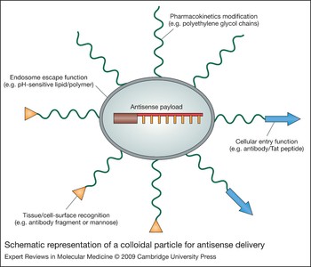
Attempts to overcome the biopharmaceutical barriers to systemic administration of antisense therapeutics have therefore developed towards rational design of multifunctional antisense oligonucleotide delivery systems – either as colloidal particles held together by electrostatic attraction or using medicinal chemistry advances to produce novel modified oligonucleotides. The goals in each case are to provide one or more of the following: improved pharmacokinetic profiles, and selective targeting to particular tissues through modification of the particle surface with ligands that allow targeting, cellular uptake and endosomal release. A schematic of such a colloidal construct is shown in Figure 2.
Schematic representation of a colloidal particle for antisense delivery. This diagram shows an ideal construct that carries antisense oligonucleotides (protected from degradation), is able to circulate without reticuloendothelial system uptake, and has targeting, cell-penetrating and endosomal escape functionalities.

Polymer-based nanoparticles are a colloidal system that has received recent attention for delivery of antisense; of particular interest are particles prepared using biodegradable polymers such as poly(d,l-lactide-co-glycolide) (Ref. Reference Kilic78), which can be used to release therapeutic agent slowly as the polyester is degraded by hydrolysis. Chitosan, a cationic biopolymer that can be prepared in nanoparticle form, has also been shown to be useful for encapsulation and cellular uptake of antisense oligonucleotides in vitro (Ref. Reference Springate79). Polycationic dendrimers are colloid-sized polymeric constructs that in some instances have been shown to be biodegradable in vivo (Ref. Reference Boyd80) and are able to induce the so-called ‘proton sponge’ endo-osmotic disruption effect. Briefly, by acting as weak bases once inside the acidic endosome environment, these agents act to increase osmotic pressure across the endosomal membrane and induce swelling and rupture of the endosome, leading to release of organelle contents. They have been employed as electrostatically associated carriers for oligonucleotides, and in vitro studies using dendrimers have shown antisense delivery and reduced luciferase expression in luciferase-expressing cell lines (Ref. Reference Yoo, Sazani and Juliano81) Further functionalised dendrimers with targeting and moieties that extend the circulatory half-life of the dendrimers (Ref. Reference Kaminskas82) indicate strong potential for dendrimers, incorporating all of the functionality of the model system in Figure 2, to overcome the barriers currently limiting extravasation and delivery of antisense in vivo. Derivitisation of polycations such as chitosan, cationic dendrimers, polyethylineimine, and cationic poly(amino acid)s is likely to be required if the intention is to use polyelectrolyte complexes as the core of a delivery system for intravenous use. Although complexes with excess cationic charge can be used to deliver antisense therapeutics in vitro, the result of injecting such complexes intravenously is substantial uptake by the reticuloendothelial system, as well as the threat of particle aggregation following interaction with blood components. When the latter occurs, the result is usually deposition of the particles in the capillary bed of the lung. These phenomena have been well documented in the field of nonviral gene therapy – that is, using cationic polymer (Refs Reference Dash83, Reference Merdan84, Reference Ward, Read and Seymour85) or lipid complexes (Refs Reference Anwer86, Reference Hofland87, Reference Liu88, Reference Mahato89) to deliver DNA expression plasmids. Although siRNA delivery is beyond the scope of this review, recent studies in this area (Ref. Reference Szoka90) suggest that small nucleic acid molecules can be delivered in neutral particles (Refs Reference Li and Szoka91, Reference Peer92) or as direct chemical conjugates with targeted neutral polymers (Refs Reference Rozema93, Reference Wolff and Rozema94), and it is likely that similar approaches can be effective using antisense oligonucleotides. An important aspect of the use of colloidal systems to deliver antisense oligonucleotides is that the toxicity of the components of the system may be a limiting factor, as has been the case in the past with cationic liposomes.
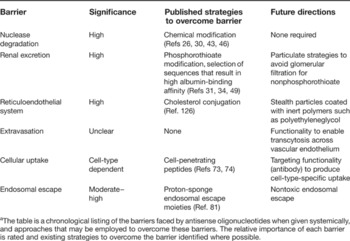
Table 2 summarises current successes in overcoming the barriers to antisense oligonucleotide delivery to the cytoplasm of target cells, along with suggested directions for future work. At the time of writing this article, Juliano and colleagues (Ref. Reference Juliano75) published a similar list of barriers to oligonucleotide delivery: there is agreement as to the problems faced – the remaining questions concern the respective solutions.
Barriers faced by antisense oligonucleotides when given systemically, and approaches for overcoming thema

a The table is a chronological listing of the barriers faced by antisense oligonucleotides when given systemically, and approaches that may be employed to overcome these barriers. The relative importance of each barrier is rated and existing strategies to overcome the barrier identified where possible.
Successful antisense knockdown after systemic administration
Although the list of barriers described earlier appears imposing, antisense oligonucleotides have been shown to reduce target expression and alter disease phenotypes in many well-controlled in vivo experiments. A great number of significant publications have emerged over the past 20 years describing antisense-mediated efficacy in vivo after systemic administration using simple injection of oligonucleotide-containing solutions. The most convincing of these combine most or all of the following properties: characterisation of the knockdown or exon skipping in vitro using robust controls [see review by Stein and Krieg (Ref. Reference Stein and Krieg95) for interpretation of antisense data], low doses of antisense oligonucleotide, and clear evidence of protein knockdown in vivo correlating with the observed phenotype change.
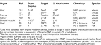
Table 3 summarises the doses used (mg/kg) and efficacy observed for selected examples of antisense efficacy in vivo across a range of target organs. It is clear that efficacy can be seen at low mg/kg doses, and that the maximum efficacy reported is greatest in the liver and kidney. The heart was chosen to represent tissues identified earlier in the review that show modest levels of tissue drug concentration. Although many groups including ourselves (Refs Reference Nguyen96, Reference Nguyen and White97) have shown antisense efficacy in the myocardium, the ceiling on the maximum possible level of target suppression appears to be around 50%, even when delivery strategies such as liposome encapsulation are used (Refs Reference Kagiyama98, Reference Zhang99). Similarly, correction of aberrant splicing in skeletal muscle has required large doses of antisense to produce relatively low levels of correction (Refs Reference Alter5, Reference Lu100), although this may be enough to cause phenotypic improvement, and in these impressive studies many muscle fibres show evidence of correction. Correction of aberrant splicing in the liver by the same investigators was more successful, with 85% correction (Ref. Reference Roberts72) at lower doses than used in skeletal muscle. Maximum levels of knockdown in cardiac and skeletal muscle therefore appear to be lower than that for the liver, in which up to 90% silencing has been shown in vivo (e.g. Ref. Reference Crooke101), and the kidney, in which 83% knockdown in the proximal tubules has been shown (Ref. Reference Guha102). Thus, the literature appears to suggest that significant enhancement of tissue and cellular uptake using delivery systems is required for high-efficacy silencing in tissues such as the heart and skeletal muscle.
Representative examples of the determination of maximal efficacy of in vivo knockdowna

a Data were collected from original research articles, across a range of target organs showing doses used and the percentage decrease in expression of target mRNA or protein (% knockdown).
bThe first reported measurement in this study was 25 days after initiation of therapy.
cApproximate; authors used 2 mg per mouse.
Abbreviations: ApoB, apolipoprotein B; B1 AR, β1 adrenoceptor; CTGF, connective tissue growth factor; Dmd, dystrophin; EGFR, epidermal growth factor receptor; EGFP, enhanced green fluorescent protein; LNA, locked nucleic acid; MOE, 2'-O-methoxyethyl; PMO, phosphorodiamidate morpholino; PS, phosphorothioate.
While a comprehensive coverage of successful employment of antisense oligonucleotides as research tools is beyond the scope of this review, it is pertinent to consider some key examples of convincing in vivo data in which the experimental controls were rigorous and the efficacy noteworthy. We direct the reader to examples of local or systemic injections of a range of antisense oligonucleotides (PS, OMe and MOE gapmers, PMO oligonucleotides and LNA antisense) which were shown to produce specific target mRNA knockdown (or exon skipping) (see Refs Reference Sazani51, Reference Nguyen96, Reference Lu100, Reference Crooke101, Reference Tyler103, Reference Lu104, Reference Alter105, Reference Yin, Lu and Wood106, Reference Reich107, Reference Xing108, Reference Sazani109, Reference Cao110, Reference Myers111, Reference Lau112) in a range of tissues (liver, gastrointestinal tract, brain, skeletal muscle, vasculature) (see Refs Reference Sazani51, Reference Nguyen96, Reference Crooke101, Reference Tyler103, Reference Lu104, Reference Reich107, Reference Cao110) and against a range of targets (enzymes, intracellular signaling molecules) (see Refs Reference Iversen9, Reference Lu104, Reference Reich107, Reference Xing108, Reference Lau112) in order to reverse disease phenotype.
Outstanding examples of impressive antisense effects in vivo using simple injection not surprisingly relate to organs with naturally leaky vascular beds – in particular the liver and in tumours (Ref. Reference Xing113). Apolipoprotein B knockdown has been reported in carefully controlled studies in animals (Ref. Reference Crooke101) and in dyslipidaemic humans (Ref. Reference Kastelein114) using moderately low doses of ISIS antisense oligonucleotides, and the future of this strategy appears to be promising. Tumour regression and inhibition of metastasis by targeting a kinase suppressor of Ras (Ref. Reference Xing113) has been elegantly demonstrated – a case in which specific intracellular signalling molecules are critical for tumour development and where gene-silencing approaches may be more useful than traditional small-molecule strategies. Conferring of positive charge on PNAs, using lysine (K) residues at the C-terminus, greatly improves the drug distribution to a range of tissues after intraperitoneal injection (Ref. Reference Sazani51). In this study, ‘PNA-4K’ antisense oligonucleotides demonstrated efficacies (correction of aberrant splicing in this case) equal to or greater than MOE antisense oligonucleotides in a range of organs, whereas uncharged PNA or PMO antisense oligonucleotides were relatively ineffective.
Direct measurement of in vitro target mRNA knockdown (or exon skipping) and/or subsequent target protein knockdown is often not possible in humans due to the inability to excise and collect target tissues for measurement of mRNA levels. As a result, reversal of disease phenotype is often the only measure of antisense drug efficacy and this is, at best, only an indirect measure of target mRNA knockdown. This is demonstrated by the first antisense drug to reach the market, fomivirsen, for which clear evidence of clinical efficacy (Refs Reference van Deventer, Tami and Wedel115, Reference Group116) but no direct evidence of antisense-mediated target knockdown in humans can be found in the literature. Furthermore, only a limited number of antisense-oligonucleotide-based therapeutics have entered clinical trials, with the vast majority being of PS chemistry and with most studies reported being Phase I trials in which safety, rather than efficacy, was the primary endpoint (Refs Reference Sewell34, Reference Cunningham117, Reference Cunningham118, Reference Marshall119, Reference Adjei120, Reference Mani121, Reference Stevenson122, Reference Bishop123). Later-phase studies with a PS oligonucleotide targeting BCL2 mRNA returned promising results as a cancer therapy adjuvant in humans (Refs Reference Bedikian124, Reference O'Brien125); however, these studies did not directly measure target mRNA or protein knockdown. One excellent example of antisense target knockdown with a MOE gapmer antisense oligonucleotide was reported by Kastelein and colleagues (Ref. Reference Kastelein1). Following on from animal studies (Ref. Reference Crooke101; discussed earlier in this section), these workers reported that simple intravenous administration of an antisense oligonucleotide targeting apolipoprotein B mRNA in humans produced dose-dependent reductions in circulating apolipoprotein B protein levels and LDL–cholesterol levels. Although not a direct measure of antisense target mRNA knockdown, the ability to measure changes in circulating levels of apolipoprotein B protein, and the clear dose-dependency of the protein knockdown and phenotype changes (reduced LDL–cholesterol) in this study provide convincing evidence of successful antisense targeted apolipoprotein B mRNA knockdown.
Conclusions and outstanding research questions
The large body of literature describing successful antisense use in vivo is ample evidence that this method of gene silencing is robust and reproducible. The antisense platform has enjoyed a considerable ‘head-start’ on other gene-silencing strategies such as those employing RNA interference. Lessons learnt from this period, in addition to those from extensive development of siRNA agents as therapeutics, provide great hope that antisense drugs will emerge in the near term. In several cases, the nature and location of the target cell type and target gene product will allow the use of simple injection of antisense oligonucleotides as potent and safe therapeutic agents; this may well be the case for ISIS 301012. For the liver, renal proximal tubules and the spleen, simple systemic administration may be sufficient to produce safe and effective therapeutic gene silencing. Where other cell types are targeted, delivery systems that overcome the ‘trapping’ of oligonucleotide within the liver, kidney and spleen and resultant low tissue concentrations are very likely to improve therapeutic indices. While in some instances this has already been achieved using liposomal delivery, endosomal escape agents, and targeting moieties, a step-change in antisense oligonucleotide therapeutics calls for further progress in antisense delivery systems.
Acknowledgements and funding
The authors thank the reviewers of this article for their insightful comments, in particular with regard to the scope of the article.