INTRODUCTION
In the past few decades there has been an increasing interest in the effects of water temperature change, due to global warming, on the structure and diversity of marine invertebrate populations (Eckert et al., Reference Eckert, Engle, Kushner and Browne2000; Perez et al., Reference Perez, Garrabou, Sartoretto, Harmelin, Francour and Vacelet2000; Baulch et al., Reference Baulch, Schindler, Turner, Findlay and Paterson2005). Temperature changes can directly influence marine organisms by inducing physiological stress, or by disturbing natural reproductive cycles (Lawrence & Soame, Reference Lawrence and Soame2004), or they can affect populations indirectly by modifying trophic interactions and competitive relationships (Stenseth et al., Reference Stenseth, Mysterud, Ottersen, Hurrell, Chan and Lima2002). Recent reviews have emphasized the potential importance of parasites and pathogens on populations under global warming (Harvell et al., Reference Harvell, Mitchell, Ward, Altizer, Dobson, Ostfeld and Samuel2002; Mouritsen & Poulin, Reference Mouritsen and Poulin2002).
The starfish Astropecten jonstoni lives on sandy bottoms in a depth of 1 to 10 m (Ribi et al., Reference Ribi, Burla and Ochsner1976). Except for a few hours per day when they actively wander over the sand searching for food, A. jonstoni spend most of the time buried within the sediment (Ferlin-Lubini & Ribi, Reference Ferlin-Lubini and Ribi1978). In the 1970s starfish of the genus Astopecten were present in large numbers along two sandy coasts in the south-east of Sardinia (Italy) (Ribi et al., Reference Ribi, Burla and Ochsner1976). When investigations were resumed in 1999 fewer active individuals could be found at the study sites than in the 1970s, with direct field observations suggesting an increase in sightings of dying animals on the sand surface (Maag, Schärer, Steinmann, personal observations). Despite high abundances of juvenile A. jonstoni detected since 2000 (Steinmann, unpublished data), observations of adults walking over the sediment during periods of warm water temperatures have remained low. In September 2005 when bottom water temperatures in the completely mixed water column of the Costa Colostrai were 23±1°C we observed no active starfish. During these surveys we came across a greater number of dead or apparently dying A. jonstoni than had been seen during the 1970s (anecdotal observations of three personnel involved in both sets of surveys i.e.1970s and 2000s). In September 2005 the Costa Rei had a stratified water column and in the cooler water below the thermocline many active starfish could be seen. In April 2006 at the Costa Colostrai, when water temperatures had cooled to 16±1°C active starfish could again be found.
We were additionally concerned that during warm periods, such as the extremely hot summer of 2003, Astropecten transferred to underwater enclosures died within a single day. During our sampling in 2005 all A. jonstoni kept in basins ashore also succumbed to a distinctive wasting disease. Such handling was used routinely in the 1970s, where it had no observably deleterious effect on the animals (G. Ribi & R. Schaerer, unpublished data).
These observations caused us to investigate the effect of water temperature, salinity and individual size on A. jonstoni survival. Although there is a mass of work on the effects of raised water temperatures causing bleaching in tropical corals, the strength of this paper is that it presents evidence for the effects of increased water temperatures on other, mobile, marine invertebrates in non-tropical waters. Such information is important if we are to track and potentially predict species' reactions to both recent and predicted temperature increases.
MATERIALS AND METHODS
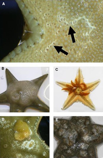
Fieldwork was conducted on Astropecten jonstoni in the south-east of Sardinia in 2005 at three different sites: Costa Colostrai North (39°20′11″N 9°36′09″E), Costa Colostrai South (39°19′51″N 9°36′20″E) and Costa Rei (39°14′34″N 9°34′29″E). In April 2006, 100 A. jonstoni uncovered by harrow and collected by SCUBA divers (see Ribi et al., Reference Ribi, Burla and Ochsner1976) from 8 m at the Costa Colostrai North (water 16°C and 35 ppt) were measured and immediately transferred into individual containers at 13±1°C for transportation to the University of Zurich (Switzerland). Three days after collection, the 99 surviving animals were moved into covered and aerated 3-l plastic jars containing sterile sand and artificial seawater, cooled by partial immersion in 12°C fresh water. The 16 d, fully factorial, experiment had three temperature and two salinity treatments. Animals, which naturally differed in size, were randomly assigned to treatments. A habituation phase of 8 d maintained 39 starfish at 12°C but gradually raised the water temperature of the remaining 60 starfish to 20°C. The first temperature treatment maintained the 39, 12°C habituated, A. jonstoni at 12°C throughout the 16 d experiment. The second temperature treatment kept 30 of the 20°C habituated animals at 20°C. The third temperature treatment initially maintained the remaining thirty 20°C habituated animals at 20°C for 7 d then increased the temperature to 25°C over 2 d and maintained it there for the remaining seven days. These temperatures are inline with those recorded by an underwater data logger between August 2003 and July 2004 at the Costa Colostrai. The unbalanced sample size was due to restricted space in the 20 and 25°C temperature basins. Animals in all temperature treatments were randomly assigned to one of two salinity treatments at 28 and 35 (±1) ppt. Each day the starfish were visually inspected to determine their state of health, based on the description by Eckert et al. (Reference Eckert, Engle, Kushner and Browne2000) and Farto et al. (Reference Farto, Armada, Montes, Guisande, Perez and Nieto2003) of wasting diseases among multiple species of starfish and the octopus Octopus vulgaris respectively (see Figure 1). Survival analysis was performed by a backward stepwise Cox Regression (SPSS 13.0 version for MS).
Manifestation of disease in Astropecten jonstoni. (A) First sign of disease was indicated by small white lesions (arrows) and a loss of the body tension. Most animals produced a lot of slime and some swelled up with a single, large blister under the dorsal skin; (B) animals could evert intact (C – Pylorus glands egested at the oral side) or partially dissociating tissues and body organs (D) through ruptures in the body wall and the white lesions then spread across the body surface (D and E). In most cases animals were largely destroyed after two days, although they were still moving their ambulacral feet. In general the animals had fully disintegrated within six days.

To get an initial overview of bacteria living in, on and around A. jonstoni, smears were taken from A. jonstoni, other invertebrates and sand during the April 2006 field collections and the laboratory experiment in Zurich. Swabs were initially maintained on trypticase soy agar (TSA) or broth at 20±1°C for 2±1 days. Colonies were transferred to new Petri dishes provided with TSA and incubated over night at 37°C. One or two examples of each macro morphological shape were sub-cultured for sequencing. DNA from 30 pure cultures was extracted by a 2 h incubation at 56°C in lysis buffer (0.1 M Tris-HCL pH 8.0, 0.05% Tween 20, 0.24 mg/ml proteinase K). The 16S rDNA was PCR amplified using standard PCR conditions (1 × 95°C for 15 min; 33 × 94°C for 30 s, 58°C for 45 s, 72°C for 60 s; 1 × 72°C for 5 min), Qiagen HotStarTaq Polymerase Master Mix and the primers 5′ CAG AGT TTG ATC CTG GCT CAG 3′ and 5′ TAC GG(CT) TAC CTT GTT ACG ACTT 3′. The PCR products were purified (Qiagen PCR purification kit) and sized by electrophoresis before being commercially sequenced using the same primer pairs by MWG Biotech (Ebersberg, Germany). Sequences were aligned to databank entries using the Fasta aligorithm (http://www-bio.unizh.ch) and then allocated to species if they shared >90% similarity with sequences held in the NCBI databases.
RESULTS
Figure 1 describes the characteristic wasting disease seen in the starfish.
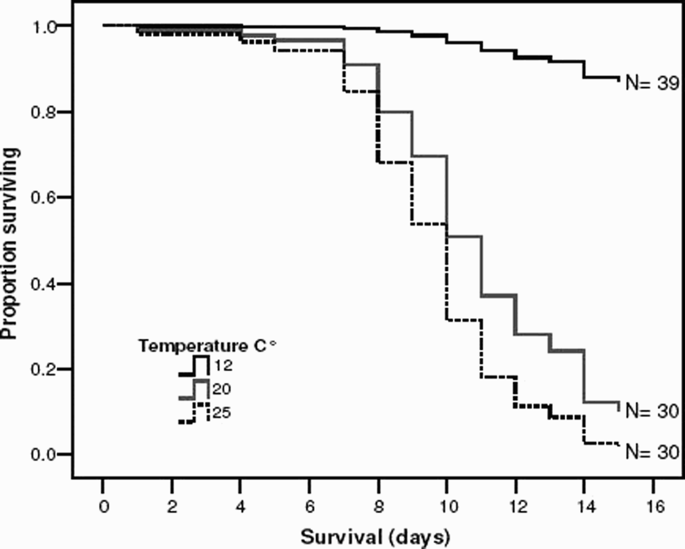
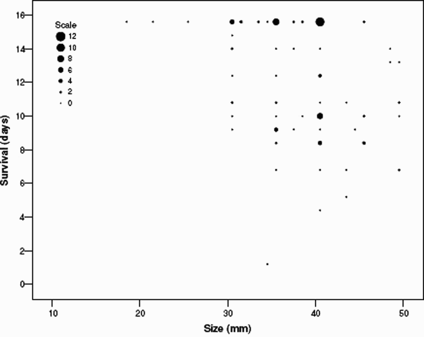
Results of the laboratory study showed that the best fit model included both water temperature treatment and starfish size (Cox Regression: chi-square = 73.632, df = 3, P < 0.001). The proportion of surviving starfish clearly differed between 12 and 20°C (Wald statistic = 26.183, df = 1, P < 0.001) as well as between 20 and 25°C (Wald statistic = 3.931, df = 1, P = 0.047) (Figure 2). Smaller starfish survived significantly longer than larger animals (Wald statistic = 4.25, df = 1, P = 0.039) (Figure 3). Salinity had no significant effect on survival.
Survival plot of Astropecten jonstoni maintained at different temperatures in the laboratory.

Scatter plot showing that larger Astropecten jonstoni died faster than smaller ones regardless of salinity and temperature. See Results for more details.

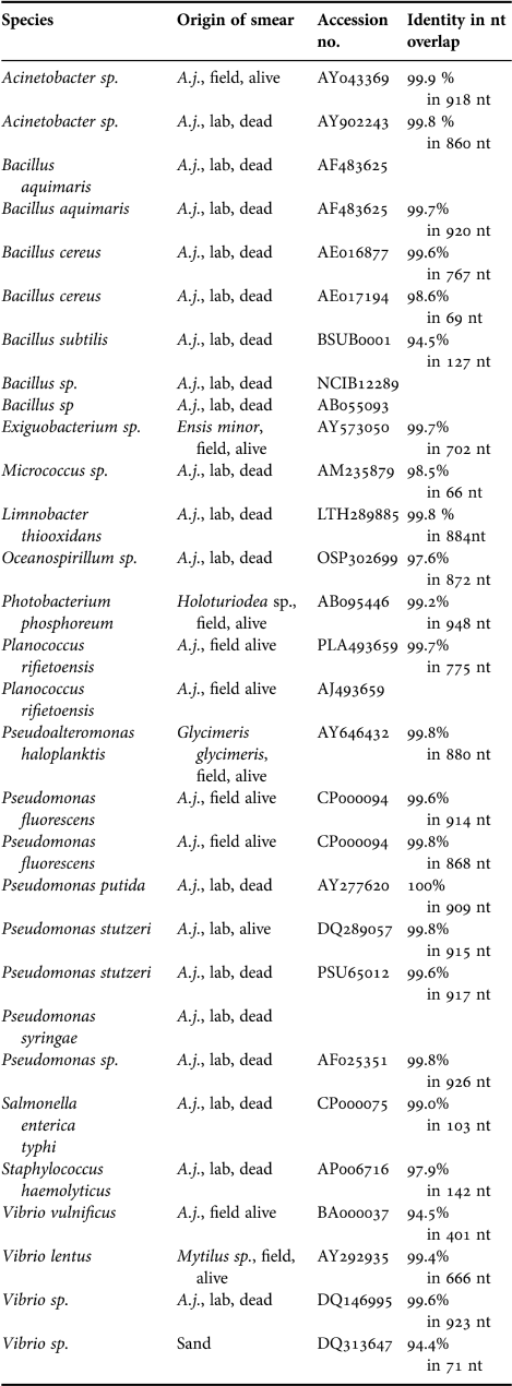
Bacterial analyses revealed that, as well as other species, bacteria of the genus Vibrio were present both on the starfish and in the environment (Table 1).
Bacteria sequences identified from swabs taken from over 100 Astropecten jonstoni, other invertebrates and sand during the April 2006 field collections and the laboratory experiment in Zurich.

A.j., Astropecten jonstoni
DISCUSSION
In the past few decades investigations on diseases in marine invertebrates and their potential direct or indirect linkage to abnormally high sea surface temperature have increased (e.g. Lawrence, 2004; Sabaté, 2006; Ward et al., Reference Ward, Kim and Harvell2007; Webster, Reference Webster2007). A majority of these studies deal with mass mortalities observed in different coral species and other sessile invertebrates (Perez et al., Reference Perez, Garrabou, Sartoretto, Harmelin, Francour and Vacelet2000; Kushmaro et al., Reference Kushmaro, Banin, Stackebrandt and Rosenberg2001; Boyett et al., Reference Boyett, Bourne and Willis2007; Remily & Richardson, Reference Remily and Richardson2006; Ward et al., Reference Ward, Kim and Harvell2007). From these various investigations we know that temperature anomalies on tropical coral reefs routinely exceed coral stress thresholds, with temperature considered to be a critical variable in coral hostpathogen systems. For example one large scale investigation along the Great Barrier Reef showed a highly significant relationship between periods of increased water temperature and an emergent disease in Pacific reef-building corals (Bruno et al., Reference Bruno, Selig, Casey, Page, Willis, Harvell, Sweatman and Melendy2007). Furthermore increasing water temperatures in reef environments allow pathogens to expand their ecological niche, for example by colonizing low pH environments (Remily & Richardson, Reference Remily and Richardson2006). Although thermal stress can induce higher levels of resistance in the host, the growth rate of pathogenic bacteria can increase, and an infection become established before host resistance is maximal (Ward, 2007).
In contrast to the numerous tropical reef studies of sessile species, relatively little information exists about thermal induced diseases and potential pathogens in temperate mobile invertebrate populations (Eckert, 2000; Farto et al., Reference Farto, Armada, Montes, Guisande, Perez and Nieto2003; Becker et al., Reference Becker, Gillan, Lanterbecq, Jangoux, Rasolofonirina, Rakotovao and Eeckhaut2004). Observations of the mass mortality of Astropecten observed along the Costa Colostrai and the Costa Rei in September 2005 and the results of the laboratory experiment presented here, serve as an example that mobile invertebrates are also sensitive to increased sea surface temperature. Several long-term records suggest that Mediterranean Sea surface temperatures have increased in the last decades. Along the Mediterranean coast of Spain sea surface temperatures are clearly higher after 1994 than in the preceding decades to 1950 (Sabaté et al., Reference Sabaté, Martin, Lloret and Raya2006). Also in the Gulf of Marseille abnormally long periods of high water temperatures have been measured in the last few years (Salat & Pascual, Reference Salat and Pascual2002).
The Mediterranean Sea anemone, Actinia equina, has been shown to reduce its growth rate during periods of abnormal high water temperature (Chomsky et al., Reference Chomsky, Kamenir, Hyams, Dubinsky and Chadwick-Furman2004). The low activity of A. jonstoni during the warm summer months, could be an indication of similar increased physiological stress. We could not find any satisfactory explanation from the literature for our finding that smaller and middle sized A. jonstoni survived longer than larger individuals in our laboratory study (Figure 3). Such findings would be expected if the immune system of larger and therefore older individuals is weaker than in juveniles, but for now this remains speculation.
Although the microbiological analysis presented here was not quantitative it revealed that, amongst other species (Table 1), bacteria of the genus Vibrio were present in the field and in laboratory animals (Figure 1E). Although we do not have evidence for a causal link, the manifestation of the disease is strikingly similar to that previously described for Vibrio-implicated mass mortalities of various marine invertebrate groups (Eckert et al., Reference Eckert, Engle, Kushner and Browne2000; Kushmaro et al., Reference Kushmaro, Banin, Stackebrandt and Rosenberg2001; Becker et al., Reference Becker, Gillan, Lanterbecq, Jangoux, Rasolofonirina, Rakotovao and Eeckhaut2004), with Vibrio abundance often coupled to increased water temperatures (Kaspar & Tamplin, Reference Kaspar and Tamplin1993; Pfeffer et al., Reference Pfeffer, Hite and Oliver2003). Future work is needed to investigate a causal link between the disease and individual bacteria found in this study. We urge other workers to look out for similar behaviour changes and disease.
CONCLUSION
Although there is a mass of work on the effects of raised water temperatures causing bleaching in tropical coral reefs and by implication the survival of coral reef systems, this paper is one of few that presents evidence for thermal induced behavioural effects and stress-induced disease on a temperate, mobile, marine invertebrate.
ACKNOWLEDGEMENTS
Heinz Maag, Samuel Tanner and Mathias Kempf assisted with fieldwork and experimental set-up. Luc Bussière and Andrew Pemberton helped with preparation of the manuscript.