Approximately 50–80% of hepatitis C virus (HCV) infections result in chronic hepatitis; of these, 10–20% of patients will develop liver cirrhosis and up to 5% of patients with HCV-related cirrhosis will develop hepatocellular carcinoma (HCC) (Ref. Reference Pawlotsky1). According to recent World Health Organisation (WHO) estimates, the global prevalence of HCV is approximately 2%, representing 123 million people infected worldwide (Ref. Reference Perz2). Epidemiological studies in different regions of the world show that the prevalence of HCV varies widely, although it is clear that HCV infection rates are higher in less developed countries. In European countries such as Germany, England and France, HCV prevalence is low, with figures ranging from 0.35% to 0.84% (Refs Reference Palitzsch3, Reference Mutimer4, Reference Meffre5). Slightly higher rates have been reported in the USA (1.6%) (Ref. Reference Armstrong6) and Japan (2%) (Ref. Reference Higuchi, Tanaka and Kiyosawa7). The highest reported prevalence rates are from countries in Africa and Asia. China, with one fifth of the world's population has a seroprevalence of 3.2% (Refs Reference Lei, Shigeko and Deng8, Reference Xia9) and Egypt with an estimated population of 73 million has a reported prevalence of approximately 20% (Ref. Reference Frank10). HCV is a leading cause of liver disease and the need for transplantation worldwide (Ref. Reference Schmeding11), and thus represents a major burden on public health.
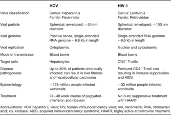
Owing to common routes of transmission, approximately 30% of patients infected with human immunodeficiency virus (HIV) in Europe and the USA are coinfected with HCV (Refs Reference Denis12, Reference Jones, Dunning and Nelson13). Although both viruses are spread via the same routes, they are not transmitted with the same efficiency (See Table 1 for a summary of virus characteristics). As a result, the prevalence of HCV/HIV coinfection varies among HIV subpopulations ranging from <15% of homosexual men to >80% of intravenous drug users (Ref. Reference Sulkowski and Thomas14). Chronic HCV progresses slowly in monoinfected individuals, with approximately 20% of patients developing cirrhosis after 20 years of infection (Ref. Reference Seeff15). By contrast, HCV/HIV coinfection has been associated with a more rapid progression to liver disease and the development of cirrhosis and HCC (Refs Reference Garcia-Samaniego16, Reference Soto17). Since the introduction of highly active antiretroviral therapy (HAART) in 1996, the clinical prognosis for HIV patients has markedly improved (Ref. Reference Palella18), and as a result, HCV infection is now a leading cause of morbidity and mortality in HIV-infected individuals (Ref. Reference Weber19).
Summary of virus characteristics

Abbreviations: HCV, hepatitis C virus; HIV, human immunodeficiency virus; nm, nanometre; RNA, ribonucleic acid; kb, kilobase; AIDS, acquired immunodeficiency syndrome; HAART, highly active antiretroviral treatment.
HCV and HIV structural biology
Molecular biology of HCV
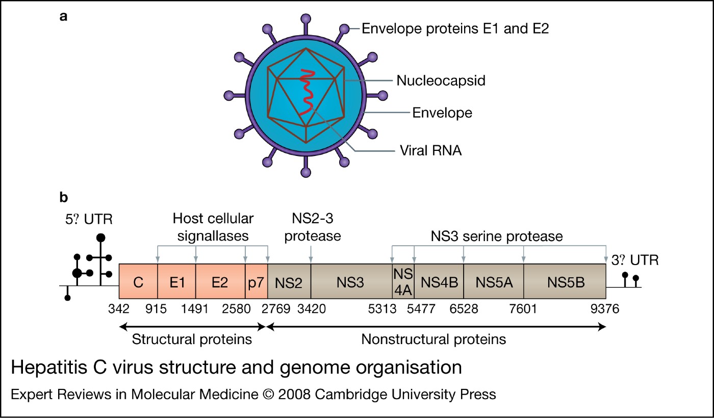
HCV has been classified as a member of the Hepacivirus genus within the Flaviviridae family (Ref. Reference Robertson20). HCV virions are spherical particles approximately 55–65 nm in diameter. The nucleocapsid of the viral particle is 30–35 nm, exhibits an icosahedral structure and is surrounded by a lipid-containing envelope with surface-spike-like projections containing viral envelope glycoproteins (Ref. Reference Kaito21) (Fig. 1a).
Hepatitis C virus structure and genome organisation. (a) Hepatitis C virions are spherical particles consisting of a nucleocapsid surrounded by a lipid-containing envelope with surface-spike-like envelope projections. (b) HCV genome organisation. The HCV genome is a positive-sense, single-stranded RNA of about 9.6 kilobases that comprises one long open reading frame encoding a protein of ~3000 amino acids. The polyprotein precursor is co- and post-translationally processed by both cellular and viral proteases to yield ten structural and nonstructural proteins. The grey arrows denote the points where the proteases cleave the polyprotein precursor. Abbreviations: HCV, hepatitis C virus; RNA, ribonucleic acid; UTR, untranslated region; NS, nonstructural.

The HCV genome is a positive, single-stranded RNA of about 9.6 kilobases (kb), which comprises one long open reading frame (ORF) encoding a protein of approximately 3000 amino acids (Ref. Reference Rice22) (Fig. 1b). The ORF is flanked by 5′ and 3′ untranslated regions (UTRs); the 5′ UTR is highly conserved and contains an internal ribosome entry site (IRES) responsible for mediating polyprotein translation (Ref. Reference Tsukiyama-Kohara23). The polyprotein precursor is co- and post-translationally processed by both cellular and viral proteases to yield ten structural and nonstructural proteins (Ref. Reference Grakoui24). The structural proteins are found at the N-terminal end of the precursor protein in the following order: core protein (C), then two envelope glycoproteins, E1 and E2. The core protein is a highly basic, RNA-binding protein responsible for forming the viral nucleocapsid (Ref. Reference Santolini, Migliaccio and La Monica25). E1 and E2 are transmembrane glycoproteins and they have an important role in host-cell entry. Located at the junction between the structural and nonstructural region, p7 is a 63 amino acid polypeptide; recent reports have indicated that p7 functions as an ion channel (Refs Reference Griffin26, Reference Pavlovic27) and is involved in the assembly and release of infectious virions (Ref. Reference Steinmann28).
The nonstructural (NS) proteins NS2, NS3, NS4A, NS4B, NS5A and NS5B are responsible for coordinating viral replication. The function of NS2 in its mature form has not been established; however, prior to cleavage from the polyprotein, it is known to play a role in the protease-mediated cleavage at the NS2–NS3 junction (Ref. Reference Grakoui29). NS3 is a multifunction protein with a serine protease domain at the N-terminus and a RNA-helicase/NTPase domain located at the C-terminus. The serine protease is responsible for the downstream cleavage of the nonstructural region: this is essential for the generation of components of the viral RNA replication complex (Ref. Reference Lindenbach and Rice30). The NTPase/helicase activity is thought to be responsible for unwinding double-stranded RNA intermediates and eliminating RNA secondary structures during replication. NS4A acts as an essential cofactor for NS3 protease activity and directs NS3 to intracellular membranes (Ref. Reference Brass, Moradpour and Blum31). The role of NS4B has not been established, but it may be involved in mediating membrane association (Ref. Reference Elazar32) and the formation of membranous structures supporting RNA replication (Ref. Reference Egger33). NS5A is a phosphorylated zinc metalloprotein whose main role in replication is not clear. NS5A initially gained wide interest after it was postulated to be involved in resisting the antiviral effect of interferon (IFN) (Ref. Reference Tan and Katze34). NS5B has been identified as the RNA-dependent RNA polymerase (RdRp) and so promotes the synthesis of new RNA genomes (Ref. Reference Behrens, Tomei and De Francesco35).
Molecular biology of HIV-1
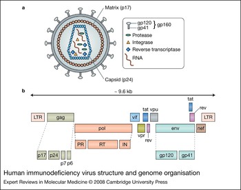
HIV-1 is a member of the Lentivirus genus classified within the Retroviridae family. Viral particles are spherical and measure ~100 nm in diameter (Fig. 2a). Each virion consists of an inner core surrounded by a lipid bilayer envelope. Two identical single-stranded RNA genomes and viral proteins necessary for replication and particle formation are contained within the inner core (Ref. Reference Cleghorn36).
Human immunodeficiency virus structure and genome organisation. (a) HIV particles are spherical and consist of an inner core surrounded by a lipid bilayer envelope. Two single-stranded RNA molecules and viral proteins necessary for replication and particle formation are contained within the inner core. (b) The genome of HIV is encoded on one single-stranded positive-sense RNA molecule approximately 9.6 kb in length. Each viral genome is composed of nine genes encoding three structural, two envelope, and six regulatory proteins, in addition to three enzymes. Abbreviations: HIV, human immunodeficiency virus; RNA, ribonucleic acid; gp, glycoprotein; kb, kilobases; LTR, long terminal repeat; PR, protease; RT, reverse transcriptase; IN, integrase.

The genome of HIV-1 is approximately 9.6 kilobases in length, contains nine ORFs, and is flanked on both ends by a long terminal repeat (LTR) (Fig. 2b). The major genes encoding structural proteins are located in the 5′ end of the genome in the order Gag, Pol and Env. The Gag gene encodes a 55 kDa gag precursor protein, which is cleaved during maturation into several proteins including p17 (matrix protein, MA), p24 (capsid protein, CA), p7 (nucleocapsid protein, NC) and p6 (Refs Reference Cleghorn36, Reference Frankel and Young37). The matrix protein is located on the inner surface of the viral membrane and has a role in viral assembly and the nuclear import of viral reverse transcripts (Ref. Reference Hearps and Jans38). p7 interacts with viral RNA and is thought to be responsible for the incorporation of the genome into the viral particle (Ref. Reference Frankel and Young37). The capsid protein forms the inner core or the virion (Ref. Reference Cleghorn36) and p6 is involved in the incorporation of the accessory protein Vpr in viral particles (Ref. Reference Paxton, Connor and Landau39). The Pol gene encodes three viral enzymes: protease (PR), reverse transcriptase (RT) and integrase (IN). PR cleaves the gag and gag-pol polyprotein precursors, IN mediates the integration of the HIV proviral DNA into the host-cell genome, and RT is an RNA-dependent DNA polymerase capable of synthesising a double-stranded copy of DNA from single-stranded viral RNA by reverse transcription (Ref. Reference Hill, Tachedjian and Mak40). The Env gene encodes a 160 kDa envelope protein, which is cleaved by a cellular protease to generate the external gp120 and the transmembrane gp41. The gp120 protein initiates viral entry by binding to specific host-cell-surface receptors, whereas gp41 facilitates fusion of the viral and cellular membranes (Ref. Reference Frankel and Young37). In addition to the structural genes, HIV-1 also encodes the regulatory proteins tat and rev, and the accessory proteins vif, vpu, vpr and nef. Tat (transactivator of transcription), a small nuclear protein, is the major regulator of HIV-1 transcription and is essential for replication (Refs Reference Arya41, Reference Fisher42). Rev (regulator of expression of virion protein) is responsible for the transport of viral RNA from the nucleus to the cytoplasm (Ref. Reference Felber43). Vif (viral infectivity factor) enhances the infectivity of the HIV-1 viral particle, and nef (negative factor) together with vpu, has been shown to play a role in the downregulation of cell-surface CD4-enhancing viral production (Refs Reference Garcia and Miller44, Reference Willey45). Vpr mediates the transportation of the preintegration complex to the host-cell nucleus (Ref. Reference Heinzinger46).
HCV/HIV disease progression
Impact of HCV on HIV infection
Studies on the influence of HCV on HIV progression have generated conflicting results. One study of 3111 HIV-infected patients, of which 1157 (37%) were HCV sero-positive, found that HCV/HIV coinfection was associated with an accelerated progression to acquired immunodeficiency syndrome (AIDS) and AIDS-related death (Ref. Reference Greub47). Lack of response to HAART was not associated with HCV serostatus, but coinfected patients receiving HAART had impaired CD4+ T-cell recovery compared with HIV-monoinfected patients. The authors postulate that the lower CD4+ levels could be responsible for the accelerated disease progression. Smaller studies also reported similar results (Refs Reference De Luca48, Reference Piroth49). By contrast, another large study performed on 5957 HIV-infected patients, 1960 (33%) of whom were HCV sero-positive, over a 6-year period, found no increase in the rate of progression to AIDS or death, nor was there an increased progression to AIDS in the intravenous drug user population of the HCV/HIV-coinfected group. A higher rate of death due to liver disease was observed in HCV/HIV-coinfected patients, and treatment responses to HAART were not associated with HCV serostatus. In addition, mortality did not differ between coinfected patients and HIV-monoinfected patients receiving HAART, and no differences were found in CD4+ T-cell counts during the administration of HAART (Ref. Reference Rockstroh50). Another study reported similar results, finding no survival difference between HCV/HIV-coinfected patients and HIV-monoinfected patients (Ref. Reference Sulkowski51). The risk of developing an AIDS-defining illness and the rate of progression to a CD4 count of less than 200 cells/μl also did not differ between groups. On the whole, the majority of reports suggest that HCV does not directly affect the progression of HIV disease (Refs Reference Rockstroh50, Reference Sulkowski51, Reference Bonacini52, Reference Dorrucci53, Reference Lincoln, Petoumenos and Dore54, Reference Staples, Rimland and Dudas55); however, coinfection with HCV does appear to have a negative influence on HAART tolerability. Up to 30% of HIV-infected patients discontinue antiretroviral treatment because of abnormal liver enzyme levels, and studies have shown that HCV increases the risk of this HAART-associated hepatotoxicity, resulting in higher discontinuation rates in coinfected patients (Refs Reference Melvin56, Reference Sulkowski57).
Impact of HIV on HCV infection
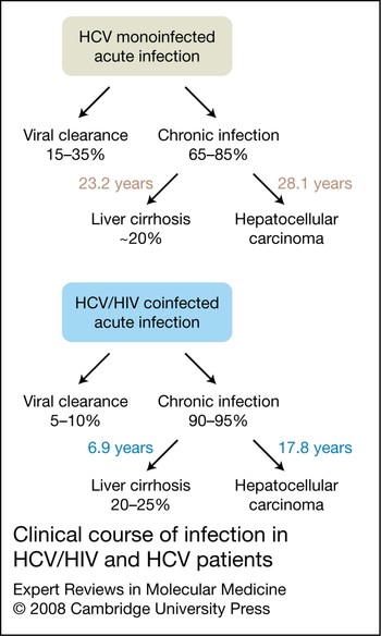
The immune suppression resulting from HIV appears to enhance HCV replication, impair HCV clearance, or both. Several studies have shown that HCV/HIV-coinfected patients have higher HCV viral loads compared with those infected with HCV alone (Refs Reference Bonacini58, Reference Sherman59). Some studies have also found an inverse relationship between HCV viral load and CD4+ levels (Refs Reference Soto17, Reference Daar60, Reference Eyster61). The standard treatment for HCV is a 24–48 week course of pegylated interferon-α (PEG-IFN) and ribavirin. This results in a sustained virological response (SVR; loss of detectable HCV RNA for at least six months and after completion of treatment) in approximately 85% in HCV genotype 2 and 3 patients but only 42–52% in patients with HCV genotype 1 (Refs Reference Hadziyannis62, Reference Manns63). HCV/HIV-coinfected patients have a lower treatment response rate compared with HCV-monoinfected patients. Initially, the SVR rates for coinfected patients ranged from 30% to 43% with only 17–29% of HCV genotype 1 patients achieving a favourable treatment outcome (Refs Reference Carrat64, Reference Chung65, Reference Torriani66). However, most of these studies used fixed low doses of ribavirin, which are currently viewed as suboptimal. Recent results from the Peg-interferon Ribavirin Espana Co-infection (PRESCO) trial, where ribavirin dosages were administered based on body weight, have reported higher treatment response rates in coinfected patients with HCV genotype 2 or 3, who achieve an SVR of 72.4% compared with 35% in patients with HCV genotype 1 or 4 (Ref. Reference Nunez67). Coinfected patients appear to have a faster progression to liver disease and development of HCC (Fig. 3). Several studies support a faster rate of liver fibrosis in coinfected patients, and this increase is correlated with a significant decrease in CD4+ cells (Refs Reference Benhamou68, Reference Canchis69). The onset of cirrhosis after HCV infection is estimated at 6.9 years in HCV/HIV-coinfected patients compared with 23.2 years in those with HCV alone (Ref. Reference Soto17). Similarly, HCC is estimated to occur after an average of 17.8 years of infection in HCV/HIV-coinfected patients compared with 28.1 years in HCV-monoinfected individuals. As a result, HCV/HIV-coinfected patients develop HCC at a younger age, with a mean age of 42 years for coinfected patients compared with a mean age of 69 years for those infected with HCV alone (Ref. Reference Garcia-Samaniego16). It is however interesting to note that after successful HAART treatment, mortality due to liver disease and the rate of fibrosis progression is significantly reduced in coinfected patients (Refs Reference Brau70, Reference Qurishi71). The reasons for the accelerated progression to liver disease in HCV/HIV-coinfected patients have not been fully clarified, but there is increasing evidence to suggest that the abnormalities in cell-mediated immune responses and an increase in rates of apoptosis may be at least partly responsible.
Clinical course of infection in HCV/HIV and HCV patients. Coinfected patients appear to have a faster progression to liver disease and hepatocellular carcinoma (HCC). The onset of cirrhosis after HCV infection is estimated at 6.9 years in HCV/HIV-coinfected patients compared with 23.2 years in those infected with HCV alone. Similarly, HCC is estimated to occur after an average of 17.8 years of infection in HCV/HIV-coinfected patients compared with 28.1 years in HCV-monoinfected individuals. Abbreviations: HCV, hepatitis C virus; HIV, human immunodeficiency virus.

Cell-mediated immunity in HCV/HIV coinfection
HCV-specific immune responses
Patients exhibiting spontaneous viral clearance mount vigorous multispecific CD4+ and CD8+ T-cell responses. By contrast, patients who progress to chronic disease tend to have weak narrowly focused responses (Refs Reference Diepolder72, Reference Lechner73, Reference Wedemeyer74), and studies in chimpanzees have shown that the in vivo depletion of CD4+ or CD8+ cells prevents HCV viral clearance (Refs Reference Grakoui75, Reference Shoukry76). These observations suggest that there is an association between viral clearance and specific cell-mediated immunity. CD4+ cells secrete cytokines capable of priming specific CD8+ cells; without CD4+ cells, the induction of immune responses is impaired and cytotoxic T lymphocyte (CTL) memory cannot be maintained (Refs Reference Grakoui75, Reference Shedlock and Shen77). Clearance of HCV infection is associated with the development of HCV-specific CD4+ Th1 responses; patients who progress to chronic disease have attenuated CD4+ Th1 responses and increased CD4+ Th2 responses with predominant production of IL-4 and IL-10 (Refs Reference Gerlach78, Reference Tsai79). Other immune deficiencies have also been shown to play a role in disease progression; for example, there is a high rate of HCV infection in patients with primary hypogammaglobulinaemia, and these patients had a severe and rapidly progressive course of infection (Ref. Reference Bjoro80). It is clear that HCV is capable of evading the cell-mediated immune response, and mutational escape from the T-cell response is considered to be responsible. Amino acid substitutions resulting in inhibition of HCV-specific T-cell recognition have been observed in chronically infected patients (Refs Reference Chang81, Reference Frasca82). Numerous studies have demonstrated that mutations within CTL epitopes promote HCV persistence (Refs Reference Erickson83, Reference Timm84, Reference Weiner85). T-cell failure and exhaustion is another potential method of viral evasion, because patients who progress to chronic disease may lose their specific T-cell responses (Refs Reference Gerlach78, Reference Thimme86). Viral escape may also occur as a result of impaired antigen presentation by dendritic cells; this functional impairment disappears once viral clearance occurs (Ref. Reference Bain87). Viral proteins are capable of subverting the cell-mediated immune response, and it has been shown that core and NS3/4A proteins interfere with monocytes and their cytokine production, thereby inhibiting dendritic cell activation and the production of Th1 cytokines (Refs Reference Brady88, Reference Dolganiuc89). There is also evidence to suggest that viral proteins can escape cell-mediated immunity by preventing TNF-α- and CTL-induced apoptosis (Refs Reference Disson90, Reference Frelin91).
The strength and breadth of HCV-specific CD4+ and CD8+ T-cell responses are reduced in HCV/HIV-coinfected patients when compared with those infected with HCV alone. (Refs Reference Capa92, Reference Dutoit93, Reference Graham94, Reference Valdez95). This suggests that the immunosuppression induced by HIV compromises immune responses to HCV. However, previous studies have shown that CD4 cell counts may not correlate with reduced T-cell responses (Refs Reference Capa92, Reference Graham96) and it is possible that a qualitative defect in CD4+ cells resulting from HIV infection is responsible for the diminished HCV-specific T-cell responses in HCV/HIV-coinfected patients. We recently found decreases in expression levels of the chemokine receptor CXCR3 on CD4+ and CD8+ T cells in HCV/HIV-coinfected patients compared with those infected with HCV only (B. Roe, unpublished results). Decreased CXCR3 expression on CD8+ T cells has also been shown during the advanced stages of HIV infection (Ref. Reference Brainard97). Since CXCR3 is expressed predominantly on Th1 cells, these findings suggest that coinfected patients may be depleted in Th1 cells, contributing to the reduced functional capacity of HCV-specific immune responses observed in HCV/HIV-coinfected patients. In addition, a lack of CXCR3 expression on lymphocytes could result in the dysfunctional recruitment of Th1 cells to the liver, resulting in lower proportions of Th1 cells, further contributing to the defective HCV-specific immunity in coinfected patients. The immune response plays an important role in the clearance of HCV after PEG-IFN and ribavirin treatment (Ref. Reference Lohr98). Although HIV targets and kills CD4+ cells, most studies have found no correlation between CD4 levels and an SVR (Ref. Reference Thomas99). It is possible that the reduced number of CD4+ T cells has a negative effect on some other aspect(s) of the immune system, resulting in a poor treatment response. In order to maximise therapeutic efficacy, the immunological defects responsible for the diminished cellular immune response to HCV in coinfected patients need to be determined.
Liver damage as a result of cell-mediated immune responses
HCV is not directly cytopathic, but there is increasing evidence to suggest that the pathological changes observed in the liver of HCV-infected patients are a direct result of the intrahepatic antiviral immune response (Refs Reference Koziel100, Reference Tseng101). Liver-infiltrating lymphocytes cause lysis of viral-infected cells, but cytokines and chemokines released by T-helper cells, and the cytotoxic effect of CTLs can also exert harmful effects on bystander hepatocytes, resulting in hepatocellular damage (Ref. Reference Jacobson Brown and Neuman102).
Chemokines
Chemokines and their receptors play an important role in the pathogenesis of HCV and HIV infection. Two chemokine receptors, CCR5 and CXCR4, facilitate cellular entry of HIV. CCR5 is the major coreceptor for macrophage-tropic strains of HIV (Ref. Reference Alkhatib103), whereas CXCR4 facilitates entry of T-cell-tropic HIV strains (Ref. Reference Feng104). The chemokine receptors CXCR3 and CCR5 are preferentially expressed on Th1 cells (Refs Reference Kim105, Reference Sallusto106); CXCR3 binds to the 10 kDa IFN-γ-inducible protein (IP-10), whereas CCR5 binds to macrophage inflammatory protein-1α (MIP-1α), macrophage inflammatory protein-1β (MIP-1β) and RANTES (regulated upon activation, normal T-cell expressed and secreted). These Th1-associated chemokine/receptor pairs appear to predominate in HCV infection (Refs Reference Apolinario107, Reference Shields108). IP-10 is involved in recruiting activated T cells to sites of infection (Ref. Reference Taub109). Previous studies have reported increased serum and intrahepatic IP-10 levels in individuals infected with HCV genotype 1 (Refs Reference Butera110, Reference Mihm, Schweyer and Ramadori111). Related studies found elevated IP-10 levels associated with increased liver damage (Refs Reference Harvey112, Reference Romero113), and these and other reports have also shown that serum IP-10 concentrations are higher in nonresponders to HCV therapy compared with those who achieve an SVR (Refs Reference Romero113, Reference Diago114). Previous studies have also reported elevated IP-10 levels in HIV-monoinfected individuals (Refs Reference Cinque115, Reference Stylianou116) and have shown that IP-10 promotes HIV replication (Ref. Reference Lane117). These have also shown that nonresponders to HAART have higher IP-10 levels compared with patients responding to treatment (Ref. Reference Stylianou116). To date, there are limited reports focusing on the role of IP-10 levels in HCV/HIV coinfection. We recently reported that IP-10 levels were significantly higher in HCV/HIV-coinfected patients compared with monoinfected and uninfected control groups. Since elevated IP-10 levels in HCV-monoinfected patients are correlated with increased liver disease, this further increase in IP-10 levels observed in HCV/HIV-coinfected patients may partly explain why there is an accelerated progression to liver disease upon coinfection. HCV/HIV-coinfected patients with low CD4+ counts (<400 cells/μl) had increased IP-10 levels compared with those with higher CD4+ cell counts. In HCV infection, IP-10 recruits CD4+ lymphocytes to the liver as part of the antiviral response. As HCV-infected patients with concurrent HIV infection have reduced CD4+ levels, it is possible that IP-10 expression is upregulated in the liver in an attempt to recruit increased numbers of CD4+ cells in response to HCV infection and as a result, liver damage inadvertently occurs (Ref. Reference Roe118).
Other chemokines have also been implicated in the accelerated progression to liver disease in coinfected patients. Hepatocytes exposed to the HCV envelope protein E2 produced the chemokine IL-8 in a dose-dependent manner; IL-8 production was increased further in the presence of HCV E2 protein and the HIV envelope glycoprotein gp120. This suggests there may be an enhanced inflammatory response in hepatocytes upon simultaneous exposure to HCV E2 and HIV gp120, and it is possible that HCV and HIV structural proteins may injure uninfected hepatocytes through an ‘innocent bystander’ mechanism (Ref. Reference Balasubramanian, Ganju and Groopman119). Another study found that HCV/HIV-coinfected patients had increased intrahepatic mRNA levels of RANTES and MIP-1α (Ref. Reference Kuntzen120), providing further evidence for the presence of an enhanced intrahepatic inflammatory response in coinfected patients.
Cytokines
Cytokines are capable of inducing broad-spectrum antiviral activity by regulating host immune responses and directly inhibiting viral replication. Cytokines can also accelerate disease progression, and both persistence of HCV and liver disease progression have been attributed in part to cytokine production. Specifically, several studies have shown that increased concentrations of proinflammatory cytokines in HCV-infected patients are associated with an increase in liver disease (Refs Reference Farinati121, Reference Zekri122). Cytokine dysregulation has also been shown to play a role in the pathogenesis of HIV infection. Serum IL-1, IL-6 and TNF-α levels are increased in HIV-infected individuals (Refs Reference Breen123, Reference Molina124), and these act on HIV-infected cells by upregulating virus replication (Refs Reference Folks125, Reference Poli126, Reference Poli, Kinter and Fauci127). Several studies have compared intrahepatic cytokine mRNA levels in HCV/HIV-coinfected patients with those infected with HCV alone (Ref. Reference Abbate128, Reference Blackard129, Reference Gonzalez130, Reference Sitia131). Coinfected patients have increased IFN-γ and TNF-α levels, and the increase in IFN-γ mRNA is associated with higher histopathological grading scores (Ref. Reference Sitia131). Another study found that HCV/HIV-coinfected patients had increased intrahepatic IFN-α mRNA levels, which correlated with an increase in HIV viral load. mRNA levels of the IFN-α receptor IFNAR-1 and the main IFN-α effector protein PKR were below the level of detection, indicating that even though there is strong IFN-α expression, there is an impaired immune response to IFN-α activation (Ref. Reference Abbate128). This inability to mount an efficient IFN-α immune response may also contribute to the acceleration of liver fibrosis.
HCV/HIV coinfection and apoptosis
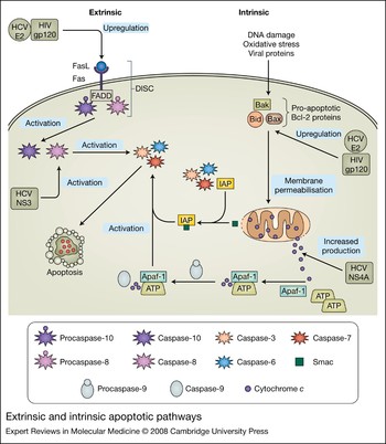
Apoptosis is necessary for the maintenance of cellular homeostasis; however, during viral infections the balance between cell growth and cell death is often disrupted. Intracellular apoptotic signalling is initiated through two different pathways: the intrinsic or mitochondrial pathway and the extrinsic or death-receptor pathway (Fig. 4).
Extrinsic and intrinsic apoptotic pathways.(See previous page for figure.) The extrinsic pathway is initiated with the binding of a death ligand, such as FasL, to its receptor Fas. This results in the recruitment of Fas-associated death domain protein (FADD) and caspase-8 and caspase-10, forming the death-inducing signal complex (DISC). Caspase-8 and caspase-10 are activated, which in turn activates caspase-3, causing cell death. The intrinsic pathway is triggered by intracellular or extracellular signals, resulting in the activation of proapoptotic proteins of the Bcl-2 family. The action of these proteins increases the permeability of the outer mitochondrial membrane, allowing cytochrome c and second mitochondria-derived activator of caspases (Smac) to be released in to the cytosol. Smac binds to inhibitor of apoptotic proteins (IAPs) preventing caspase inhibition, and cytochrome c binds to apoptotic protease activating factor-1 (Apaf-1) forming the apoptosome. Procaspase-9 is recruited to the apotosome, converted to active caspase-9, resulting in the subsequent activation of caspase-3, caspase-6 and caspase-7. The effects on HCV monoinfection and HCV/HIV coinfection on apoptosis are illustrated in grey. Abbrevations: DNA, deoxyribonucleic acid; ATP, adenosine triphosphate; FasL, Fas ligand, FADD, Fas-associated death domain protein; DISC, death-inducing signal complex; Smac, second mitochondria-derived activator of caspases; IAPs, inhibitor of apoptotic proteins; Apaf-1, apoptotic protease activating factor-1; HCV, hepatitis C virus; HIV, human immunodeficiency virus; gp, glycoprotein; NS, nonstructural.

Intrinsic/extrinsic apoptotic pathways
The intrinsic pathway is triggered by extra- or intracellular signals, including DNA damage, oxidative stress, growth factor deprivation and viral proteins (Refs Reference Guicciardi and Gores132, Reference Jin and El-Deiry133). These stress signals activate proapoptotic members of the Bcl-2 family such as Bax, Bak and Bid, thereby increasing the permeability of the outer mitochondrial membrane. As a result, mitochondrial proteins, such as cytochrome c and second mitochondria-derived activator of caspases (Smac) are released into the cytosol (Ref. Reference Jin and El-Deiry133). Caspases are a group of proteolytic enzymes that degrade cellular constituents, resulting in apoptosis. Under normal conditions, caspases are controlled by a group of proteins known as inhibitors of apoptotic proteins (IAPs); Smac binds to and interacts with IAPs, preventing caspase inhibition (Ref. Reference Rupinder, Gurpreet and Manjeet134). Cytochrome c, which is also released from the mitochondria, binds to apoptotic protease activating factor-1 (Apaf-1) forming a complex in the presence of ATP, which is known as the apoptosome. Procaspase-9 is recruited to the apoptosome and converted into the active caspase-9 through oligomerisation, resulting in the activation of caspase-3, caspase-6 and caspase-7, and subsequent cell death (Ref. Reference Riedl and Salvesen135). The extrinsic pathway is initiated at the plasma membrane where death ligands such as Fas ligand (FasL)/CD95L, tumour necrosis factor-α (TNF-α), and tumour necrosis factor related apoptosis-inducing ligand (TRAIL) bind to their corresponding receptors – TNF receptor-1 (TNF-R1), Fas/CD95 and TRAIL receptor-1 and -2 (TRAIL-R1 and TRAIL-R2). These receptor–ligand interactions are responsible for the recruitment of adaptor proteins such as Fas-associated death domain protein (FADD) and caspase-8 and caspase-10, resulting in the formation of the death-inducing signal complex (DISC). Activation of caspase-8 and caspase-10 in turn activate caspase-3, causing cell death.
Liver damage as a result of apoptosis
Hepatocytes are subjected to a high rate of cell injury and destruction owing to the continuous intrahepatic inflammation caused by chronic HCV infection. Apoptosis has been shown to play a role in this HCV-mediated liver damage through the induction of apoptotic pathways, primarily as a result of host immune responses and also, to some extent, by HCV viral proteins. Enhanced apoptosis of hepatocytes has been described during chronic HCV infection and correlates with the degree of liver damage (Refs Reference Bantel136, Reference Bantel137, Reference Calabrese138). An increase in caspase activation has also been demonstrated in the HCV-infected liver (Ref. Reference Bantel136). Previous studies have shown that caspase-3-knockout mice are largely resistant to liver damage (Ref. Reference Woo139) and suppression of hepatic injury using caspase inhibitors has been achieved (Refs Reference Kunstle140, Reference Rouquet141). The role of HCV proteins in initiating apoptosis has not been fully clarified, with conflicting reports describing both a pro- and anti-apoptotic role, depending on the experimental conditions employed. HCV core protein has been shown to enhance TRAIL-mediated apoptosis and induce mitochondrial injury and oxidative stress in hepatocytes, making them more susceptible to apoptosis (Refs Reference Chou142, Reference Okuda143). By contrast, inhibition of TNF-α-mediated apoptosis and increased production of Bcl-xl by core protein has also been reported (Refs Reference Otsuka144, Reference Ray145). NS3 has been shown to induce caspase-8-mediated apoptosis in hepatocytes (Ref. Reference Prikhod'ko146) but anti-apoptotic effects have also been described, involving cleavage of the adaptor molecule Cardif, which inhibits the proapoptotic effects of the RNA helicase RIG-1 (Ref. Reference Meylan147). NS4A causes mitochondrial damage resulting in the release of cytochrome c with caspase-3-mediated apoptosis. NS5A contains a Bcl-2 homology domain and can inhibit apoptosis by interacting with FKP38, Bax and p53 (Refs Reference Chung, Sheu and Yen148-Reference Lan150). These antiapoptotic mechanisms are further mediated through the NS5A activation of STAT3, NF-κB, and a PI3-kinase/AKT survival pathway (Refs Reference Gong151, Reference Street152). By contrast, NS5A has been shown to directly induce apoptosis in mature dendritic cells (Ref. Reference Siavoshian153).
In recent years, several reports have suggested that increases in the levels of apoptosis may play a role in the accelerated progression to liver disease in HCV/HIV-coinfected patients. The effects of HCV and HIV envelope proteins on hepatocytes have been studied using an in vitro model of coinfection (Ref. Reference Munshi154). Following costimulation with HCV E2 and HIV gp120, an enhanced level of apoptosis was observed and apoptosis increased when cells were exposed simultaneously to both envelope proteins. Upregulation of FasL and dephosphorylation of the antiapoptotic molecule AKT was also noted. These results suggest that HCV and HIV envelope proteins may act collaboratively in injuring uninfected hepatocytes through an ‘innocent bystander’ mechanism, as discussed earlier. A subsequent study observed an increased expression of FasL and its association with Fas following stimulation with HCV and HIV envelope proteins. Increased associations of FADD with Fas and caspase-8 were also observed, suggesting that these viral envelope proteins induce the formation of DISC. An increase in expression of the proapoptotic molecule Bid, caspase-2 and caspase-7 was found (Ref. Reference Balasubramanian155). It was subsequently demonstrated that FasL expression and apoptosis were induced by the STAT1-dependent pathway (Ref. Reference Balasubramanian, Ganju and Groopman156). There have been only limited clinical studies describing intrahepatic apoptosis in HCV/HIV-coinfected patients; one report found that Fas expression in hepatocytes was increased in coinfected patients and the level of intrahepatic apoptosis was enhanced when compared with that in HCV-monoinfected patients (Ref. Reference Macias157). Taken together, these results suggest that coinfection with HCV and HIV induces apoptosis in hepatocytes via a Fas–FasL-dependent pathway and indicate that this could account for the acceleration of liver disease in coinfected patients.
Research in progress and outstanding research questions
It is clear that coinfection with HIV impacts the course of HCV infection, increasing the likelihood of persistent infection and accelerating the progression of liver disease. The reasons for this are probably due to an array of factors, which include suboptimal immune responses and increased levels of apoptosis. The strength of HCV-specific immune responses is compromised in HCV/HIV-coinfected patients, and it seems likely that this is clinically reflected in increased HCV viral loads. Since decreased CD4 counts are not always associated with reduced T-cell responses, a qualitative defect in certain aspects of the immune response resulting from HIV infection may be responsible, and future studies are required to resolve this. However, delineation of potential defects is likely to prove difficult, because a large proportion of HIV-infected patients are in receipt of HAART and with the resulting immune restoration, the influence of HIV infection may not be fully appreciated. Various studies have shown different immunological responses when comparing HAART-naive patients with those in receipt of HAART (Refs Reference Autran158, Reference Pontesilli159, Reference Tilton160), and it is possible that coinfected patients receiving HAART may not accurately reflect the immunological status of untreated HIV-infected patients. Ideally, studies should be performed using HAART-naive patients, but establishing cohorts with sufficient patient numbers could prove a challenge and this would also have significant ethical implications. It is generally accepted that mutations within CTL epitopes promote HCV persistence (Refs Reference Erickson83, Reference Timm84, Reference Weiner85); there are currently no published reports on the presence of these mutations in HCV/HIV-coinfected patients or on the effects they may have on disease progression; future studies aiming to resolve this issue could provide some interesting results. Enhanced hepatocyte apoptosis has been shown to affect the progression of liver disease during both HCV monoinfection and HCV/HIV coinfection. Preliminary results from a study testing the efficacy of anti-caspase therapy have been positive, and patients exhibited significantly decreased liver enzyme levels with no apparent drug-related side effects (Ref. Reference Pockros161). Treatments aimed at decreasing apoptosis levels could prove to be of clinical importance, particularly for HCV/HIV-coinfected patients who fail to respond to regimens of PEG-IFN plus ribavirin. Future studies elucidating the molecular and immunological mechanisms responsible for HCV persistence and progression of liver disease will also lead to additional therapeutic strategies for the management of coinfected patients.
Acknowledgments
This work is supported by the National Virus Reference Laboratory, University College Dublin, Ireland (Grant 8090).