The membranous septum is a small area in the normal heart, and defects in this region usually extend variably into the inlet, trabecular, or outlet portions of the muscular septum. Defects situated in this region have the area of tricuspid–aortic–mitral continuity – central fibrous body – as part of their rim and constitute 80% of all ventricular septal defects. The conduction axis is invariably situated postero-inferior to these defects.Reference Soto, Becker, Moulaert, Lie and Anderson 1 – Reference McDaniel and Gutgesell 3
It has been over two decades since perimembranous ventricular septal defects have been closed using transcatheter techniques. The proximity of such defects to the aortic and tricuspid valves and the conduction system have seen a number of devices being developed for their closure. Although specific devices for perimembranous ventricular septal defects have been developed, they are not without disadvantages. The morphology of perimembranous defects is such that ductal occluders can be used for their successful closure. We sought to analyse the feasibility and complications associated with their use.
Patients and methods
The present study is a data analysis of a prospective study carried out at two institutes from August 2010 to April 2013. A total of 126 patients, comprising 69 males and 57 females, underwent transcatheter closure of perimembranous ventricular septal defects. The primary operator was the same at both institutes. The study was approved by the institutional ethics committee and a written informed consent was obtained from all patients or their parents (for children <18 years) after explaining the procedure and its complications.
Inclusion criteria
-
∙ Weight⩾7 kg
-
∙ Restrictive perimembranous ventricular septal defects with a prior history of infective endocarditis.
-
∙ Restrictive perimembranous ventricular septal defects with aortic cusp prolapse and development of aortic regurgitation on serial follow-up.
-
∙ Patients with symptoms suggestive of left ventricular volume overload, such as dyspnoea, fatigue, palpitations, poor weight gain, or recurrent respiratory infections, defined as more than two episodes of lower respiratory tract infections in a year. Demonstrable left atrial and left ventricular enlargement on echocardiography was mandatory in all symptomatic patients. Left atrial/left ventricular dilatation was defined by a left ventricular end-diastolic dimension z score of >2, and a left atrial: aortic ratio of ⩾1.2 on parasternal long-axis view using M-mode measurements.
-
∙ Asymptomatic, moderate-sized perimembranous ventricular septal defects with evidence of left atrial, and ventricular volume overload on echocardiography.
-
∙ Perimembranous ventricular septal defects with mean pulmonary artery pressures less than two-thirds of the mean systemic pressure.
-
∙ Postoperative residual perimembranous defects with left atrial and ventricular volume overload.
Patients with predominant right-to-left shunt, aortic cusp prolapse causing moderate and severe aortic regurgitation, large non-restrictive defects, and those with NYHA–IV symptoms were excluded. Non-restrictive defects were defined as those with a transseptal gradient of <20 mmHg.
Transthoracic echocardiograms were used initially for patient selection. The size of the defect, the presence of restrictive physiology (gradient of at least 20 mmHg), distance from the aortic cusps, the presence of aortic regurgitation, and tricuspid regurgitation were analysed before device closure. During the procedure, transthoracic echocardiograms were used to check the position of the device before the final release and to look for new-onset aortic and tricuspid valve regurgitation. We did not use transoesophageal echocardiography in any patient.
Haematological and biochemical investigations at baseline included a complete blood count and renal function tests. An electrocardiogram was recorded on the morning of the procedure and post-procedurally, immediately after the procedure, daily for the next 2 days, and at follow-up. Aspirin, 5 mg/kg/day for children and 150 mg/day for those over 18 years, was initiated a day before the procedure and continued post-procedurally for 6 months.
Arterial and venous access was obtained in all patients except in four children who only had an arterial access. The pulmonary artery was entered using a Berman or Judkins right catheter and pressures were recorded. Simultaneous aortic pressures were recorded using a pigtail catheter placed in the ascending aorta. Oximetry run and shunt calculation were not routinely performed on all patients.
Left ventricular angiograms were obtained in the left anterior oblique view with appropriate cranial angulations to best visualise the defect. The defect was crossed using right Judkins, multipurpose, or internal mammary artery diagnostic catheters (Cordis Corporation, Miami, Florida, United States of America) over a guide wire. This was then exchanged for an exchange length wire, which was subsequently snared in the superior or inferior vena cava and exteriorised through the venous sheath. Snaring of the wire was only occasionally performed in the pulmonary artery. In most cases, the Terumo glide wire (Terumo Medical Corporation, Japan) was allowed to track retrogradely on its own across the defect, into the right ventricle, right atrium, and superior vena cava, where it was snared. This was to avoid entrapment of the snare in the trabeculae of the right ventricle.
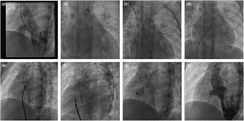
Although transthoracic echocardiograms gave us an idea of defect size, measurements obtained on left ventricular angiograms were used to select the appropriate device. The widest portion of the neck of the defect on left ventricular angiogram was measured on the left ventricular side (Fig 1a). Device sizes were chosen to not exceed this diameter by 2 mm. For example, if a defect measured 6 mm on the left ventriculogram, we would use a 6/8 mm Patent ductus arteriosus occluder. In case of tubular defects where Amplatzer Duct Occluder-II was used, we might be able to close the defect completely using an Amplatzer Duct Occluder-II device of the same diameter as the defect, as the device would conform to the tubular morphology of the ventricular septal defect. We have only closed defects that had a subaortic rim of at least 2 mm. The disc of the duct occluder is opened completely on the left ventricular side of the defect and uses the aortic rim above and the trabecular septum below for support. Defects that were immediately beneath the aortic cusps with no superior rim were excluded, as constant contact with the aortic cusps might lead to cusp erosion or aortic regurgitation.
Steps in closure of pmVSD using first-generation duct occluder. ( a ) Left ventriculogram in LAO view showing a large tubular pmVSD, opacifying the right ventricle. ( b ) VSD crossed using Terumo wire and Judkins right catheter. ( c ) Terumo wire being snared in the superior vena cava. ( d ) Mullins sheath being passed from the venous end over the exchange length wire. ( e ) Disc of the Lifetech Patent ductus arteriosus occluder being opened on the left ventricular side. ( f ) Device positioned across the defect. ( g ) Device in position after release. ( h ) Left ventricular angiogram shows no shunt across the occluded VSD. LAO=left anterior oblique; pmVSD=perimembranous ventricular septal defect.

The venous sheath was exchanged for a Mullins sheath, and the devices were deployed from the venous end after confirming their position on angiography and transthoracic echocardiography (Figs 1a-h). There were no problems encountered while passing the delivery sheath across the tricuspid valve. The Lifetech Patent Ductus Arteriosus Closure Systems (Lifetech Scientific Co., Ltd, Shenzhen, China) are available in sizes ranging from 4 to 24 mm. Devices with a waist diameter up to 10 mm have retention discs that are larger by 6 mm, and the larger devices have retention discs exceeding the waist diameter by 8 mm. Depending on the size of the device, they are delivered using 5–14 F sheaths and are recommended for use in children over 6 kg. The waist length varies from 7 to 10 mm. Amplatzer Duct Occluder-II, used in 45 patients, was deployed retrogradely from the arterial end through a 5 or 6 F Judkins Right guide catheter (Figs 2a-f). Post-deployment angiograms were obtained to look for residual defects and aortic regurgitation. The arterial and venous sheaths were removed immediately post-procedure, and haemostasis was achieved using manual compression.
Steps in closure of pmVSD using ADO-II. ( a ) Left ventricular angiogram in LAO view showing opacification of the right ventricle through the pmVSD. ( b ) VSD crossed using Judkins right guide catheter and Terumo wire. ( c ) ADO-II device loaded within the guide catheter. ( d ) One disc of the device being released on the right ventricular side of the defect. ( e ) Waist and disc on left ventricular side opened completely. ( f ) The ADO-II device positioned across the VSD defect after the final release. ADO-II=Amplatzer Duct Occluder-II; LAO=left anterior oblique; pmVSD=perimembranous ventricular septal defect.

Patients were followed up with electrocardiograms and echocardiograms for the evaluation of rhythm disturbances and complications such as residual shunts, aortic, or tricuspid regurgitation. These were performed during hospital stay and on scheduled follow-up visits at 1 month, 3 months, 6 months, and a year post-procedure. Subsequent follow-up was carried out annually. Patients without complications were discharged 48–72 hours post-procedure.
Results
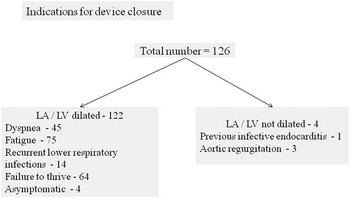
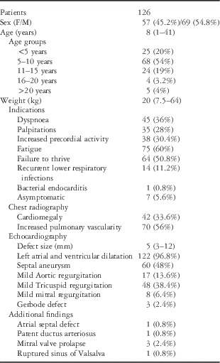
The baseline characteristics of patients are presented in Table 1. The median age of our population was 8 years with a range from 1 to 41 years. A total of 64 children weighed less than the fifth percentile for age according to the National Center for Health Statistics growth charts. Of these children, 52 completed follow-up at 1 year. A total of 23 patients showed an increase in weight following device closure of their defects and were no longer below the fifth percentile for age. Figure 3 shows indications for closure in the patient population based on their symptoms. Ventricular septal aneurysm was noted in 60 patients. Of these patients, three had a perforation in the septal tricuspid leaflet, which had undergone aneurysm transformation, with colour flow jets directed into the right atrium – Type 1 Gerbode defect – in addition to the right ventricle. These defects were also closed successfully using Lifetech Patent ductus arteriosus occluders. There was one case of ruptured right sinus of Valsalva into the right ventricle. This patient had an associated perimembranous ventricular septal defect. He underwent device closure of the ruptured sinus of Valsalva and the ventricular septal defect, using a total of three devices. The ventricular septal defect was closed using a 4/6 mm Patent ductus arteriosus occluder (Lifetech Scientific Co., Ltd).
Flow chart showing indications for device closure.

Baseline characteristics of patients (median with range).

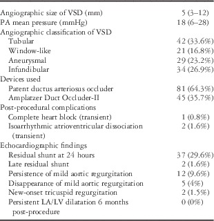
Amplatzer Duct Occluder-II devices were used in 45 patients (St Jude Medical Inc., Minnesota, United States of America), and Patent ductus arteriosus occluders in 81 patients (64.3%). Patent ductus arteriosus occluders ranged in size from 4/6 to 14/16 mm, and Amplatzer Duct Occluder-II devices ranged from 3 to 6 mm in diameter. The 8/10 mm Patent ductus arteriosus occluder was used most often, in a total of 30 patients across all weight ranges. The 4 mm diameter Amplatzer Duct Occluder-II was used most often, in 22 patients. The largest devices measured 14/16 mm and were deployed in two boys aged 7 and 8 years, weighing 20 and 22 kg, respectively (Table 2). A larger defect size per se was not a criterion for exclusion if they were at least mildly restrictive (gradient of 20 mmHg).
Procedural characteristics and post-procedural findings (median with range).

LA=left arial; LV=left ventricle; PA=pulmonary artery; VSD=ventricular septal defect.
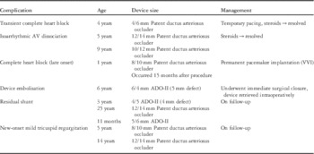
Complications in relation to device size.

ADO-II=Amplatzer Duct Occluder-II; AV=atrioventricular.
Qin et alReference Qin, Chen and Zhao 4 have proposed an angiographic classification of perimembranous defects and have stated that tubular and infundibular variants are easier to close using septal occluders. Tubular defects have a uniform diameter on the left and right ventricular side of the defect. Aneurysmal types of defects have an aneurysmal pouch, which is well delineated on left ventricular angiograms. Infundibular variants have a narrow opening on the right ventricular side and are conical in shape. In the window-like type, the shunt is scattered immediately on the right ventricular side. Tubular (33.6%) and infundibular (26.9%) variants were more common in our series of patients, but all the four varieties have been successfully closed using duct occluders.
Procedural success, as defined by the ability to deploy the device without embolisation or heart block during the procedure was achieved in all but one of our patients (99.2%). We attempted closure of a 5 mm defect with a 6/4 mm Amplatzer Duct Occluder-II device. The device embolised into the pulmonary artery soon after deployment. As the defect was in close proximity to the aortic valve, we did not attempt closure with a larger first-generation duct occluder. The device was retrieved and the patient underwent successful surgical closure of the defect. There was mild residual shunt noted in 37 patients immediately after the procedure. Colour Doppler evidence of residual shunt was seen in three patients, and this has persisted even after a year of follow-up in two of these patients. One of these patients had an extension of the defect in the high muscular region, and we probably underestimated the defect size. There has been no haemolysis in either of these patients.
Complete heart block was noted in a patient, a day after deployment of a 4/6 mm Patent ductus arteriosus occluder. She had an episode of syncope while being monitored in the post-cardiac catheterisation unit and needed emergent transvenous pacing. She was treated with intravenous methylprednisolone (30 mg/kg/day) followed by oral Prednisolone (2 mg/kg), tapered over a period of 1 month. Heart block resolved within 48 hours of initiating treatment with steroids and there was no recurrence. The pacing wire was removed after 48 hours. A 5-year-old boy had transient isoarrhythmic atrio-ventricular dissociation, 12 hours after closure of his defect with a 12/14 mm Patent ductus arteriosus occluder. The lowest ventricular rate recorded was 56 beats/minute and he remained haemodynamically stable. He was in sinus rhythm 96 hours after initiating steroids and has not had any further recurrence. A Holter recording was carried out on this patient a week after the procedure, and only few supraventricular ectopics were noted. Another boy aged 9 years had transient isoarrhythmic dissociation with ventricular and atrial rates of 74 per minute, noted within 24 hours of the procedure. This resolved within the subsequent 48 hours after initiation of steroids. A 1-year-old boy, weighing 7.5 kg at the time of procedure, was noted to have complete heart block (late onset), 15 months after the procedure. He was asymptomatic and underwent permanent pacemaker implantation. In all, 17 patients had mild aortic regurgitation at the time of initial diagnosis and it persisted in 12 patients after the closure of their defects. There were no instances of new-onset aortic regurgitation. New-onset mild tricuspid regurgitation occurred in two patients immediately after the procedure and has not progressed during follow up (Table 3). The number of patients who followed up at specific time frames since the completion of the procedure is presented in Table 4.
Follow-up data of patients post-procedure (August 2010–April 2013)

Discussion
Although a number of devices have been used for closure of ventricular septal defects, there is limited data on the use of ductal occluders for their closure. Salient findings from our study include a high success rate of the procedure (99.2%), the ability to close moderately large defects up to 12 mm using first-generation duct occluders and few transient complications. There was only one instance of transient complete heart block and two cases of transient isoarrhythmic atrioventricular dissociation. Late-onset complete heart block was noted on routine follow-up in one child, 15 months after the procedure, needing permanent pacemaker implantation. During the initial 2 years of our experience with the closure of perimembranous ventricular septal defects, we tended to use a larger number of perimembranous ventricular septal occluders. However, several encouraging reports from other centres and our own experience have brought about a change in practice, with almost all defects currently being closed using duct occluders.
Long-term results of transcatheter closure of perimembranous ventricular septal defects using the Amplatzer perimembranous ventricular septal occluder and muscular septal occluder were initially published by Butera et al,Reference Butera, Carminati and Chessa 5 with successful closure in over 96% of patients. They reported heart block in 5.7% of the subjects and identified younger age (<6 years) as a risk factor. Although the exact mechanisms underlying this complication remain speculative, trauma, inflammation, and fibrosis have been implicated. Larger device sizes have been reported by some authors to be a risk factor.Reference Butera, Carminati and Chessa 5 The patient who developed late-onset complete heart block in our series weighed 7.5 kg and had an 8/10 mm Lifetech Patent ductus arteriosus occluder used for closure. The incidence of complete heart block requiring permanent pacemaker implantation in our series is much less (0.8%). Interestingly, we did not experience any heart block in our patients with the largest devices used (14/16 mm). Both children weighed <25 kg and were 7 and 8 years old, respectively.
Earlier studies have stressed on an adequate margin of at least 2 mm from the aortic valve.Reference Fu, Bass and Amin 6 However, asymmetric Amplatzer septal occluders, with a 0.5 mm rim of the left disc towards the aortic valve, have allowed closure of defects with minimal rims on the aortic side. Qin et al have described the use of a modified asymmetric septal occluder with no rim on the aortic end of the left disc. This not only minimises the risk of aortic regurgitation, but has also been shown to support the aortic cusp and decrease regurgitation in some cases.Reference Qin, Chen and Zhao 4 Aortic cusp prolapse has traditionally been considered a contraindication to device closure of ventricular septal defect owing to concerns of worsening aortic regurgitation. However, we have not noted any increase in those with mild aortic regurgitation.
Aneurysm formation is one of the natural mechanisms of closure for perimembranous defects. However, formation of a complete aneurysmal pouch on the right ventricular side is not necessary while attempting closure using ductal occluders. The presence of at least some tissue in the region of the defect makes the defect restrictive. We do not feel that large defects with laminar flows across the defect are ideally suited for transcatheter closure using ductal occluders. Although, angiographically, a large number of defects closed in our study were of the aneurysmal type, we have not placed devices entirely inside the aneurysmal pouch. This often tends to leave residual leaks if all the openings on the right ventricular side have not been occluded by the device and might even require much larger devices, as the aneurysmal tissues usually tend to be flimsy and expand considerably, much beyond the true defect size as measured on the left ventricular side. Said et al have placed Amplatzer Duct Occluder-I devices entirely within the aneurysmal pouch in an attempt to avoid the conduction system. In a series of 19 patients followed up for a median period of 1.9 years, they did not observe any instance of atrioventricular block.Reference El Said, Bratincsak, Gordon and Moore 7 Defects of the tubular variety, where Amplatzer Duct Occluder-II devices have been deployed, have at times been pulled entirely into the defect with no margin of the disc taking support from the superior rim beneath the aortic valve. The tubular nature of the defect and the conformation of the Amplatzer Duct Occluder-II device to this morphology would enable complete closure of such defects without residual leaks.
Duct occluders for the closure of perimembranous defects were initially reported from Vietnam by Hieu et alReference Hieu 8 in 2002. In a series of 133 patients over a 7-year period, they reported successful closure in 97% of the patients, with no significant arrhythmias. The defects measured between 3 and 8 mm, and patient weight range was between 6.5 and 58 kg.
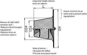
The main challenges in transcatheter closure of perimembranuos ventricular septal defects are proximity of the defect to the aortic and tricuspid valves and the conduction system. Devices designed for the closure of these defects have sought to minimise these complications. Although not designed for the closure of ventricular septal defects, ductal occluders have several advantages (Fig 4). The Lifetech Patent ductus arteriosus occluder has a retention skirt only on one side and has a long waist length (7–10 mm) with a tapered configuration. Stability of a device is determined mainly by pressure gradients between the two chambers. Hence, a retention skirt on one side should suffice to hold the device in place, as in restrictive perimembranous defects. Appropriate sizing of the defect and choosing the correct device size are important to prevent device embolisation. Ensuring a minimal distance of 2–3 mm from the aortic valve avoids the chances of device-induced aortic regurgitation. We have seen that the most perimembranous defects have some aortic rim to allow safe placement of these devices. The absence of a right-sided disc minimises the chances of tricuspid regurgitation, although there would be some tricuspid regurgitation in cases where the septal leaflet of the tricuspid valve contributes significantly to aneurysm transformation of the defect. All devices that have been designed for the closure of perimembranous ventricular septal defects have retention skirts on either side of the device, which could cause considerable strain and friction on the ventricular septum with each cardiac contraction. These have been postulated as mechanisms for heart block. A long waist length and lack of a right-sided retention skirt help the device to align itself appropriately after deployment and also avoid a constraining effect on the septum, with a lesser incidence of heart block. The waist of the device fits snuggly into the aneurysmal pouch, if present, or occupies the region of the defect to ensure complete closure. Bentham et alReference Bentham, Gujral, Adwani, Archer and Wilson 9 have highlighted on the instability of the device, with exaggerated motion, as a risk factor for the development of complete heart block. A longer waist length affords greater stability of the device with little motion during phases of the cardiac cycle with probably lesser trauma to the conduction system. Ductal occluders are also cost-effective compared with septal occluders.
First-generation duct occluder and its advantages in closure of perimembranous ventricular septal defects.

The Amplatzer Duct Occluder-II has retention discs on either side that exceed the waist diameter by 6 mm. The central waist elongates and conforms itself to the shape of tubular ducts for which it was originally designed and also conforms to the shape of tubular perimembranous ventricular septal defects. The device may also be used for the closure of other angiographic types of perimembranous defects. As the waist is stretchable and the device has a lower profile, it causes lesser strain on the ventricular septum, despite having a disc on either side. The Amplatzer Duct Occluder-II occluder can be used to close defects <5.5 mm and may be advantageous in smaller children as smaller sheath sizes (5–6 F) suffice. They can also be deployed retrogradely, avoiding the need for formation of an arteriovenous rail road and may minimise fluoroscopic times as reported by Koneti et al.Reference Koneti, Penumatsa, Kanchi, Arramraj and Somaraju Bhupathiraju 10 , Reference Koneti, Sreeram, Penumatsa, Arramraj, Karunakar and Trieschmann 11 However, we have not found significant difference in procedural times with the Amplatzer Duct Occluder-II occluder. Assessment of aortic regurgitation is difficult with the retrograde technique. The stretched waist length might contribute to lesser degrees of heart block, but this needs to be tested in long-term studies with this device. The fabric-free design makes it more flexible and might reduce chances of tricuspid or aortic leaflet entrapment, but might increase the chances of embolisation.
The purpose of this study was not to compare the two devices. The decision to use one type of device over another was mainly dictated by the size of the defect and its morphology. We chose Amplatzer Duct Occluder-II devices only if the defect measured <6 mm on echocardiography and angiography. They were also more suited for tubular configurations, as the waist tends to elongate and conform to the shape of the defect. Although tubular configurations may be suited for closure by Amplatzer Duct Occluder-II devices, size of the defect is an important determinant. Any defect measuring more than 6 mm in diameter (even if tubular) can only be closed with the conventional duct occluder. The presence of an aneurysmal pouch, larger defects, Gerbode defects, and multiple jets in the right ventricle favoured the use of first-generation Patent ductus arteriosus occluders.
Conclusion
Most defects that are amenable to transcatheter closure are restrictive moderate-sized defects with volume overload of the left heart. Although various devices have been used for transcatheter closure of perimembranous ventricular septal defects, heart block, aortic, and tricuspid regurgitation have continued to be major concerns. The present study demonstrates the effective use of ductal occluders for closure of most types of perimembranous defects with a low incidence of heart block, tricuspid regurgitation, and aortic regurgitation. They are also cost-effective and can be used in smaller children. The newer Amplatzer Duct Occluder-II may be an option in children with smaller body sizes and relatively small defects. The present study sample is not very large, and long-term follow-up data are needed to address the issue of late heart block.
Acknowledgements
None.
Financial Support
This research received no specific grant from any funding agency, commercial or not-for-profit sectors.
Conflicts of Interest
None.
Supplementary Material
To view supplementary material for this article, please visit http://dx.doi.org/10.1017/S1047951114001255.