Introduction
Nematodes of the genus Anisakis are ubiquitous in the marine environment, have a worldwide distribution and reproduce in the digestive tract of marine mammals, mainly cetaceans (Klimpel & Palm, Reference Klimpel and Palm2011). The life cycle of these nematodes involves planktonic crustaceans as first intermediate hosts, fishes and cephalopods as paratenic (transport) hosts, in which no larval development occurs but are crucial for the successful transfer to the definitive hosts, and cetaceans as definitive hosts (Mattiucci & Nascetti, Reference Mattiucci and Nascetti2008; Klimpel & Palm, Reference Klimpel and Palm2011). In cetaceans, the third larval stage (L3) moults to pre-adult (L4) and then to adult, which typically attaches to the mucosa of the stomach, often generating granulomatous ulcers (Smith, Reference Smith1989; Abollo et al., Reference Abollo, Lopez, Gestal, Benavente and Pascual1998a; Hrabar et al., Reference Hrabar, Bočina, Kurilj, Đuras and Mladineo2017). In severe infections, profuse haemorrhage and stomach perforation can occur, causing peritonitis and, eventually, the death of the host (Jaber et al., Reference Jaber, Pérez, Arbelo, Zafra and Fernández2006).
Humans can develop anisakiasis when they ingest raw or undercooked fish or squid infected with L3 of Anisakis spp. (Mattiucci et al., Reference Mattiucci, Cipriani, Levsen, Paoletti and Nascetti2018). The larvae can attach to the stomach or the intestine, or penetrate the gut and migrate through the viscera (Baird et al., Reference Baird, Gasser, Jabbar and Lopata2014). Allergic reaction can also result from anisakiasis, or, even more frequently, from the ingestion of dead worms in food fish (Mattiucci et al., Reference Mattiucci, Cipriani, Levsen, Paoletti and Nascetti2018). Anisakiasis is currently a serious emerging human health problem, which also generates negative effects on consumer confidence (Bao et al., Reference Bao, Pierce, Strachan, Martínez, Fernández and Theodossiou2018). Since the early 1980s, more than 20,000 cases of anisakiasis have been reported worldwide and 2000–3000 cases are reported annually only in Japan (EFSA-BIOHAZ, 2010; Ivanović et al., Reference Ivanović, Baltić, Bošković, Kilibarda, Dokmanović, Marković, Janjic and Baltić2017). There have also been marked increases in medical case reports of anisakiasis in other countries over the last decade, which have been attributed to several factors – in particular, improved public health diagnoses and a growing preference for raw or poorly cooked food (Bao et al., Reference Bao, Pierce, Strachan, Martínez, Fernández and Theodossiou2018; Mattiucci et al., Reference Mattiucci, Cipriani, Levsen, Paoletti and Nascetti2018).
However, whether there has also been a long-term increase in Anisakis infections due to ecological factors affecting the population of this parasite is an open question. Increases in the population sizes of intermediate, paratenic and especially definitive hosts; changes in the hosts' distribution or diet, enhancing transmission rates; and/or abiotic factors (e.g. temperature) affecting the survival of the free-living stage (see Mattiucci et al., Reference Mattiucci, Cipriani, Levsen, Paoletti and Nascetti2018 and references therein) could have also occurred at local or regional scales over the last decades. Unfortunately, we lack appropriate time series of epidemiological data on the different hosts in the cycle to investigate trends. During the period 1991–1996, a long-term study was conducted to report on the abundance, the prevalence and the extension of ulcerative lesions caused by Anisakis (as a surrogate of parasite abundance) in ten cetacean species that were found stranded along the Galician coast (north-west Spain) (Abollo et al., Reference Abollo, Lopez, Gestal, Benavente and Pascual1998a). Here, we carried out a similar analysis during the period 2017–2018 and compared infection parameters with the previous survey.
Material and methods
Sample collection
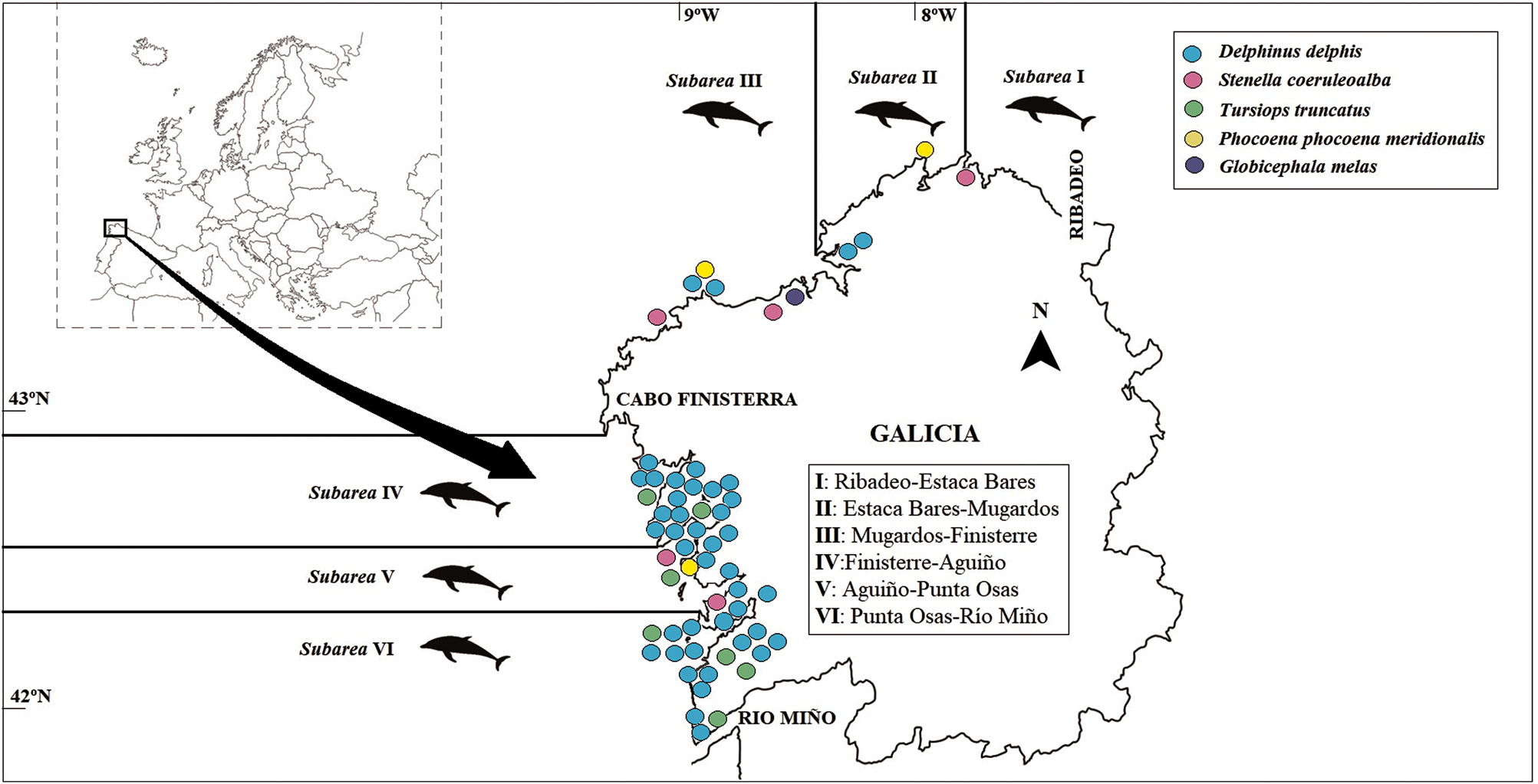
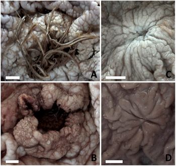
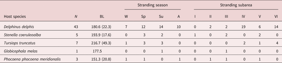
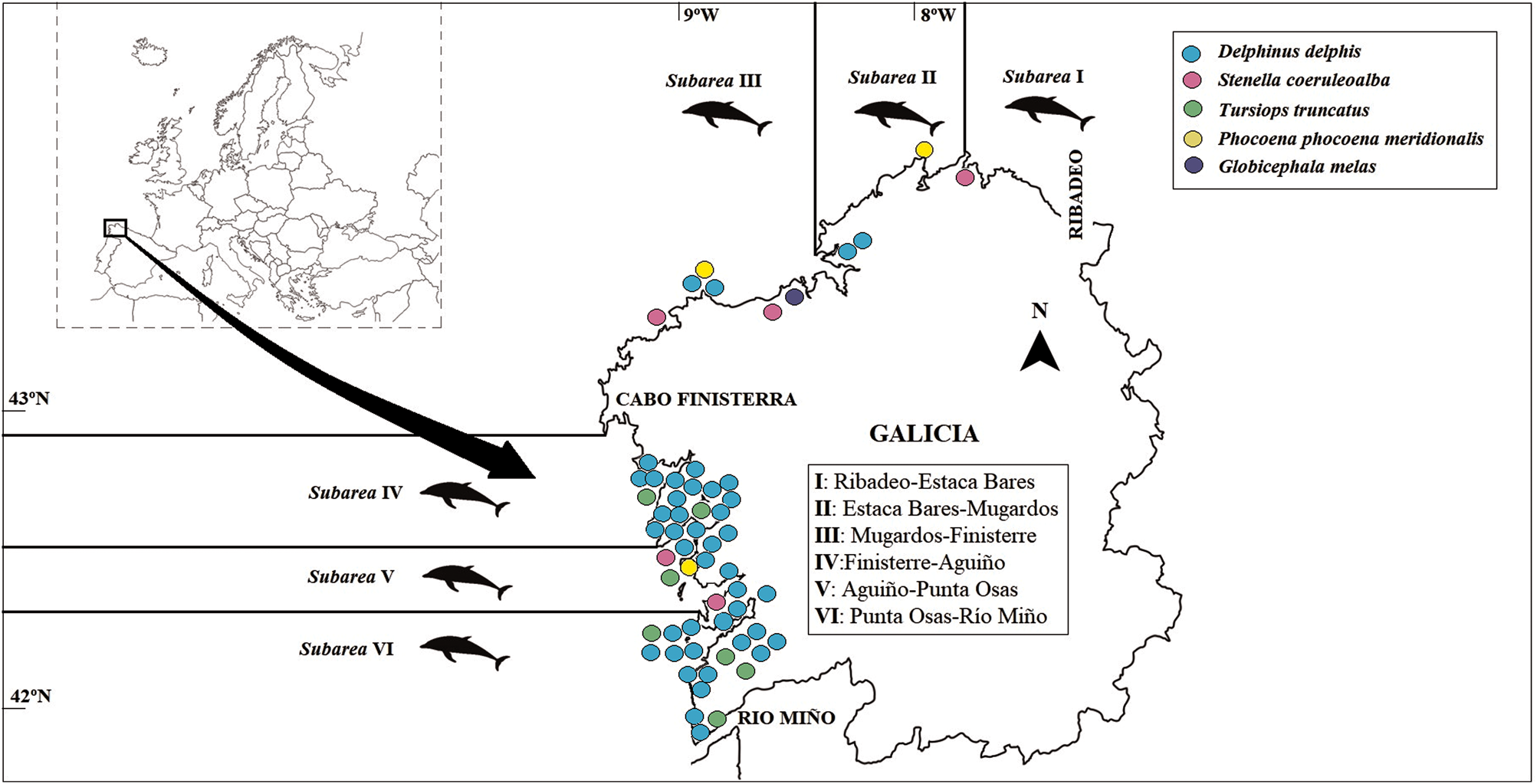
A total number of 59 stomachs belonging to short-beaked common dolphin (Delphinus delphis), striped dolphin (Stenella coeruleoalba), bottlenose dolphin (Tursiops truncatus), harbour porpoise (Phocoena phocoena meridionalis) and long-finned pilot whale (Globicephala melas) stranded on the Galician coast (north-west Spain) were collected from March 2017 to September 2018 (table 1) by the Galician Stranding Network carried out by the Coordinadora para o Estudio dos Mamíferos Mariños. Total body length, quarter of the year (table 1) and sex were recorded. Stranding areas were also recorded according to the delimitation (in subareas) of the Galician coast established by Abollo et al. (Reference Abollo, Lopez, Gestal, Benavente and Pascual1998b) (table 1 and fig. 1). Stomachs were collected during the necropsy and were brought to the Vigo Center of the Instituto Español de Oceanografía and frozen at −20°C. After thawing, stomachs were opened and the fore-, main and pyloric stomach were examined separately for macroscopic lesions caused by Anisakis spp. According to their macroscopic appearance, ulcerative lesions were classified following Smith (Reference Smith1989) as (1) open ulcers (OUs) (fig. 2a, b), with hemispherical appearance and a prominent central ‘crater’ with eroded epithelium; and (2) scars (Ss) (fig. 2d) with raised, apparently fibrotic tissue but no visible ‘crater’. Scarring tissue with traces of an old OU (fig. 2c) was considered as a third category of ulcerative lesion. The maximum diameter of OUs was measured in situ with a ruler (to the nearest 0.5 cm).
Stranding locations of cetaceans sampled in 2017–2018 for ulcers of Anisakis spp. along the Galician coast.
Putative healing stages of ulcerative lesions caused by Anisakis spp. in the forestomach of short-beaked common dolphins, Delphinus delphis, in Galician waters. (A) Open ulcer with a cluster of worms attached to eroded tissue; (B) open ulcer with ‘crater’ after natural worm's disappearance; (C) scarring tissue with traces of an old open ulcer; (D) scar with healed epithelium. Scale bars: 10 mm.

Hosts species, number of hosts individuals (N), body length (BL, mean ± standard deviation) and number of host individuals per stranding quarters (W, winter; Sp, spring; Su, summer; A, autumn) and subarea (I–VI) of cetaceans stranded along the Galician coast during 2017–2018. Stranding subareas follow the delimitation established by Abollo et al. (Reference Abollo, Lopez, Gestal, Benavente and Pascual1998b) (see the text for details).

Prevalence (percent of hosts with ulcerative lesions) and mean abundance (average number of ulcerative lesions per host) were calculated considering all sets of ulcerative lesions. Prevalence and mean abundance were also calculated only for OUs to allow comparison with data from the survey by Abollo et al. (Reference Abollo, Lopez, Gestal, Benavente and Pascual1998a). The 95% confidence interval (CI) for prevalence was estimated with Sterne's exact method, and for mean abundance based on 20,000 bootstrap replications, using the software Quantitative Parasitology 3.0 (Rózsa et al., Reference Rózsa, Reiczigel and Majoros2000)
We used Fisher's tests to compare prevalence among geographical subareas (table 1 and fig. 1) for both common dolphins and the remaining cetacean species pooled. In the case of common dolphins, the effect of host body length, sex, year (2017 vs. 2018) and quarter of the year on these variables were investigated with Spearman's correlation, Mann–Whitney test and Kruskal–Wallis test, respectively.
Differences of prevalence of OUs per species between the periods 1991–1996 and 2017–2018 were tested with Fisher's tests when host sample size was ≥5. Statistical significance was set at P < 0.05. All analyses were conducted with SPSS v. 24 (IBM SPSS Statistics, Reference IBM Corp2016).
Morphological and molecular identification of Anisakis spp.
A random sample of 90 L3s and 18 adult worms (ten larvae and two adults per host individual) that had been collected from the gastric lumen, or detached from ulcers of six common dolphins, two striped dolphins and one harbour porpoise from the host sample, were available for identification. All specimens had been fixed and stored in 70% ethanol.
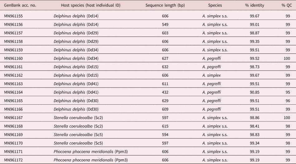
Larvae were cleared in lactic acid and examined under a stereomicroscope (X 80) to be identified as Type I or Type II according to morphological criteria – that is, presence/absence of mucron and shape of ventriculus (Berland, Reference Berland1989). For molecular identification, DNA was isolated from the 18 adults using the DNeasy® Blood and Tissue Kit (QIAGEN, Hilden, Germany) (table 2). The mitochondrial cytochrome c oxidase II (cox2) gene was amplified using the specific primers 211F 5′TTT TCT AGT TAT ATA GAT TGR TTY AT-3′ and 210R 5′-CAC CAA CTC TTA AAA TTA TC-3′ (Nadler & Hudspeth, Reference Nadler and Hudspeth2000) spanning the mtDNA nucleotide position 10,639–11,248, as defined in Ascaris suum (GenBank X54253). The polymerase chain reaction (PCR) reaction mixtures had a final volume of 20 µl, with 2 µl DNA, 4.8 µl PCR water, 1.6 µl of each primer at a concentration of 5 pmol/μl and 10 µl MyFiTM DNA Polymerase and Mix (BioLine, Meridian Life Science Inc., Taunton, USA). After an initial heat-activation step of 94°C for 3 min, the reaction consisted of 34 cycles of 94°C for 30 s, 46°C for 1 min and 72°C for 1 min 30 s, followed by a final step at 72°C for 10 min. Both positive and negative (no DNA) controls were used in the PCR.
GenBank accession number (GenBank acc. no.), host species, sequence length and putative species inferred from mtDNA cox2 sequences of 18 adult Anisakis spp. collected from three cetacean species in Galicia (north-west Spain) between 2017 and 2018. Percent of identity (% identity) and query cover (% QC), showing the similarity of the query sequence with the target sequence and the coverage of the query sequence by the target sequence, respectively, are also given.

Amplicons were purified with the Nucleospin® PCR and Gel Purification Clean-up Kit (Machery-Nagel, Düren, Germany) and were sent off to Macrogen Europe (Amsterdam, Netherlands) for sequencing with the same primers used for the PCR reactions. Nucleotide sequences from both strands were used to assemble consensus sequences with Geneious R7 (https://www.geneious.com) and were analysed compared with published sequences for identification using the National Institute of Health's National Centre for Biotechnology Information Basic Local Alignment Search Tool (http://blast.ncbi.nlm.nih.gov).
Results
Ulcerative lesions (fig. 2) were detected only on the wall of the forestomach. Clusters of Anisakis spp. at various stages of development (L3, L4 and adult forms) were found frequently attached to OUs (fig. 2a), but their prevalence was not recorded precisely. Based on pictures of open stomachs, at least eight short-beaked common dolphins harboured clusters of worms in their OUs (fig. 2a).
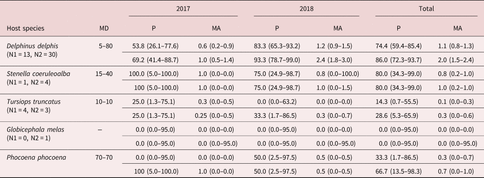
Ulcerative lesions were found in 86.0% of common dolphins, and the prevalence on OUs was just somewhat lower (74.4%) (table 3). The mean abundance of ulcerative lesions and OUs per dolphin were 2.0 and 1.1, respectively, and the maximum diameter of OUs ranged from 5 to 80 mm (table 3). Areas of scarring tissue with traces of old OUs (fig. 2c) co-occurred with OUs in two out of three dolphins that harboured them. Extensive areas of raised, fibrotic tissue with no visible ‘crater’ (maximum diameter: 50–90 mm) were detected with and without attached Anisakis and, in three out of five dolphins in which those areas were present, appeared together with OUs and Ss.
Prevalence (P) and mean abundance (MA), with confidence intervals in parenthesis, for open ulcers (first row) and all set of ulcerative lesions (second row) per each cetacean species stranded on the Galician coast during 2017–2018. Maximum diameter (MD) of open ulcers is also given in millimetres. N1 and N2 are samples sizes for 2017 and 2019, respectively.

In the other cetacean species, ulcerative lesions and OUs were detected from 0 (in long-finned pilot whale, and harbour porpoises) to 80.0% (in striped dolphins) of individuals, with mean abundance ranging from 0 to 1.0 for ulcerative lesions, and from 0 to 0.8 for OUs (table 3). Maximum diameter of OUs ranged from 10 to 70 mm (table 3). Areas of scarring tissue (fig. 2c) were not found, but extensive areas of fibrotic tissue were noticed in 1/3 harbour porpoises, 2/5 striped dolphins and 1/7 bottlenose dolphins (maximum diameter: 55–210 mm); the forestomach of the only individual of long-finned pilot whale examined did not harbour any kind of macroscopic lesion.
We found no significant differences in the prevalence of ulcerative lesions among geographical subareas in common dolphins (Fisher's test P > 0.85) and other cetaceans pooled (P > 0.85). In common dolphins, differences between 2017 and 2018 in the prevalence of OUs was close to significance (Fisher's test, P = 0.057), as was the mean number of lesions in infected dolphins (Mann–Whitney test, U = 66.5, n = 35, P = 0.056), but not the maximum diameter of OUs (U = 83.0, n = 35, P > 0.2). There were not seasonal differences in the occurrence of ulcerative lesions (Chi-square test, χ2 = 1.649, 3 d.f., P > 0.7). The relationship between body length and number of ulcerative lesions was positive and significant at 90% CI (if we consider, commonly used in ecology, α = 0.1) (Spearman correlation, r s = 0.277, n = 43, P = 0.073). Furthermore, the total area affected by ulcerative lesions (measured as the sum of their diameters) increased significantly with body length (r s = 0.345, n = 43, P = 0.024).
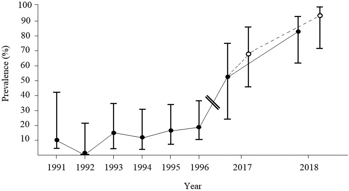
Compared with the period 1991–1996, the prevalence of OUs in common dolphins (Fisher's test, P < 0.00001, fig. 3) and striped dolphins (P = 0.024) increased significantly in 2017–2018. The mean abundance of ulcers parallels those of prevalence – that is, 1.1 vs. 0.22 and 0.8 vs. 0.5, respectively (table 3). Nevertheless, prevalence of OUs remained apparently unchanged in the case of bottlenose dolphins (P = 0.654), and mean abundance turned out to be lower in the period 2017–2018 (0.1 vs. 0.69). The small sample size for long-finned pilot whales and harbour porpoises precluded any statistical comparison.
Prevalence of open ulcers (black dots) caused by Anisakis spp. in short-beaked common dolphins, Delphinus delphis, during the periods 1991–1996 and 2017–2018. For the later period, values are also given for all of the set of ulcerative lesions described in fig. 2 (empty dots). Bars represent 95% confidence intervals.

Morphological and molecular identification of Anisakis spp.
All 90 larvae were morphologically identified as belonging to Type I. The mtDNA cox2 gene was successfully amplified and sequenced from all 18 adult specimens, generating sequences ranging from 432 to 632 bp in length (table 2). Twelve individuals were identified as Anisakis simplex sensu stricto (s.s.) and six as Anisakis pegreffii. The latter species was found only in common dolphins (table 2). Specimens from both species of Anisakis co-occurred in two common dolphins (table 2).
Discussion
Species of Anisakis commonly occur in clusters in stomach of small cetaceans, often causing granulomatous ulcers (Smith, Reference Smith1989; Motta et al., Reference Motta, Pinheiro, Carvalho, Viana, Vicente and Iñiguez2008), which have been used as a surrogate for the presence of the parasite (Abollo et al., Reference Abollo, Lopez, Gestal, Benavente and Pascual1998a). Furthermore, since ulcers can be found with or without attached worms (Smith, Reference Smith1989; Abollo et al., Reference Abollo, Lopez, Gestal, Benavente and Pascual1998a; Motta et al., Reference Motta, Pinheiro, Carvalho, Viana, Vicente and Iñiguez2008), and healing ulcers presumably remain for much longer than worms themselves, ulcerative lesions can be useful to trace past infections by Anisakis (fig. 2). However, this approach has several limitations in both the survey carried out by Abollo et al., Reference Abollo, Lopez, Gestal, Benavente and Pascual1998a and the present study. First, there is the possibility of false negatives if the parasite is present (e.g. in small amounts) but does not cause ulcers. Second, the finding of ulcers may indicate the presence of the parasite, but the number and/or extension of ulcerated areas likely provide, at best, a coarse proxy for the intensity of infection. Finally, at least three Anisakis spp. have been found occurring in both delphinids and phocoenids (Abollo et al., Reference Abollo, Gestal and Pascual2001; Mattiucci et al., Reference Mattiucci, Abaunza, Ramadori and Nascetti2004; Cipriani et al., Reference Cipriani, Mattiucci and Paoletti2017) in Galician waters; thus, the question arises as to whether these Anisakis species cause ulcers in the mentioned cetacean families to a comparable extent. Although we were fully aware of these problems, in this study we made a post-hoc use of ulcer data because it was also the only information available from the previous survey (Abollo et al., Reference Abollo, Gestal and Pascual2001) and, therefore, this was the only way to make a meaningful comparison. However, in the present study we also provide relevant data on the identity of the species of Anisakis involved that will be useful for more detailed analyses in the future.
All 90 larvae collected from three cetacean species belonged to Type I, a clade that includes six species typically infecting ziphiids (Anisakis ziphidarum, Anisakis nascetti), delphinids (Anisakis typica) or a wide array of delphinoid odontocetes and mysticetes (species of the A. simplex complex, i.e. A. simplex s.s., A. berlandi and A. pegreffii) (Mattiucci et al., Reference Mattiucci, Cipriani, Webb, Paoletti, Marcer, Bellisario, Gibson and Nascetti2014 and references therein). The adults analysed in this study were identified as A. pegreffii and A. simplex s.s. Both species have been reported in Galician waters infecting the same cetacean species analysed in this study (Mattiucci et al., Reference Mattiucci, Cipriani, Levsen, Paoletti and Nascetti2018). Therefore, we hypothesize that most, if not all, type I larvae found in our sample also belong to any of these two species. Interestingly, there is preliminary evidence A. pegreffii and A. simplex s.s. may differ in their pathogenic potential. Arizono et al. (Reference Arizono, Yamada, Tegoshi and Yoshikawa2012) found that larvae of A. simplex s.s. tolerated artificial gastric juice better and penetrated the agar at significantly higher rates than A. pegreffii, also being the only species that was detected infecting humans in Japan. Thus, whether the severity of lesions in the stomach of dolphins also differ between these two species cannot be ruled out. On the other hand, we found that A. pegreffii and A. simplex s.s. co-occurred in the same host individuals. This raises the intriguing question of whether they can aggregate in interspecific clusters attached to the same ulcers.
In the only host species with a suitable sample size – namely, the short-beaked common dolphin (D. delphis) – Abollo et al. (Reference Abollo, Lopez, Gestal, Benavente and Pascual1998a) detected a weak but significant increase in the number of ulcers with host length. Likewise, we find a similar tendency in the number and extension of ulcerative lesions. This pattern can hardly be accounted by age-related immunity differences between hosts (i.e. young individuals should be expected to develop ulcers more readily) and would rather be related to two non-exclusive factors. First, larger (=older) dolphins may have had more opportunities to get infected with Anisakis, and, thus, traces of ulcerative lesions would tend to accumulate with the number of past infections. Second, since larger hosts need to eat higher amounts of food; this would increase the recruitment rate of a ubiquitous trophically transmitted parasite such as Anisakis (Abollo et al., Reference Abollo, Gestal and Pascual2001).
The results of this study indicate a highly significant increase in the prevalence of Anisakis ulcers in two out of three cetacean species with a reasonable sample size – that is, common dolphin and striped dolphin (S. coeruleoalba), collected in 2017–2018 in comparison with those from the early 1990s. A preliminary question that arises concerns the extent to which this temporal pattern could be confounded by other factors. First, this may occur if there are spatial differences in infection rates of Anisakis within the study area, and the host sampling area greatly differs between periods. This seems unlikely because, in both studies, dolphins were collected in the same subareas from Galicia (see Abollo et al., Reference Abollo, Lopez, Gestal, Benavente and Pascual1998a, Reference Abollo, Lopez, Gestal, Benavente and Pascualb for details). In addition, results from the present study indicated that the prevalence of ulcerative lesions did not significantly differ among subareas. Second, the size/age composition of host samples could be heterogeneous between periods. Abollo et al. (Reference Abollo, Lopez, Gestal, Benavente and Pascual1998b) provided average lengths (with standard deviation) for each cetacean species in their sample and, assuming a normal distribution, the length difference with animals from our sample was not be statistically significant for any species (P > 0.05).
Alternatively, two biological factors could most likely account for the increase of ulcer prevalence in two of the three species: (1) common and striped dolphins suffer more from environmental stress, thus being more susceptible to the pathological effects of Anisakis; and/or (2) they have been more exposed to infections by A. pegreffii, A. simplex s.s. or both, in recent years. In the study area, striped dolphins occur typically over the continental slope and in oceanic waters (Mèndez-Fernández et al., Reference Mèndez-Fernandez, Bustamante and Bode2012), whereas common dolphins favour more inshore waters (Mèndez-Fernández et al., Reference Mèndez-Fernandez, Bustamante and Bode2012; Santos et al., Reference Santos, German, Correia, Read, Cedeira, Caldas, López, Velasco and Pierce2013; Saavedra et al., Reference Saavedra, Gerrodette, Louzao, Valeiras, García, Murcia, Cerviño, Pierce and Santos2015, Reference Saavedra, Gerrodette, Louzao, Valeiras, García, Cerviño, Pierce and Santos2018), and bottlenose dolphins (T. truncatus) are even more coastal (Mèndez-Fernández et al., Reference Mèndez-Fernandez, Bustamante and Bode2012; Louzao et al., Reference Louzao, Valeiras and García-Barcelona2019). However, both common and striped dolphins consume more pelagic prey than bottlenose dolphins, which rely more on both demersal fish and cephalopods (Santos et al., Reference Santos, German, Correia, Read, Cedeira, Caldas, López, Velasco and Pierce2013). Interestingly, Abollo et al. (Reference Abollo, Gestal and Pascual2001) noted that, in the study area, the life cycle of A. simplex s.s., but not that of A. pegreffii, seems to be linked to the demersal realm; thus, an open question is whether the increase of the Anisakis population may involve predominantly the latter species. Another important point is whether a higher prevalence of ulcerative lesions detected in recent years implies that populations of Anisakis are also larger. Some cetacean species (e.g. common dolphins) are abundant in the study area due to the presence of a rich ecosystem supported by an upwelling system, even having probably increased over the last decades (e.g. Hammond et al., Reference Hammond, Lacey and Gilles2017; Saavedra et al., Reference Saavedra, Gerrodette, Louzao, Valeiras, García, Cerviño, Pierce and Santos2018). Thus, a greater availability of definitive hosts would favour parasite reproduction and enhance the completion of the life cycle as it has been documented previously (Mattiucci & Nascetti, Reference Mattiucci and Nascetti2008; Mattiucci et al., Reference Mattiucci, Cipriani, Levsen, Paoletti and Nascetti2018). In this context, we cannot rule out that oceanic changes and/or shifts in the abundance of prey species may have occurred in the region since the 1990s, thus impacting the life cycles of Anisakis spp. Finally, there is the possibility that fish gutting onboard has become more common in Galician waters lately, thus increasing the number of larval stages that would otherwise be removed from the trophic web (González et al., Reference González, Gracia, Miniño, Romón, Larsson, Maroto, Regueira and Pascual2018), such as it has also been described in the Mediterranean Sea (see Mattiucci et al., Reference Mattiucci, Cipriani, Levsen, Paoletti and Nascetti2018). Given the zoonotic condition of Anisakis, we recommend a close surveillance of infections in both fish and cetaceans from Galician waters in the coming years.
Acknowledgements
Our thanks to the Coordinadora para o Estudio dos Mamíferos Mariños (CEMMA) volunteers that helped with field work to provide stomach samples and biometric data, and Dr Elvira Abollo for useful comments and advise during the elaboration of a previous draft of this manuscript.
Financial support
The Galician Stranding Network has the support of the General Directorate of Natural Heritage of the regional government Xunta de Galicia. AL is supported by national funds (OE), through FCT – Fundação para a Ciência e a Tecnologia (SFRH/BPD/108007/2015 to R.P., 094-88-ARH/2018), I.P., in the scope of the framework contract foreseen in the numbers 4, 5 and 6 of article 23, Decree-Law 57/2016 and Law 57/2017.
Conflicts of interest
None.
Ethical standards
The whole sample related to this study was collected by CEMMA organization and underwent all the legal permits provided by environmental administrations.