Implications
Erhualian and Large White are two typical breeds of pig, showing strikingly different metabolic characteristics and stress-coping. Hippocampal glucocorticoid receptor (GR) is critical to energy balance and stress response. Our results, demonstrating breed-specific dissimilarities in the expression of hippocampal GR and the DNA methylation status of GR proximal promoter region, provide fundamental information for further investigations into the function of GR in mediating the effects of glucocorticoids on hippocampal development in the pig.
Introduction
The glucocorticoid receptor (GR), a member of the nuclear receptor subfamily 3, mediates the action of glucocorticoid to regulate the transcription of numerous target genes (Pujols et al., Reference Pujols, Mullol, Roca-Ferrer, Torrego, Xaubet, Cidlowski and Picado2002), thereby contributing to metabolic homeostasis or energy balance (Peters, Reference Peters2011; Rose and Herzig, Reference Rose and Herzig2013). GR is expressed ubiquitously in almost all the living tissues/cells of the body, in a tissue-specific manner (Lu and Cidlowski, Reference Lu and Cidlowski2006). GR is highly expressed especially in metabolic-active tissues, such as liver, muscle, and brain (Pujols et al., Reference Pujols, Mullol, Roca-Ferrer, Torrego, Xaubet, Cidlowski and Picado2002).
The hippocampus is an important brain area serving as a regulatory center for energy balance and stress response (Joels et al., Reference Joels, Verkuyl and Van Riel2003; Peters, Reference Peters2011). Coincidently, hippocampus is also the main region in the brain to express high abundance of GR (van Steensel et al., Reference van Steensel, van Binnendijk, Hornsby, van der Voort, Krozowski, de Kloet and van Driel1996). In fact, GR serves as a major component of the ‘set-point’ machinery in the hippocampus to regulate the activity and the reactivity of hypothalamus–pituitary–adrenal (HPA) axis under basal and stressful situations (Gadek-Michalska et al., Reference Gadek-Michalska, Spyrka, Rachwalska, Tadeusz and Bugajski2013). Therefore, basic understanding on the GR expression in hippocampus is essential for the elucidation of the central control of energy homeostasis and stress responses.
GR expression is predominantly regulated at the level of transcription. Human GR gene contains nine exons (Russcher et al., Reference Russcher, Vash, de Jong, Brinkmann, Hofland, Lamberts and Koper2007), among which the un-translated exon 1 is alternatively transcribed driven by tissue-specific promoters. The promoters of GR gene are rich of cytosine-guanine dinucleotides (CpGs) which are susceptible to DNA methylation. Alternative usage of GR promoters results in numerous mRNA transcript variants in different mammalian species (Turner et al., Reference Turner, Schote, Macedo, Pelascini and Muller2006; Reyer et al., Reference Reyer, Ponsuksili, Wimmers and Murani2013). Moreover, the level of DNA methylation on some specific CpG sites in GR promoter has been reported to change in response to maternal nutrition (Lillycrop et al., Reference Lillycrop, Slater-Jefferies, Hanson, Godfrey, Jackson and Burdge2007) or maternal care (Weaver et al., Reference Weaver, Cervoni, Champagne, D’Alessio, Sharma, Seckl, Dymov, Szyf and Meaney2004) in rats. Alterations of DNA methylation on specific CpG site are reported to modulate the binding of transcription factors, such as transcription factor (NGFI-A), to the GR promoter and thus regulate GR transcription (Oberlander et al., Reference Oberlander, Weinberg, Papsdorf, Grunau, Misri and Devlin2008). Nevertheless, the relationship of the methylation status of different regions or single CpG site of GR promoter and the expression of GR exon 1 mRNA variants appears to be tissue- and species-specific.
The domestic pig serves as a good model for human metabolic studies as it shares high similarities in anatomy and physiological–biochemical characteristics. Different breeds of pigs demonstrate distinct phenotypes in growth, metabolism, as well as stress-coping characteristics (Cassady, Reference Cassady2007), which is associated with dramatic difference in serum cortisol concentrations (Desautes et al., Reference Desautes, Sarrieau, Caritez and Mormede1999). Previous studies described the breed-dependent pattern of total GR expression in hippocampus (Wei et al., Reference Wei, Xia, Li, Xiao, Bao, Parvizi and Zhao2010), liver (Yang et al., Reference Yang, Liu, Albrecht, Dong, Maak and Zhao2012) and muscle (Li et al., Reference Li, Yang, Shan, Shi, Xia, Wegner and Zhao2009) in pigs showing different stress coping style, hepatic gluconeogenesis, and intramuscular fat deposition, respectively. Recently, we cloned a 5.3 kb proximal promoter sequence of porcine GR gene, and identified seven un-translated alternative first exons each preceded by a distinct promoter (Zou et al., Reference Zou, Li, Jia, Yang, Ni, Cong, Soloway and Zhao2012). Transcript variants of the porcine GR gene were reported (Reyer et al., Reference Reyer, Ponsuksili, Wimmers and Murani2013), and GR exon 1 to 4 and 1 to 5 transcripts were found to be differentially expressed in the liver between Large White (LW) and Erhualian (EHL) pigs (Zou et al., Reference Zou, Li, Jia, Yang, Ni, Cong, Soloway and Zhao2012). However, the breed-dependent expression of GR and its 5′ exon 1 mRNA variants in porcine hippocampus has not been described. Moreover, it remains unknown whether the profile of CpG methylation on porcine GR promoter differ between breeds, and how the methylation status is correlated with the abundance of total GR and its exon 1 mRNA variants.
Therefore, here we use newborn LW and EHL piglets as model to investigate the breed differences in the hippocampal expression of GR and its exon 1 mRNA variants; to quantitatively measure the methylation ratios on each CpG site within the GR proximal promoter region using Sequenom’s MassARRAY system; and to analyze the possible correlation between the expression of GR and its mRNA variants and the methylation status of CpGs on porcine GR promoter in the hippocampus of two breeds of pigs.
Material and methods
Animals
Purebred male LW and EHL piglets (six per breed) were selected within 6 h after birth from two neighboring pig breeding farms in Changzhou, Jiangsu Province, China. Piglets were weighed before exsanguination. The blood was taken from the precaval vein and the serum samples were collected and stored at −20°C. The hippocampus was separated from the brain, weighed and frozen in liquid nitrogen immediately after slaughter. The slaughter and sampling procedures in this study complied with the ‘Guidelines on Ethical Treatment of Experimental Animals’ (2006) No. 398 set by the Ministry of Science and Technology, China and the ‘Regulation regarding the Management and Treatment of Experimental Animals’ (2008) No. 45 set by the Jiangsu Provincial People’s Government. The specific protocol of this experiment was approved by the Animal Ethics Committee of Nanjing Agricultural University.
Quantitation of mRNA by real-time PCR
Frozen hippocampus was ground in liquid nitrogen before total RNA extraction. Total cellular RNA was extracted with TRIzol reagent (15596026; Invitrogen Life Technologies,Carlsbad, CA, USA) and treated with DNase I (D2215; Takara, Dalian, Japan) to eliminate possible contamination of genomic DNA. Two micrograms of total RNA were reverse transcribed with M-MLV reverse transcriptase (Promega, Madison, WI, USA) following the standard protocol. Two microliters of diluted cDNA (1 : 20) were used for real-time PCR analysis which was performed on Mx3000P (Stratagene, La Jolla, CA, USA) with SYBR Premix Ex TaqTM (TaKaRa, Dalian, China), using β-actin as a reference gene. All the primers used in the present study were listed in Supplementary Table S1. The method of 2−ΔΔC t was used to analyze the real-time PCR data (Livak and Schmittgen, Reference Livak and Schmittgen2001). The abundance of total mRNA was presented as the fold change relative to the average level of the LW piglets, while GR variants mRNA was presented as the percentage of total GR mRNA.
Western blotting analysis for total GR protein
Total protein was extracted from hippocampus samples as previously described (Zou et al., Reference Zou, Li, Jia, Yang, Ni, Cong, Soloway and Zhao2012). The concentrations were determined with a Pierce BCA Protein Assay kit (23225; Thermo, Rockford, USA). Anti-GR antibody (sc-1004, 1 : 500) was purchased from Santa Cruz Biotechnology, Santa Cruz, USA; anti-ACTB antibody (AP0066, 1 : 10 000) was purchased from Bioworld, Louis Park, MN, USA. The signals were detected by enhanced chemiluminescence (ECL) using the LumiGlo substrate (Super Signal West Pico Trial Kit; Pierce, Rockford, USA). ECL signals were captured using VersaDoc 4000MP system (Bio-Rad, Hercules, CA, USA) and analyzed with Quantity One software (Bio-Rad). The protein content was presented as the fold change relative to the average content of the LW piglets.
Sequenom’s MassARRAY
Sequenom’s MassARRAY (CapitalBio, Beijing, China) was employed to determine the methylation status of porcine GR proximal promoter in hippocampus of newborn LW and EHL piglets. Genomic DNA was extracted and treated with sodium bisulfite using EZ-96 DNA methylation kit (Zymo Research, Irvine, CA, USA). Bisulfite-converted DNA was amplified by PCR with primers, of which 10-mer T7 promoter tag was added to each reverse primer for in vitro transcription. Transcripts generated from shrimp alkaline phosphatase-treated PCR products were subjected to uracil-specific cleavage to produce small fragments. All these fragments were automatically detected by MALDI-TOF mass spectrometry, and the quantitative data were processed by EpiTYPER software to analyze the methylation status for each CpG on GR promoter region in each genomic DNA sample.
Statistical analysis
The breed differences were tested with t-test for independent samples by SPSS 13.0 for Windows (SPSS Inc., Chicago, IL, USA). The mRNA abundances of the alternative GR first exons are expressed as the percentage of the total GR mRNA. Protein abundances are expressed as the fold-change relative to LW.
Methylation status of each detectable CpG is presented as percentage. Correlation analysis between the methylation status of each CpG site on GR promoter and the mRNA abundance of individual GR transcript variants was performed in R program environment, which provided the correlation coefficient (R) and significance (P-value). The significant correlation level was defined at P<0.05.
Results
BW, hippocampus weight, and hippocampal expression of neurotropic and apoptotic factors
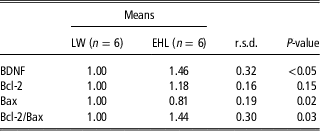
The BW of EHL piglets (0.75±0.03 kg) was significantly lower (P<0.01) than that of LW piglets (1.31±0.04 kg), while the hippocampus index of EHL piglets (0.34±0.01 g/kg) was significantly higher (P<0.01) than that of LW piglets (0.22±0.01 g/kg), indicating that EHL piglets had higher hippocampus weight relative to BW. Hippocampal mRNA expression of brain-derived neurotrophic factor (BDNF) was significantly higher (P<0.05) in the hippocampus of EHL piglets. Expression of Bax, a pro-apoptotic gene, was significantly lower (P<0.05) in EHL piglets. No significant breed difference was observed for Bcl-2, an anti-apoptotic gene. As a result, the Bcl-2/Bax ratio was significantly higher in the hippocampus of EHL piglets (Table 1).
Expression of neurotrophic and apoptotic genes in the hippocampus of newborn piglets

BDNF=brain-derived neurotrophic factor; LW=Large White; EHL=Erhualian.
Hippocampal expression of total GR and its exon 1 mRNA variants
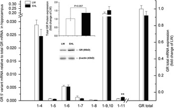
There was no significant breed difference in total GR mRNA expression in hippocampus. However, the total protein content of GR in hippocampus tended to be higher (P=0.057) in EHL than in LW. Seven GR exon 1 mRNA variants, including GR exon 1 to 4, 1 to 5, 1 to 6, 1 to 7, 1 to 8, 1 to 9, 10 and 1 to 11, were detected in the hippocampus of both breeds of piglets. GR exon 1 to 9, 10 was the most abundant transcript, followed by 1 to 4, 1 to 6, 1 to 11, 1 to 7, 1 to 5 and 1 to 8, in a descending order. Among all these mRNA variants, only GR exon 1 to 11 mRNA variant showed breed difference, being significantly higher (P<0.01) in EHL than in LW (Figure 1).
Breed differences in hippocampal expression of total glucocorticoid receptor (GR) mRNA, GR exon 1 mRNA variants and GR protein. RT-PCR demonstrated that hippocampal expression of GR exon 1 to 11 mRNA was higher in Erhualian (EHL) than Large White (LW), while there was no significant breed difference for GR 1 to 4, 1 to 5, 1 to 6, 1 to 7, 1 to 8, 1 to 9, 10 and total GR mRNA. Western blotting analysis showed that total GR protein content in hippocampus tended to be higher in EHL than in LW. **Indicates significant difference between breeds at P<0.01.

Methylation status of porcine GR promoter
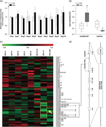
Each CpG site in the proximal region of GR promoter was annotated in Supplementary Figure S1. The methylation status of each CpG in 10 segments of GR promoter was quantified by Sequenom’s MassARRAY. We failed to amplify some regions containing extremely high CGs, so these regions were left as gaps undetected. The overall methylation levels of all the detected CpGs on GR promoter did not differ between breeds in the hippocampus of newborn piglets (Supplementary Table S2), nor did the overall methylation level for each amplified segment (Figure 2a). When comparative analysis was performed for each individual CpG site, CpG266.267 and CpG334 demonstrated significant breed differences (Figure 2b), however in a reversed manner: CpG266.267 was hypermethylated (P<0.05), while CpG334 was hypomethylated (P<0.05), in EHL piglets.
Methylation status of CpGs on porcine glucocorticoid receptor (GR) promoters. (a) Comparison of methylation status of CpGs in the segments of PCR products between Large White (LW) and Erhualian (EHL) piglets. (b) Breed differences in the methylation status of individual CpG on GR promoters. The methylation status of CpG266.267 and CpG334 was significantly different between LW and EHL. *Indicates significant difference between breeds at P<0.05. (c) Correlation analysis between methylation status of CpG sites in GR promoter region and the expression of GR mRNA variants in hippocampus of newborn piglets. The color legend at the top represents the level of correlation, red indicating positive correlation and green indicating negative correlation. The codes on the legend are correlation coefficients. The methylation status of CpG248, 259, 260, 268 and 271 within the CpG242 to 299 region (marked with yellow dot line and arrow) was negatively correlated to GR 1 to 11 mRNA expression. *Indicates significant correlation with GR 1 to 11 transcription at P<0.05. (d) Location of CpG242–299 and CpG334 on GR promoters.

Correlation of CpG methylation status and GR exon 1 mRNA abundance
As shown in Figure 2c, hippocampal expression of GR exon 1 to 11 mRNA variant was significantly (P<0.05) correlated with the methylation status of CpG248 (R=−0.67), CpG259 (R=−0.65), CpG260 (R=−0.74), CpG268 (R=−0.64) and CpG271 (R=−0.68).
Discussion
Hippocampus is one of the most important brain areas involved in memory integrity, stress response and energy balance. The structural and functional development of hippocampus is susceptible to prenatal or perinatal nutrition (Cintra et al., Reference Cintra, Granados, Aguilar, Kemper, DeBassio, Galler, Morgane, Duran and Diaz-Cintra1997; Morgane et al., Reference Morgane, Mokler and Galler2002) or stress. Different strains of mice from different rearing condition and domestication background demonstrated differences in hippocampal development (Schaefers, Reference Schaefers2013). Moreover, antenatal glucocorticoid treatment reduced BW and hippocampal volume in mice at birth (Noorlander et al., Reference Noorlander, Tijsseling, Hessel, de Vries, Derks, Visser and de Graan2014). Similarly, posttraumatic stress resulted in smaller hippocampal volumes in adulthood in human (Carrion et al., Reference Carrion, Weems and Reiss2007). However, we observed a positive correlation between serum cortisol level and the relative hippocampal weight in neonatal piglets. EHL piglets, which had two-folds higher serum cortisol level (Zou et al., Reference Zou, Li, Jia, Yang, Ni, Cong, Soloway and Zhao2012), demonstrated significantly higher hippocampus weight relative to BW as compared with LW piglets. It is noted that the higher serum cortisol level in EHL pigs is a breed characteristic that is evolved over generations of domestication and selection for this breed. The effect of such breed-specific high cortisol on hippocampal development could be different from that of stress-induced or experimentally administered cortisol. The functional disparity of cortisol on hippocampal development is largely determined by the expression and the function of GR in hippocampus.
Differences in hippocampus volume/weight imply different cell proliferation and survival patterns in hippocampus (Schaefers, Reference Schaefers2013). It has been shown that hippocampal expression of genes involved in cell proliferation and apoptosis differs across rat strains (Neeley et al., Reference Neeley, Berger, Koenig and Leonard2011). BDNF, which is essential for the hippocampal development (Bartrup et al., Reference Bartrup, Moorman and Newberry1997), was reported to be downregulated during hippocampal neuronal apoptosis (Hwang et al., Reference Hwang, Choi, Kim, Ko, Shin, Kim, Kim, Jin, Chung and Yi2013) in response to glucocorticoid treatment in vivo (Schaaf et al., Reference Schaaf, de Jong, de Kloet and Vreugdenhil1998) or in vitro (Yu et al., Reference Yu, Lee, Lee and Son2004). In this study, higher BDNF expression, together with higher Bcl-2/Bax ratio, was detected in the hippocampus of EHL piglets. This finding is in line with the higher hippocampus weight relative to BW observed in EHL piglets, yet appears to be contradictory to the higher serum cortisol level detected in this breed. Again, GR plays a major role for mediating the effects of glucocorticoids, acute or chronic, on hippocampal expression of neurotrophic and apoptotic genes.
GR is expressed in a tissue-specific manner with multiple GR exon 1 mRNA variants in human (Turner et al., Reference Turner, Schote, Macedo, Pelascini and Muller2006), rodents (Yau et al., Reference Yau, Noble, Chapman and Seckl2004) and pigs (Zou et al., Reference Zou, Li, Jia, Yang, Ni, Cong, Soloway and Zhao2012). Promoter analysis revealed tissue-specific activities of alternative GR promoters (Jiang et al., Reference Jiang, Qian, Zou, Jia, Ni, Yang and Zhao2014). In this study, the abundance of total GR mRNA did not differ between breeds. Among all the GR exon 1 mRNA variants detected, GR 1 to 11 mRNA variant demonstrated breed-dependent expression in the hippocampus of piglets. Although the most abundant GR exon 1 mRNA variants, GR 1 to 4 and GR 1 to 9, 10, did not differ between breeds, GR protein content tended to be higher in the hippocampus of EHL piglets with higher GR 1 to 11 mRNA expression. Interestingly, GR 1 to 4 and 1 to 5 mRNA variants were reported to be differentially expressed in the liver of LW and EHL piglets (Zou et al., Reference Zou, Li, Jia, Yang, Ni, Cong, Soloway and Zhao2012), indicating the tissue-specific usage of GR alternative promoters in the pig.
It is reported that the status of DNA methylation in GR promoters is closely associated with the transcription of GR exon 1 mRNA variants (Turner et al., Reference Turner, Alt, Cao, Vernocchi, Trifonova, Battello and Muller2010) in human brain (Cao-Lei et al., Reference Cao-Lei, Suwansirikul, Jutavijittum, Meriaux, Turner and Muller2013) and hippocampus (McGowan et al., Reference McGowan, Sasaki, D’Alessio, Dymov, Labonte, Szyf, Turecki and Meaney2009). In this study, the methylation status of individual CpG site on GR promoters was profiled. To our disappointment, the overall level of methylation across all the measurable CpG sites did not differ between breeds. Among all the measureable CpG sites, CpG266.267 and CpG334 were differentially methylated, however in a reversed manner, between these two breeds. The significance of these differentially methylated CpG sites in the regulation of breed-dependent GR transcription in porcine hippocampus remains unknown. Nevertheless, correlation analysis revealed that GR 1 to 11 expression was negatively correlated with the methylation status of some CpG sites located in the proximal promoter region of GR exon 1 to 11 (Figure 2d). This finding agrees with a previous report that the transcription of GR mRNA variants was regulated by the DNA methylation at single CpG site (Weaver et al., Reference Weaver, Cervoni, Champagne, D’Alessio, Sharma, Seckl, Dymov, Szyf and Meaney2004). We might speculate that the breed differences in the methylation of CpG266.267 or CpG334 may be involved in the breed-specific GR 1 to 11 transcription. Transcription factor prediction analysis indicated that there are SP1 binding sites CGCGG (−2395 to ~−2391) at CpG266.267 and CCTGCCCTC (−1771 to ~−1763) at CpG334, suggesting that single CpG methylation may affect SP1 binding to the promoter and thereby regulate the expression of GR mRNA variants. Nevertheless, functional analysis is required to verify the effect of differentially methylated CpGs on GR exon 1 to 11 mRNA transcription.
In summary, here we demonstrate the breed-specific expression pattern of GR exon 1 mRNA variants in the hippocampus of LW and EHL newborn piglets, as well as the profile of methylation status of individual CpG on proximal GR promoters. Our results indicate that hippocampal expression of GR exon 1 mRNA variants is not directly correlated with the overall methylation status of the promoters. However, methylation changes on some individual CpG sites in GR promoter may play a role to fine-tune GR transcription in the hippocampus of neonatal piglets. These findings provide fundamental information for further investigations into the function of GR in mediating the effects of glucocorticoids on hippocampal development in the pig.
Acknowledgment
This work was supported by the National Basic Research Program of China (2012CB124703), the Major National Science & Technology Program (2009ZX08009–138B), the Special Fund for Agro-scientific Research in the Public Interest (201003011, and the Fundamental Research Funds for the Central Universities (KYZ200913). The funders had no role in study design, data collection and analysis, decision to publish, or preparation of the manuscript.