Introduction
Glaucoma, which leads to a degeneration and loss of the retinal ganglion cells (RGCs), is a major cause of blindness worldwide (Resnikoff et al., Reference Resnikoff, Pascolini, Etya’ale, Kocur, Pararajasegaram, Pokharel and Mariotti2004). Glaucoma is made up of a variety of disorders, including primary open-angle and primary angle closure glaucoma, which have different causes and risk factors involved with the most well studied and defined being elevated intraocular pressure (IOP). Many studies are currently being performed looking at possible treatment strategies for the disease, and in numerous cases, rodent models are used to test their validity in the first instance. Previous studies in glaucoma patients (Kiyosawa et al., Reference Kiyosawa, Bosley, Kushner, Jamieson, Alavi, Savino, Sergott and Reivich1989; Miki et al., Reference Miki, Nakajima, Takagi, Shirakashi and Abe1996; Sugiyama et al., Reference Sugiyama, Utsunomiya, Ota, Ogura, Narabayashi and Ikeda2006; Duncan et al., Reference Duncan, Sample, Weinreb, Bowd and Zangwill2007a,Reference Duncan, Sample, Weinreb, Bowd and Zangwillb) and in primate models (Imamura et al., Reference Imamura, Onoe, Shimazawa, Nozaki, Wada, Kato, Nakajima, Mizuma, Onoe, Taniguchi, Sasaoka, Hara, Tanaka, Araie and Watanabe2009) of the disease have suggested that there may be changes in the function of central visual areas due to the disease (Smith et al., Reference Smith and Bedi1993). Although many studies use rodent models of the disease, only a few previous studies have looked at the function of the superior colliculus in rat models of glaucoma. The superior colliculus is the major central target of RGCs in rodents, and there is also a greater knowledge of the afferent and efferent projections to and within the superior colliculus than for the lateral geniculate nucleus (LGN) in rodents. King et al. (Reference King, Sarup, Sauve, Moreland, Carpenter and Sharma2006) used an ocular hypertension (OHT) model of glaucoma where three episcleral veins were cauterized leading to elevated IOP and made extracellular multiunit recordings in the superior colliculus. They found that the visual receptive field was significantly larger in the superior colliculus receiving input from glaucomatous eyes compared to contralateral unoperated eyes and unoperated animals. They suggested that this may represent the possibility for plasticity and compensation in the retina and superior colliculus after RGC degeneration. However, the mechanisms behind the changes seen in receptive field size are still to be investigated, and it is unclear as to whether they represent central or retinal changes.
Glutamate is the major excitatory neurotransmitter in the visual pathway from the retina to the superior colliculus (Binns & Salt, Reference Binns and Salt1998; Binns, Reference Binns1999; Lo & Mize, Reference Lo and Mize1999), and the postsynaptic response of superior colliculus neurons to retinal input is largely dependent on ionotropic glutamate receptors (Mize & Butler, Reference Mize and Butler1996; Jeon et al., Reference Jeon, Hartman and Mize1997a,Reference Jeon, Gurski and Mizeb), including the N-methyl-D-aspartate (NMDA) receptor. We have recently found changes in the NMDA receptor component to retinocollicular transmission in a rat glaucoma model (Georgiou et al., Reference Georgiou, Guo, Cordeiro and Salt2010) when using an in vitro slice preparation of the superior colliculus. We found that 32 weeks after IOP elevation, there was an increase in NMDA receptor contribution to the synaptic response in superior colliculus slices receiving input from the glaucomatous eyes. The findings suggested that there is possible functional compensation in the superior colliculus after RGC degeneration induced through elevated IOP that may be represented by the functional changes seen in the NMDA receptors.
In addition to the ionotropic glutamate receptors, metabotropic glutamate receptors (mGluRs) have been found in the superior colliculus (Martin et al., Reference Martin, Blackstone, Huganir and Price1992; Shigemoto et al., Reference Shigemoto, Nakanishi and Mizuno1992; Baude et al., Reference Baude, Nusser, Roberts, Mulvihill, McIlhinney and Somogyi1993; Fotuhi et al., Reference Fotuhi, Sharp, Glatt, Hwang, von Krosigk, Snyder and Dawson1993; Nakajima et al., Reference Nakajima, Iwakabe, Akazawa, Nawa, Shigemoto, Mizuno and Nakanishi1993; Ohishi et al., Reference Ohishi, Shigemoto, Nakanishi and Mizuno1993a,b, Reference Ohishi, Shigemoto, Nakanishi and Mizuno1998; Shigemoto et al., Reference Ohishi, Neki and Mizuno1993; Tanabe et al., Reference Shigemoto, Nomura, Ohishi, Sugihara, Nakanishi and Mizuno1993; Okamoto et al., Reference Tanabe, Nomura, Masu, Shigemoto, Mizuno and Nakanishi1994; Kinzie et al., Reference Okamoto, Hori, Akazawa, Hayashi, Shigemoto, Mizuno and Nakanishi1995; Romano et al., Reference Kinzie, Saugstad, Westbrook and Segerson1995; Neki et al., Reference Romano, Sesma, McDonald, O’Malley, Van den Pol and Olney1996; Petralia et al., Reference Neki, Ohishi, Kaneko, Shigemoto, Nakanishi and Mizuno1996; Bradley et al., Reference Petralia, Wang, Niedzielski and Wenthold1998; Hudtloff & Thomsen, Reference Bradley, Rees, Yi, Levey and Conn1998; Thomsen & Hampson, Reference Hudtloff and Thomsen1999; Mutel et al., Reference Thomsen and Hampson2000; Cirone et al., Reference Mutel, Ellis, Adam, Chaboz, Nilly, Messer, Bleuel, Metzler, Malherbe, Schlaeger, Roughley, Faull and Richards2002b; Puente et al., Reference Cirone, Sharp, Jeffery and Salt2005), and these have been shown to be involved in the modulation of visual transmission to and within the superior colliculus both in vivo (Cirone & Salt, Reference Cirone, Pothecary, Turner and Salt2000, Reference Cirone and Salt2001; Cirone et al., Reference Cirone and Salt2002a) and in vitro (Cirone et al., Reference Cirone and Salt2002a; Pothecary et al., Reference Pothecary, Thompson and Salt2002; White et al., Reference White, Kylanpaa, Christie, McIntosh, Irving and Platt2003; Thompson et al., Reference Thompson, Neale and Salt2004; Lacey et al., Reference Lacey, Pothecary and Salt2005; Neale & Salt, Reference Neale and Salt2006). The mGluRs can be divided into three groups on the basis of sequence homology, pharmacology, and coupling to second messenger systems. Group I receptors (mGluR1 and mGluR5) are predominantly coupled to postsynaptic inositol phosphate mechanisms, whereas Group II receptors (mGluR2 and mGluR3) and Group III receptors (mGluR4, mGluR6, mGluR7, and mGluR8) typically inhibit forskolin-stimulated cyclic adenosine monophosphate (cAMP) production and are often involved in presynaptic modulation of transmitter release. High levels of group III mGluRs are expressed in the superficial layers of the superior colliculus as seen in an autoradiographic study by Hudtloff and Thomsen (Reference Hudtloff and Thomsen1998). At the ultrastructural level, mGluR4 is specifically located on retinal terminals within the superior colliculus (Puente et al., Reference Puente, Hermida, Azkue, Bilbao, Elezgarai, Diez, Kuhn, Donate-Oliver and Grandes2005), and we have shown that agonists of this receptor can modulate retinocollicular transmission in vivo and in vitro (Cirone & Salt, Reference Cirone, Pothecary, Turner and Salt2000, Reference Cirone and Salt2001; Pothecary et al., Reference Pothecary, Thompson and Salt2002; Thompson et al., Reference Thompson, Neale and Salt2004; Lacey et al., Reference Lacey, Pothecary and Salt2005). The broad-spectrum agonist of group III mGluRs, L-AP4, is a phosphonic analog of glutamate, which has been shown to cause depression of synaptic transmission through presynaptic mechanisms (Evans et al., Reference Evans, Francis, Jones, Smith and Watkins1982; Lanthorn et al., Reference Lam, Kaufman, Gabelt, Eleanor and Matsubara1984; Forsythe & Clements, Reference Forsythe and Clements1990; Thomsen, Reference Thomsen1997). In particular, L-AP4 can depress retinocollicular transmission presumably via activating presynaptic mGluR4, although it is possible that there is also some contribution from mGluR8 (Saugstad et al., Reference Saugstad, Kinzie, Shinohara, Segerson and Westbrook1997; Thomsen & Hampson, Reference Thomsen and Hampson1999; Cirone & Salt, Reference Cirone, Pothecary, Turner and Salt2000, Reference Cirone and Salt2001; Pothecary et al., Reference Pothecary, Thompson and Salt2002; Thompson et al., Reference Thompson, Neale and Salt2004; Lacey et al., Reference Lacey, Pothecary and Salt2005; Puente et al., Reference Puente, Hermida, Azkue, Bilbao, Elezgarai, Diez, Kuhn, Donate-Oliver and Grandes2005).
If there are central changes during the progression of experimental glaucoma, then it might be expected that changes in retinocollicular transmission and its modulation would be affected. Group III receptors and mGluR4 in particular is a likely contender in view of location and function on retinal terminals within the superior colliculus (see above). Furthermore, we have previously shown that there are changes in mGluR4/8 modulation of retinocollicular transmission during the course of development and aging and during the progression of retinal degeneration (Cirone & Salt, Reference Cirone, Pothecary, Turner and Salt2000, Reference Cirone and Salt2001; Pothecary et al., Reference Pothecary, Thompson and Salt2002; Thompson et al., Reference Thompson, Neale and Salt2004; Lacey et al., Reference Lacey, Pothecary and Salt2005). Thus, the aim of the present work was to test the hypothesis that there are changes in mGluR4 modulation of retinocollicular transmission during the progression of experimental glaucoma. To this end, we have used a rat model of glaucoma that is used by many labs, where hypertonic saline is injected into the episcleral veins to induce IOP elevation (Morrison et al., Reference Morrison, Moore, Deppmeier, Gold, Meshul and Johnson1997; Cordeiro et al., Reference Cordeiro, Guo, Luong, Harding, Wang, Jones, Moss, Sillito and Fitzke2004) and which results in apoptosis and eventual degeneration of the RGCs (Guo et al., Reference Guo, Tsatourian, Luong, Podoleanu, Jackson, Fitzke and Cordeiro2007). We have used an in vitro slice preparation comprising the superior colliculus and the optic tract (OT) as it enters the superior colliculus (Pothecary et al., Reference Pothecary, Thompson and Salt2002, Reference Pothecary, Thompson and Salt2005; Lacey et al., Reference Lacey, Pothecary and Salt2005; Georgiou et al., Reference Georgiou, Guo, Cordeiro and Salt2010) so as to be able to assess the effects of the agonist L-AP4 on retinocollicular transmission without the complications brought about due to a retinal input (which is degenerated in this model) that would affect in vivo studies where visual stimulation is used. Furthermore, using stimulation of the OT fibers as they enter the superior colliculus allows us to better assess changes in synaptic transmission at this locus. Using these methods, we see that there are changes in the modulation of retinocollicular transmission by mGluR activation during the progression of this experimental glaucoma model.
Materials and methods
Animals
All conditions and experimental procedures were carried out in accordance with the U.K. Animals (Scientific Procedures) Act 1986 and associated guidelines and the ARVO Statement for the Use of Animals in Ophthalmic and Vision Research. Animals were obtained from Harlan UK Ltd. (Blackthorn, UK) and given at least 48 h to accommodate before any procedures were performed. Forty-four male Dark Agouti rats age 7–8 weeks (150–200 g) were housed in a 12-h light/12-h dark cycle with unlimited access to food and water. Eighteen of the animals had the OHT model induction performed with the other 26 animals being used as age-matched unoperated controls (referred to as controls henceforth).
OHT induction
For the glaucoma OHT model induction, animals were anesthetized using an intraperitoneal injection of a mixture of ketamine (100 mg/ml) (37.5%)/medetomidine (1 mg/ml) (25%)/sterile water (37.5%) solution at 0.2 ml/100 g. Unilateral elevation of IOP was induced in the left eye only of 18 animals using an adaptation of the Morrison method (Morrison et al., Reference Morrison, Moore, Deppmeier, Gold, Meshul and Johnson1997), which has been described, characterized, and validated in our lab (Cordeiro et al., Reference Cordeiro, Guo, Luong, Harding, Wang, Jones, Moss, Sillito and Fitzke2004; Guo et al., Reference Guo, Moss, Alexander, Ali, Fitzke and Cordeiro2005a,b, Reference Guo, Salt, Luong, Wood, Cheung, Maass, Ferrari, Russo-Marie, Sillito, Cheetham, Moss, Fitzke and Cordeiro2006, Reference Guo, Salt, Maass, Luong, Moss, Fitzke and Cordeiro2007). Briefly, 50 μl of hypertonic saline solution (1.8 m) was injected into two episcleral veins and ointments (Chloramphenicol 1% and Hydrocortisone 0.5%; Martindale Pharmaceuticals Ltd, Romford, UK) were then applied to the eye. Animals were then allowed to recover from anesthesia in their home cages on top of heat mats.
IOPs were measured at baseline before IOP elevation, then at weekly intervals for a month, and then monthly intervals thereafter. Animals were under isoflurane anesthesia (Merial; Animal health, Ltd., Essex, UK), and IOP measurements were made using a rebound tonometer (custom built by the John Danias group, Department of Ophthalmology, Mount Sinai School of Medicine, New York, NY). The differences between IOP values of the unoperated and IOP-elevated eyes were measured, and statistical analysis was performed using a one-way analysis of variance (ANOVA) and Tukey honestly significant difference (HSD) post hoc analysis using SPSS.
Superior colliculus in vitro slice preparations
For the superior colliculus, in vitro slice preparation animals from the OHT model group and the control group were taken at 3, 16, or 32 weeks after surgery and at the corresponding ages for the controls. Animals were anesthetized using an intraperitoneal injection of a mixture as above and left for 30 min. Animals were then decapitated and their brains were removed rapidly and placed into ice-cold oxygenated sucrose Krebs medium containing (mM): sucrose 202, KCl 2, KH2PO4 1.25, MgSO4 10, CaCl2 0.5, NaHCO3 26, glucose 10. The cerebellum and small amount of frontal cortex were removed and cuts were made on the right- and left-hand sides parallel to the midline so that a flat surface was revealed. The flat surface was then glued on one side to the cutting stage of a vibratome (Integraslice 7550 MM; Campden Instruments, Lafayette, IN), and parasagital slices of 400-μm thickness were cut through the superior colliculus. This method allows the OT and its connections to the superficial layers of the superior colliculus to be maintained in slices from both sides of the brain. Given that the retinal projection to the superior colliculus is more than 90% crossed (Dreher et al., Reference Dreher, Sefton, Ni and Nisbett1985), recordings were possible from superior colliculus slices with RGC input from both the elevated IOP and normal IOP eyes from the same animal. After cutting, slices were transferred to a bath containing oxygenated recovery Krebs medium containing (mM): NaCl 124, KCl 2, KH2PO4 1.25, MgSO4 5, CaCl2 1, NaHCO3 26, glucose 10. Slices were then left for at least 1 h before being transferred to an interface recording chamber where they were perfused at a rate of 0.7 ml/min with warmed (33–34°C) oxygenated Krebs medium containing (mM): NaCl 124, KCl 2, KH2PO4 1.25, MgSO4 1, CaCl2 2, NaHCO326, glucose 10.
Stimulation to the OT immediately prior to its entry to the superior colliculus was via a bipolar tungsten-in-glass electrode with extracellular recordings being made in the superficial gray layer of the superior colliculus via a Krebs-filled glass micropipette (5–10 μm in diameter). Responses were recorded using an Axoprobe 1A amplifier (Axon Instruments, Foster City, CA, USA), digitized at 10 kHz via a CED1401 interface, and stored on a computer using Spike 2 software (Cambridge Electronic Design, Cambridge, UK).
Each recording session was started by determining a minimum and maximum response to stimulus intensity (0.1 ms pulses) for the slice and the intensity level was then set at 75% of that which produced the maximum response. A stable baseline was then recorded for 20 min with 20-ms paired-pulse intervals, with 20 s between stimulations. mGluR group III agonist, L-AP4 (10 μM; Ascent Scientific, Bristol, UK), was then added for 10 min within the bathing medium with the effect of this on synaptic responses being assessed in the final 3 min of application. The slice was then perfused with bathing medium for at least 10 min until the L-AP4 was fully washed out.
Responses to stimuli were waveform averaged (three trials, equal to 60 s of recording) and peak amplitude of the field excitatory postsynaptic potential (fEPSP) was measured. The fEPSP amplitude measurements taken for the last 3 min of drug application were normalized and compared to baseline condition measurements. The effect of L-AP4 on peak amplitude was statistically analyzed using paired t-tests, and differences between the groups were analyzed using one-way ANOVA and a post hoc Tukey HSD test using SPSS software.
Results
IOP measurements
In control animals, there were no statistically significant differences in IOPs between the right and left eyes at any of the time points that this was measured. At baseline recordings before IOP elevation, there was no significant difference in the IOPs between the left and right eyes of the OHT model animals. However, between 1 and 4 weeks in the OHT model animals, there was a significantly greater IOP in the IOP-elevated eyes compared to the unoperated eyes, which by 8 weeks after surgery had reduced to nonsignificant levels (Table 1) (Georgiou et al., Reference Georgiou, Guo, Cordeiro and Salt2010).
IOP differences between left and right eyes in OHT model animals

Characteristics of synaptic responses in superior colliculus slices
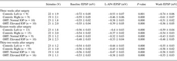
Stimulation of the OT in our parasagital superior colliculus slices typically evoked fEPSPs in the superficial gray layer of the superior colliculus with characteristics similar to those we have described previously in adult rats (Cirone et al., Reference Cirone and Salt2002a; Pothecary et al., Reference Pothecary, Thompson and Salt2002, Reference Pothecary, Thompson and Salt2005; Thompson et al., Reference Thompson, Neale and Salt2004; Lacey et al., Reference Lacey, Pothecary and Salt2005). Responses to stimulation under baseline conditions in control animals had a peak amplitude of −0.65 mV and latency to peak of 2 ms on average, and similar results were obtained irrespective of from which side of the brain slices had been obtained (Tables 2 and 3). The response to the second of each pair of stimuli was also consistently depressed compared to the first, showing paired-pulse depression. The paired-pulse ratios during baseline conditions in control animals ranged from 0.72 to 0.76 (Fig. 3).
The effect of L-AP4 on fEPSP amplitude in control and OHT model animals

There were no significant differences in the peak amplitude of the fEPSP under baseline conditions, nor the stimulus intensities used to evoke responses, between the left and right SC slices in either the control or the OHT model animals at any of the time points (P > 0.05).
Stimulus intensities to evoke responses are shown in the second column. Values are average (V or mV) ± s.e.m. of n values.
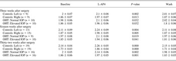
The effect of L-AP4 on latency of the peak of the fEPSP in control and OHT model animals

Values are average (ms) ± s.e.m. of n values.
In slices taken from rats with experimental glaucoma, there were no significant differences in the peak amplitude of the fEPSP in elevated IOP slices compared to normal IOP slices at any of the time points tested (Table 2), and the degree of paired-pulse depression seen in these slices was also similar to that seen in control slices. It is important to note that there were no significant differences in the stimulus intensity required to produce a 75% of maximal response between any of the experimental or control groups (Table 2).
L-AP4 effects in control animals
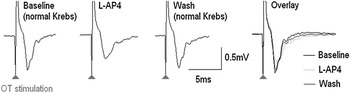
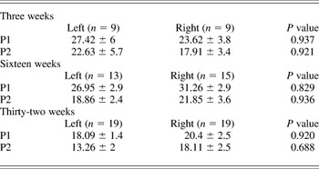
In this study as with previous ones from our lab (Pothecary et al., Reference Pothecary, Thompson and Salt2002; Thompson et al., Reference Thompson, Neale and Salt2004; Lacey et al., Reference Lacey, Pothecary and Salt2005), 10 min bath application of 10 μM L-AP4 significantly reduced the fEPSP peak amplitude, which was returned to baseline levels after 10 min wash out with normal Krebs medium (Fig. 1). In control animals, there were no significant differences in the effect of L-AP4 on fEPSP peak amplitude in slices from the left or right hemisphere of the superior colliculus at any of the time points tested (Table 4); therefore, slices from both hemispheres in these animals were grouped together in any further analysis.
The effect of application of L-AP4 to the peak amplitude of the fEPSPs. The figure shows waveform-averaged traces (six stimulations each) of the fEPSP recorded from the superficial gray layer of the superior colliculus after OT stimulation. Application of L-AP4 causes a reduction in the peak amplitude of the fEPSP compared to baseline levels. Ten minutes wash out with normal Krebs returns the peak amplitude of the fEPSP to baseline levels. This example is from a control animal at 23 weeks of age, which showed a 26% reduction in peak amplitude due to bath application of L-AP4.

The effect of L-AP4 on the fEPSP in control animals: Percentage reduction of the fEPSP

P1—first peak in response to the first of a pair of stimuli.
P2—peak response to the second of a pair of stimuli.
Values are average (% reduction) ± s.e.m. of n values.
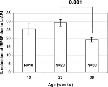
We found, as was seen in the previous study by Lacey et al. (Reference Lacey, Pothecary and Salt2005), that there was an age-related effect of L-AP4 on the amplitude of the fEPSP. The effect of L-AP4 on fEPSP amplitude was similar in superior colliculus slices from rats at 10 and 23 weeks of age (26 ± 3.5% and 29 ± 2.1% reduction, respectively, P = 0.499). However, at 39 weeks old, the effect of L-AP4 was significantly reduced compared to its effect in slices from animals at 23 weeks old (19 ± 1.4% and 29 ± 2.1% reduction, respectively, P = 0.001, Fig. 2).
Age-related effects of L-AP4 on fEPSP amplitude. The graph shows the age-dependent effect of the L-AP4 reduction of the fEPSP amplitude in control animals. Slices from the left and right hand superior colliculus were grouped together for each age group. At 39 weeks of age, there was a significant reduction in the effect of L-AP4 on fEPSP peak amplitude compared to the effect at 23 weeks of age.

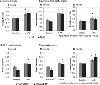
In most slices, the reduction in peak amplitude during L-AP4 application was greater in the response to the first of the pair of stimuli compared to the reduction seen in the response to the second, that is, a reduction in the paired-pulse ratio (peak 2/peak 1). The paired-pulse ratio was seen to increase overall during L-AP4 application at all time points in control animals, although this was not always to a significant extent (Fig. 3).
Paired-pulse ratios for the control (A) and OHT model (B) animals. The graph shows the paired-pulse ratios at baseline (normal Krebs) and with L-AP4 applied in the control (A) and OHT model (B) animals. L-AP4 addition caused a consistent increase in the paired-pulse ratio, although this was not always significant. There were no significant differences between the left and right superior colliculus slices in baseline or L-AP4 conditions at any time point. Error bars indicate standard error of mean.

L-AP4 effects in the OHT model of glaucoma
As with control animals, in the OHT model animals, L-AP4 application also significantly reduced the peak amplitude and increased the latency of the peak of the fEPSP in both elevated IOP and normal IOP superior colliculus slices (Tables 2 and 3).
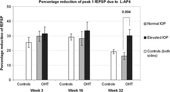
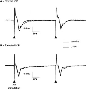
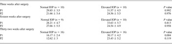
There were no significant differences in the effect of L-AP4 on peak amplitude of the fEPSP in elevated IOP slices compared to normal IOP slices at 3 weeks (32 ± 4.5% and 30 ± 3.5% reduction in peak amplitude, respectively, P = 0.992) or at 16 weeks (34 ± 5.7% and 28 ± 4.7% reduction, respectively, P = 0.813, Table 5, Fig. 4). However, at 32 weeks after surgery, there was a significantly greater effect of L-AP4 in the elevated IOP superior colliculus slices (30 ± 4.2% reduction) compared to the normal IOP slices (16 ± 2.4% reduction, P = 0.004, Table 5, Fig. 4). Example traces from a normal IOP and an elevated IOP slice from an OHT model animal at 32 weeks can be seen in Fig. 5.
The effect of L-AP4 in slices from the OHT model. The graph shows the percentage reduction of the first of a pair of fEPSPs in response to OT stimulation due to bath application of L-AP4. Effects in the control animals are shown with slices from both sides of the superior colliculus grouped together. In the OHT model animals, at 3 and 16 weeks after surgery, there were no significant differences in the effect of L-AP4 in the elevated IOP slices compared to the normal IOP slices. However, at 32 weeks after surgery, the effect of L-AP4 was significantly greater in elevated IOP slices compared to normal IOP slices. This indicates that the modulation of retinocollicular transmission through group III mGluRs is changed at this later stage of RGC degeneration in this model of glaucoma.

Examples of the effect of addition of mGluR group III agonist, L-AP4, on the fEPSP in normal IOP slices and elevated IOP slices at 32 weeks after surgery. (A) An example from a normal IOP slice showing a 17% reduction in the peak amplitude of the first peak of a pair due to addition of L-AP4. (B) An example from an elevated IOP slice showing a greater effect of the addition of mGluR group III agonist, L-AP4. In this slice, there was a 30% reduction in the peak amplitude of the first peak of the pair due to L-AP4 addition.

The effect of L-AP4 on the fEPSP in OHT model animals: Percentage reduction of the fEPSP

P1—first peak in response to the first of a pair of stimuli.
P2—peak response to the second of a pair of stimuli.
Values are average (% reduction) ± s.e.m. of n values.
Application of L-AP4 again caused a reduction in paired-pulse depression overall (as indicated by the increase in paired-pulse ratio) and in most individual slices in the OHT model animals as was seen in the control animals at all time points after surgery (Fig. 3). There were no significant differences in the degree of paired-pulse ratio at baseline or during L-AP4 application between the elevated IOP slices compared to the normal IOP slices at any time point tested.
Discussion
In this study, we have found that at 3 and 16 weeks after surgery, there are no changes in the modulation of synaptic transmission from the RGCs to the superior colliculus through group III mGluRs in the rat OHT model of glaucoma. This was indicated by the fact that there were no significant differences in the effect of L-AP4 on the peak amplitude of the fEPSP in elevated IOP slices compared to normal IOP slices. This may suggest that at these earlier stages of RGC degeneration due to the glaucoma model induction, there may not be a large enough degeneration to cause changes in synaptic transmission from the RGC terminals to the superior colliculus. However, the results suggest that at 32 weeks after IOP elevation, there is a change in the modulatory effect of the group III mGluRs on synaptic transmission from the RGC terminals to the superior colliculus. We found that the effect of L-AP4 on peak amplitude of the fEPSP was significantly increased at this time point in the elevated IOP slices compared to the normal IOP slices. This change may be some form of compensation for the degeneration of RGC terminals in this model. We can speculate that the changes seen may be through increases in expression of the group III mGluRs on RGC terminals to the superior colliculus or through changes in the function of these receptors. Other possibilities that may result in the differences in synaptic transmission that are seen such as dendritic changes may also be considered and are discussed in detail below.
Previous studies from our lab on the model of glaucoma used have shown that around 60% of RGCs are lost by 16 weeks after IOP elevation and that as peak and integral IOP increases, there is an increase in RGC loss (Cordeiro et al., Reference Cordeiro, Guo, Luong, Harding, Wang, Jones, Moss, Sillito and Fitzke2004; Guo et al., 2005a). This is in agreement with other studies using the Morrison method to induce IOP elevation, which have shown axonal loss of around 60–70% (Chauhan et al., Reference Chauhan, Pan, Archibald, LeVatte, Kelly and Tremblay2002; Jiang et al., Reference Jiang, Moore, Zhang, Klassen, Langer and Young2007) and have shown that RGC loss increases as peak and/or integral IOP increases (Morrison et al., Reference Morrison, Moore, Deppmeier, Gold, Meshul and Johnson1997; Chauhan et al., Reference Chauhan, Pan, Archibald, LeVatte, Kelly and Tremblay2002; Hanninen et al., Reference Hanninen, Pantcheva, Freeman, Poulin and Grosskreutz2002; Tezel et al., Reference Tezel, Yang and Cai2005). However, it must be remembered that the degree of loss of RGC cell bodies may not immediately be identical to the degree of optic nerve fiber loss in the superior colliculus. Indeed, we saw that OT stimulation still elicited responses in the superior colliculus in the elevated IOP slices, which suggests that the remaining RGCs still have many functional terminals in this area. This is supported by the lack of change in the stimulus intensity required to elicit 75% maximal responses in control or elevated IOP slices. Furthermore, our finding that paired-pulse depression was unchanged suggests that the release probabilities of the remaining terminals are unaffected. The fact that even at the later stage (32 weeks), there was no significant difference in the peak amplitudes of the fEPSPs under baseline conditions in the elevated IOP and the normal IOP slices using the same stimulation intensities suggests that there is still good transmission in the retinocollicular pathway even after RGC degeneration. This is in agreement with the study by Smith et al. (Reference Smith and Bedi1993) who found that in a monkey glaucoma model, although there was a reduced number of visually responsive cells recorded in the lateral geniculate nucleus in the glaucomatous eye layers, the cells that did survive showed no qualitative differences in the responses that were seen compared to those from the control eye layers. This contrasts however to the almost complete loss of responses seen in the superior colliculus after optic nerve transection in rats (Turner et al., Reference Turner, Sauve, Varela-Rodriguez, Lund and Salt2005), which causes a much quicker and more severe loss of RGCs of 80–90% in 2 weeks after surgery (Berkelaar et al., Reference Berkelaar, Clarke, Wang, Bray and Aguayo1994; Cordeiro et al., Reference Cordeiro, Guo, Luong, Harding, Wang, Jones, Moss, Sillito and Fitzke2004). This suggests that in the glaucoma model, there may be more room for compensation in the surviving RGCs and their terminals, which may aid in the ability to develop treatments and thus rescue vision in this disease.
We have also confirmed previous findings from our lab (Lacey et al., Reference Lacey, Pothecary and Salt2005) that there is an age-dependent decrease in the effect of L-AP4 and thus a reduction in the modulatory role of the group III mGluRs in neurotransmission in the retinocollicular pathway in rodents with increasing age. As was suggested by Lacey et al. (Reference Lacey, Pothecary and Salt2005), this decline in the effect of L-AP4 is likely due to a reduction in the expression of the group III mGluRs with age as has been seen in other areas of the central nervous system (Catania et al., Reference Catania, Landwehrmeyer, Testa, Standaert, Penney and Young1994; Bradley et al., Reference Bradley, Rees, Yi, Levey and Conn1998; Simonyi et al., Reference Simonyi, Miller and Sun2000) including the RGCs (Tehrani et al., Reference Tehrani, Wheeler-Schilling and Guenther2000) and layer 5 cortical neurons (Bradley et al., Reference Bradley, Rees, Yi, Levey and Conn1998) the major glutamatergic afferents to the superior colliculus. The elevated IOP superior colliculus slices at 32 weeks did not show this decline in the effect of L-AP4, which may suggest that in this model, the RGC terminals may be showing plasticity by converting to an earlier ability for modulation of the visual transmission to the superior colliculus through the group III mGluRs. This may be through an increase in expression of the mGluR4 receptors either on surviving RGC terminals or through an increase in synapses sprouted from these surviving terminals, which may increase the number of mGluR4 receptors in the area.
It is most likely that in this study, L-AP4 is activating the mGluR4 subtype on RGC terminals in the superior colliculus to cause the modulation of retinocollicular transmission seen. This is suggested through an autoradiographic study by Thomsen and Hampson (Reference Thomsen and Hampson1999) which using mGluR4 knockout mice showed that these receptors are expressed to a large degree in the superior colliculus with only residual activity seen due to mGluR8. Puente et al. (Reference Puente, Hermida, Azkue, Bilbao, Elezgarai, Diez, Kuhn, Donate-Oliver and Grandes2005) also showed that the mGluR4a subtype is expressed specifically on RGC terminals to the superior colliculus rather than on other structures in this brain area. Our data and previous data from our lab using L-AP4 in the same preparation (Lacey et al., Reference Lacey, Pothecary and Salt2005) have shown that application causes a reduction in paired-pulse depression, which suggests that it is activating presynaptic receptors. Therefore, the increases in the effect of L-AP4 seen at 32 weeks after surgery in the glaucoma model are likely to be due to changes in the mGluR4 subtype of the group III mGluRs. Interestingly, although we saw an increase in L-AP4 effect at 32 weeks after surgery, suggesting that a greater degree of presynaptic inhibition can take place, there was no difference in the reduction of paired-pulse depression induced by this agonist. This is at first sight surprising, as an increase in presynaptic inhibition would be expected to reduce transmitter (L-glutamate) output from retinal nerve terminals and thus reduce paired-pulse depression. However, the low stimulus frequency used in the present study makes it unlikely that sufficient L-glutamate is released to activate presynaptic receptors and contribute to paired-pulse depression. Our previous finding that Group III mGlu receptor antagonists such as (R,S)-α-cyclopropyl-4-phosphonophenylglycine (CPPG) have little effect on fEPSPs at low stimulus frequency (Thompson et al., Reference Thompson, Neale and Salt2004) would support this conclusion.
There are no previous studies looking at changes in mGluR expression in the superior colliculus in rodent glaucoma models. However, a study by Dyka et al. (Reference Dyka, May and Enz2004) has shown that the mRNA concentration for most of the mGluRs including mGluR4a and b is increased in the retina of the DBA2/J mouse model of glaucoma at 6 months of age when IOP levels are increased. This may support the data from the present study and suggest that in glaucoma, there may be an increase in mGluR4 in RGCs and their terminals in the superior colliculus. We saw no alteration in the paired-pulse depression during application of L-AP4 in the elevated IOP slices compared to the normal IOP slices at 32 weeks after surgery. This also suggests that the receptor function itself was not affected but rather the amount of receptors was increased causing the enhancement of the effect of L-AP4 in this model. It will be important in future studies to examine this further by performing receptor binding or expression studies in the superior colliculus in the rat OHT model of glaucoma to confirm the changes in the receptors that are occurring to cause the changes in synaptic transmission seen in the present study.
Previous studies using enucleation of the eye in rodents to produce RGC degeneration have also found remodeling and plasticity in the superior colliculus. These changes include reinnervation of the degenerated optic nerve terminals with new synapses (Lund & Lund, Reference Lund and Lund1971; Smith & Bedi, Reference Smith, Chino, Harwerth, Ridder, Crawford and DeSantis1998), reorganization of synapses in GABAergic neurons (Houser et al., Reference Houser, Lee and Vaughn1983), increases in the kainate receptors (Chalmers & McCulloch, Reference Chalmers and McCulloch1991b), and reductions in AMPA receptors (Chalmers & McCulloch, Reference Chalmers and McCulloch1991b), group II mGluRs (Kim & Jeon, Reference Kim and Jeon1999), serotonin 5-HT receptors (Segu et al., Reference Segu, Abdelkefi, Dusticier and Lanoir1986; Chalmers & McCulloch, Reference Chalmers and McCulloch1991a; Boulenguez et al., Reference Boulenguez, Abdelkefi, Pinard, Christolomme and Segu1993), and Adenosine A1 receptors (Chalmers & McCulloch, Reference Chalmers and McCulloch1991b). These data indicate that the RGC terminals and the superior colliculus may show plastic changes after extensive degeneration of the RGCs even in adult animals.
Glaucoma models do not cause as severe damage to the RGCs as enculeation models; however, previous studies in glaucoma models have also suggested that plasticity may occur in the visual system after RGC degeneration. Studies in the visual cortex in monkey glaucoma models have provided evidence that synaptic plasticity is a likely consequence in the visual system during RGC degeneration. Lam et al. (Reference Lanthorn, Ganong and Cotman2003) showed that in the glaucomatous bands of V1, there were modifications in the expression of growth-associated protein (GAP-43), GABAa receptors, synaptophysin, and calcium-/calmodulin-dependent protein kinase IIα (CAMKIIα), which are associated with axonal sprouting, synaptogenesis, and the regulation of neuronal activity. They suggest that this indicates that when metabolic activity is reduced due to RGC degeneration, the visual cortex neurons express these cortical plasticity molecules and may initiate subsequent cortical reorganization.
The data from the current study suggest that there is a change at the presynaptic RGC terminals into the superior colliculus, which may have some influence on the NMDA receptor contribution increases which we have previously shown to occur postsynaptically in the superior colliculus at the same 32-week time point after IOP elevation in this glaucoma model (Georgiou et al., Reference Georgiou, Guo, Cordeiro and Salt2010). The changes in group III mGluR modulation of retinocollicular transmission seen in the present study and the increase in NMDA receptor contribution to retinocolliculuar transmission previously reported (Georgiou et al., Reference Georgiou, Guo, Cordeiro and Salt2010) may bring some light as to the possible changes occurring in the superior colliculus in the rodent glaucoma model studied by King et al. (Reference King, Sarup, Sauve, Moreland, Carpenter and Sharma2006) to bring about the increases in visual field size seen.
A study by Crish et al. (Reference Crish, Sappington, Inman, Horner and Calkins2010) using the DBA/2 mouse model of glaucoma where IOP increases with age due to pigmentary dispersion in the anterior eye has also found that there is a substantial number of surviving RGC axons, terminals, and synaptic markers seen in the superior colliculus even in older animals and when transport from the RGCs to the superior colliculus has been lost. Ahmed et al. (Reference Ahmed, Chaudhary and Sharma2001) also found in a rat model of glaucoma that the dendritic arbors of surviving RGCs was increased within the eye suggesting that there may be a corresponding increase in the number of synapses from these surviving RGCs terminating in the superior colliculus. Although it needs to be confirmed with anatomical and expression studies with this possible increase in synapses, there may also be an increase in the numbers of presynaptic mGluR4 receptors therefore increasing the effect of L-AP4 as was seen in the present study 32 weeks after IOP elevation in a rat glaucoma model.
The present study and previous ones discussed above suggest that there is a possibility for plasticity both in the RGC terminals to the superior colliculus and their postsynaptic targets after RGC degeneration even in the adult central nervous system. The findings of the present study and the previous ones by our lab (Georgiou et al., Reference Georgiou, Guo, Cordeiro and Salt2010) and the study by King et al., (Reference King, Sarup, Sauve, Moreland, Carpenter and Sharma2006) highlight the importance for examining the functional changes in central visual areas due to RGC degeneration in glaucoma. Interestingly, we have previously found in vivo in the superior colliculus that Group III receptors (likely mGluR4) participate in physiological modulation of habituation to visual responses, and that their activation increased visual habituation (Cirone & Salt, Reference Cirone and Salt2001). A prediction from this and the present results would be that visual habituation processes in the superior colliculus might be affected in experimental glaucoma. A better understanding of these changes and their timings in the disease will allow treatment studies to be targeted to either both the eye and the central areas or to be given at an early enough time point to the eye alone.
Conclusions
The data from the present study add to the, at present, limited understanding of the changes that occur in the central targets of RGCs in rodent glaucoma models. We saw that there is an increase in the modulation of retinocollicular transmission through the group III mGluRs (most likely due to changes in the mGluR4 subtype on RGC terminals) in our rat glaucoma model. This data provides evidence of the possibility of plasticity of the surviving RGC terminals that may aid in the treatment of RGC degeneration and the limitation of vision loss due to this.
Acknowledgments
This work was supported by the Medical Research Council (UK) and the Wellcome Trust.