Introduction
Superficial angiomyxomas represent a relatively uncommon subset of cutaneous myxomatous lesions. About one-third of cases have an epithelial component, which has long been the subject of speculation regarding the origin of these tumours. The authors of the original description of superficial angiomyxoma did not distinguish it from a group of follicular tumours including myxoid perifollicular fibroma, trichodiscoma and fibrofolliculoma.Reference Allen, Dymock and MacCormac1, Reference Birt, Hogg and Dube2 More than a decade later, a clinicopathological analysis of a large series further defined the diagnostic criteria of superficial angiomyxoma, morphologically as well as immunohistochemically.Reference Calonje, Guerin, McCormick and Fletcher3
Superficial angiomyxoma occurs most commonly in the fourth decade, presenting as a slowly growing, painless mass. To our knowledge, no reports of metastasis or malignancy of this tumour have been published. According to Allen et al. and Birt et al., superficial angiomyxoma has an approximately 30–40 per cent incidence of local, non-destructive recurrence, and should be completely excised with a clear margin whenever possible.Reference Allen, Dymock and MacCormac1, Reference Birt, Hogg and Dube2 These authors excluded the possibility of follicular tumours, and considered the epithelial component to be probably derived from entrapped, pre-existing skin adnexal structures.
Here, we report the first published case of superficial angiomyxoma with an epithelial component occurring in the retropharynx.
Case report
A 28-year-old man presented with a six-month history of difficulty swallowing and increasing hoarseness. He also suffered attacks of nocturnal coughing.
Physical examination revealed a retropharyngeal mass behind the posterior wall of the mesopharynx, situated between the uvula and the hypopharynx and covered with an intact mucosal lining. The larynx was dislocated.
Magnetic resonance imaging revealed a retropharyngeal mass, 11 cm in longitudinal extension, with no definite involvement of the great vessels (Figure 1). Angiography showed no direct connection with the carotid artery.
Sagittal plane magnetic resonance imaging scan showing a 11 × 5 cm, retropharyngeal mass (arrowheads).

The retropharyngeal mass was resected via a transcervical approach, under general anaesthesia. The mass reached the skull base and spread directly to the other side of the retropharyngeal space. It could be dissected easily from the surrounding tissues. During the dissection, the surgeon took great care when handling the nerves of the cervical spinal cord and the main blood vessels of the neck, which lay close to the resected mass.
The resected material was fixed in 10 per cent buffered formalin and embedded in paraffin. Four-micrometre thin sections were stained with haematoxylin and eosin, van Gieson, periodic acid at pH 3.5 and Schiff and toluidine blue stains at pH 2.3.
Immunostaining was performed by the avidin-biotin peroxidase complex method, after heat-induced antigen retrieval and endogenous peroxidase blocking using a Techmate 1000 automated immunostainer (Ventana Medical System, Tuscon, Arizona, USA). The following primary antibodies were used: pan-cytokeratin (MNF116), vimentin (3B4), α-smooth muscle actin (1AReference Mentzel, Schärer, Kazakov and Michal4), desmin (D33), cluster of differentiation 34 glycoprotein (QBEND10), S100 protein, cluster of differentiation 68 glycoprotein (PGM-1), cluster of differentiation 15 glycoprotein (C3D1), cluster of differentiation 3 glycoprotein, cluster of differentiation 20 glycoprotein (L26), cluster of differentiation 45RO glycoprotein (UCHL-1) and Ki67 (MIB-1) (all from Dako, Glostrup, Denmark), as well as factor XIIIa (Behring Diagnostics, San Diego, California, USA). Appropriate positive and negative controls were used throughout.
On macroscopic inspection, the lesion was a well circumscribed, yellow-white, soft tumour with a diameter of 11×7 cm. The cut surface was glistening. A stellate, keratinous cyst was obvious at the tumour's centre, with a brownish discolouration. No haemorrhage or necrosis could be identified macroscopically.
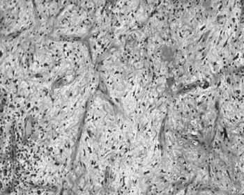
On light microscopy, the myxoid growth was sparsely cellular, with spindle-shaped cells within an extensive basophilic stroma which stained metachromatically with toluidine blue. The tumour was composed mostly of bland fibroblastic cells having scant, pale, tapering cytoplasm and bland, oval nuclei. Scattered fibroblast-like or stellate stromal giant cells with basophilic cytoplasm were also present. There was no nuclear hyperchromasia or pleomorphism. Mitotic figures were absent. The lesion had prominent vascularity with small, thin-walled blood vessels, which were focally arborising or slightly angiomatous in appearance. A mixed cellular infiltrate was present throughout the angiomyxoid stroma, composed of lymphocytes, histiocytes and neutrophil polymorphs (Figure 2). The epithelial cyst showed partial preservation of the stratified squamous epithelial lining, with no granular layer (Figure 3). It was surrounded by a dense inflammatory infiltrate composed mostly of lymphocytes and plasma cells.
Photomicrograph showing bland fibroblastic cells in a myxoid matrix containing scattered collagen fibrils (H&E; original magnification ×250).

Photomicrograph showing stratified squamous epithelial lining of the epithelial cyst, surrounded by a dense, lymphoplasmacytic infiltrate (H&E; original magnification ×100).

Immunohistochemically, the tumour cells were negative for cytokeratin, S100 protein, cluster of differentiation 34 glycoprotein, desmin and Ki67. Occasional fibroblasts, in addition to pericytes, were positive for α-smooth muscle actin. The lymphoid cells were mostly T cells positive for cluster of differentiation 3 and 45RO glycoprotein, with occasional cluster of differentiation 20 glycoprotein positive B cells. Neutrophil polymorphs were identified by positivity for cluster of differentiation 15 glycoprotein. Histiocytes demonstrated positivity for both cluster of differentiation 68 glycoprotein and factor XIIIa. The giant cells were negative for these markers. Only the stratified squamous epithelial lining was positive for cytokeratin.
Following total removal of the tumour, our patient showed no evidence of recurrence over three years of follow up.
Discussion
Superficial angiomyxoma presents a diagnostic challenge to the pathologist as it closely resembles other myxoid lesions. The differential diagnosis includes benign and malignant myxoid tumours and several non-neoplastic cutaneous and subcutaneous lesions, and is shown in Table I.
Differential diagnosis of superficial angiomyxoma

Y = years; CK = cytokeratin; CD34 = cluster of differentiation 34 glycoprotein; += positive; −= negative
• Superficial angiomyxomas represent a relatively uncommon subset of cutaneous myxomatous lesions
• This paper demonstrates that a cutaneous type of angiomyxoma with epithelial-lined structures can occur in deep soft tissue such as the retropharynx
The histopathology and immunophenotype of the presented tumour were those of superficial angiomyxoma.
The striking morphological and immunophenotypical resemblance between our patient's retropharyngeal angiomyxoma and superficial angiomyxomas of the skin suggest a similar mechanism of pathogenesis.
The significance of an epithelial component in the development of angiomyxoma is not clear. Among authors studying large superficial angiomyxoma series, there is general agreement that angiomyxoma is the primary lesion, and the majority of tumours do not exhibit epithelial components.Reference Allen, Dymock and MacCormac1, Reference Calonje, Guerin, McCormick and Fletcher3 Calonje et al. considered epithelial lesions to be trapped skin adnexal structures.Reference Calonje, Guerin, McCormick and Fletcher3 Allen et al. suggested that epithelial proliferation was present in at least some of these lesions.Reference Allen, Dymock and MacCormac1 In our case of retropharyngeal angiomyxoma, skin adnexal structures could not be a source of the observed epithelial component. An epithelial proliferation or ‘growth disorder’ induced by the adjacent angiomyxoma is more conceivable.Reference Allen, Dymock and MacCormac1 The presence of an epithelial component in superficial angiomyxomas constitutes an important diagnostic feature of the tumour. The admixture of neutrophil polymorphs into the chronic inflammatory infiltrate, which is unique to superficial angiomyxoma, may be the consequence of ruptures of epithelial elements into the angiomyxoid tissue. In our case, the giant cells were negative for cluster of differentiation 68 glycoprotein, demonstrating the absence of a foreign body reaction to a ruptured epithelial component. However, the massive chronic inflammatory exudates surrounding the epidermal cyst were probably due to a rupture in the wall of the cyst.