Implications
Evaluation of reactive oxygen species variables may be useful as part of a system for predicting fertility, although a larger sample size is needed to unravel the different effects and improve systems to predict stallion fertility.
Introduction
Over the last few years, there has been a decrease in foaling rates following artificial insemination (AI) in horses (Brinkerhoff et al., Reference Brinkerhoff, Love, Thompson, Blodgett, Teague and Varner2010; Katila et al., Reference Katila, Reilas, Nivola, Peltonen and Virtala2010). This decrease is concomitant with the increased use of transported, cooled semen doses for AI. The reason for this apparent decrease in fertility has not been determined but may be partly because of lack of an effective, objective in vitro evaluation of semen quality – the ‘fertility potential’ of a stallion (Colenbrander et al., Reference Colenbrander, Gadella and Stout2003). Alternatively, it may represent an increased use of stallions whose semen quality is initially good, but whose spermatozoa do not tolerate cooling (Aurich, Reference Aurich2008). In studies using cooled semen from Swedish warmblood stallions, a significant correlation was observed between pregnancy rate and the proportion of spermatozoa with normal morphology in the original ejaculate, as well as a significant negative correlation between pregnancy rate and sperm chromatin fragmentation (Morrell et al., Reference Morrell, Johannisson, Dalin, Hammar, Sandebert and Rodriguez-Martinez2008).
One factor not measured in the previous study is the level of reactive oxygen species (ROS) in stored semen doses. All metabolizing cells, including spermatozoa, produce ROS. Under physiological conditions, these by-products of metabolism do not cause a problem for spermatozoa, as they are mostly inactivated by antioxidants in reproductive tract secretions or their presence is required for the spermatozoa to undergo capacitation (Leclerc et al., Reference Leclerc, de Lamirande and Gagnon1997). Although ROS can adversely affect sperm function (Aitken, Reference Aitken1995), low levels appear to be a prerequisite for normal sperm functioning, being involved in cell signalling and fertilization. However, in situations where spermatozoa are stored before AI, they may be damaged by rising levels of ROS in the environment (Aurich et al., Reference Aurich, Schonherr, Hoppe and Aurich1997). Some ROSs are known to have a negative effect on sperm membrane integrity (Alvarez et al., Reference Alvarez, Touchstone, Blasco and Storey1987; Griveau et al., Reference Griveau, Dumont, Renard, Callegari and LeLannou1995), with excessive levels causing damage to sperm DNA and RNA, possibly initiating apoptosis. On this basis, our work hypothesis is that ROS-associated damage to spermatozoa could explain, at least partly, why the pregnancy rates after AI with cooled semen are lower than after AI with fresh semen.
There are several types of ROS, of which superoxide, peroxide and OH∙ radicals are particularly relevant to spermatozoa. To detect ROS in spermatozoa by flow cytometry, a fluorescent probe is used that binds to the ROS. A method has been described for boar spermatozoa, where hydrogen peroxide is detected by dichlorodihydrofluorescein diacetate (H2DCFDA), whereas superoxide is detected by hydroethidine (HE) (Guthrie and Welch, Reference Guthrie and Welch2006). It has also been shown that compounds other than superoxide might also contribute to the fluorescence from HE derivatives (Zielonka et al., Reference Zielonka, Hardy and Kalyanaranman2009). This method was successfully used in our laboratory (van Wienen et al., Reference van Wienen, Johannisson, Wallgren, Parlevliet and Morrell2011) and measures the content of ROS in the spermatozoa, rather than the actual production, although it can be seen as reflecting ROS production by the cell until the time of measurement. Menadione, which generates superoxide intracellularly as a consequence of disruption of mitochondrial NADH-ubiquinone oxidoreductase (Floreani et al., Reference Floreani, Napoli and Palatini2002), is added to investigate induced ROS production.
Previous studies have examined ROS production in frozen stallion semen (Ortega Ferrusola et al., Reference Ortega Ferrusola, Gonzáles Fernández, Morrell, Salazar Sandoval, Macías García, Rodríguez-Martinez, Tapia and Peña2009), and in cooled stored stallion season at different times during the breeding season (Morrell et al., Reference Morrell, Winblad, Georgakas, Stuhtmann, Humblot and Johannisson2013). The objectives of the present study were to investigate ROS levels occurring naturally or stimulated by menadione in cooled, stored stallion semen doses for AI, and to determine their relationship to parameters of semen quality (sperm morphology, motility, viability and chromatin integrity) and to pregnancy rate, as a step towards developing a multiparameter model for fertility prediction of stallion semen.
Material and methods
Animals and husbandry
Semen (42 ejaculates) was collected from 15 warmblood stallions, 4 to 23 years old, of known fertility at a commercial stud in Sweden (Flyinge AB). The semen was collected during April and May using an artificial vagina (Colorado or Missouri, depending on which functioned best for each stallion), removing the gel with an in-line filter. Semen was collected regularly from the stallions (up to three times per week) throughout the breeding season according to standard practice on a commercial stud. The mean interval between collections was either 4.5 or 5.5 days for all stallions. The semen was extended 1 : 3 with INRA96 (IMV Technologies, l’Aigle Cedex, France) at 35°C to give 1×10⁹ progressively motile spermatozoa in ∼20 ml. The extended semen was packaged in 20 ml syringes and placed in Styrofoam boxes with a cold pack for overnight transport to different studs and to the laboratory at Swedish University of Agricultural Sciences (SLU). On arrival at SLU, sperm concentration was measured using a Nucleocounter-SP100 (Chemometec, Allerød, Denmark), according to the manufacturer’s instructions. For the analysis of plasma membrane integrity, as well as for the analysis of ROS, the extended sperm samples were further diluted by adding 2500 µl of Cellwash (Becton Dickinson, San José, CA, USA) to 500 µl of sperm sample. The diluted samples were centrifuged (400 g, 20 min), and the supernatant was removed. Cellwash was added (500 µl) and these samples were used for analysis at 0 h, or kept at 38°C for analysis after 2 h.
Computer-assisted sperm motility analysis
Sperm motility was assessed using the SpermVision motility analyser (Minitube, Tiefenbach, Germany). Aliquots (5 µl) of extended cooled semen were placed on a microscope slide on the warm stage (38°C) of a microscope fitted with a 20× objective and a 10× eyepiece. Sperm motility of ∼1000 spermatozoa in eight fields of view was analysed using the SpermVision software program using the settings established by the manufacturer. The cell identification area was set at 18 to 90 µm2 with the following classifications: spermatozoa having an average orientation change of head of <7° were defined as immotile, those with average path velocity (VAP) >20 but <40 µm/s and straightness <50 were locally motile, whereas those with VAP >40 µm/s were progressively motile.
Morphology
Aliquots were taken for evaluation of sperm morphology as previously described (Johannisson et al., Reference Johannisson, Morrell, Thorén, Jönsson, Dalin and Rodriguez-Martinez2009). Briefly, aliquots (5 μl) were used to prepare air-dried slides for the assessment of sperm head shape of 500 spermatozoa at 1000× magnification. The following abnormalities were assessed: proximal cytoplasmic droplets, detached heads, acrosome defects, nuclear pouches, midpiece defects and tail defects (bent, coiled or double bend). The proportions were used to calculate the total pathological heads and the number of morphologically normal spermatozoa. In addition, aliquots (100 μl) were fixed in formal saline for counting of 200 spermatozoa on wet smears prepared from a 5 μl drop of the mixture (1000×), focussing on the following: pear-shaped heads, heads narrow at the base, heads with abnormal contour, undeveloped heads, detached heads, narrow (tapering) heads and heads of variable size. The mean proportion of morphologically normal spermatozoa was estimated as the remaining proportion left from total abnormal spermatozoa, including those spermatozoa with distal cytoplasmic droplets, which were considered to be normal. The morphology evaluation was carried out by skilled personnel according to the standard protocol in Swedish Sperm Reference Laboratory at SLU.
Plasma membrane integrity
Assessment of plasma membrane integrity was made using the procedure described by Johannisson et al. (Reference Johannisson, Morrell, Thorén, Jönsson, Dalin and Rodriguez-Martinez2009) at 0 and 2 h after arrival of semen at the laboratory. Briefly, 300 μl of each sample was stained with 0.6 μl of SYBR-14 (final stain concentration 0.4 μM) followed by 3 μl of PI (final stain concentration 24 μM; Live-Dead® Sperm Viability Lit L-7011; Invitrogen, Eugene, OR, USA). After incubating at 38°C for 10 min, the fluorescence was measured using a BD LSR flow cytometer (Becton Dickinson). Excitation was with an argon-ion laser (488 nm). Green fluorescence was detected with a FL1 band pass filter (530/30 nm) with the previous use of the dichroic 555 LP, whereas red fluorescence was measured using a FL3 long pass filter (>670 nm), with the previous use of dichroics 620 SP and 670 LP. A total of 50 000 sperm-specific events were evaluated and calculated as percentages after gating out debris and aggregates based on light-scattering characteristics. The cells were classified as: living (SYBR14-positive/PI-negative), dead (SYBR14-negative/PI-positive) or dying (SYBR14-positive/PI-positive). The percentage of living cells reported was calculated as 100×living/(dead+dying).
ROS
After 0 and 2 h, aliquots (300 μl) of semen were stained with 9 μl of Hoechst 33 258 (HO) (final stain concentration 1.2 μM); 9 μl of HE (final stain concentration 1.2 μM); and 9 μl of H2DCFDA (final stain concentration 60 μM). The HO was purchased from Sigma, Stockholm, Sweden and HE and H2DCFDA from Invitrogen Molecular Probes. At each time point, menadione was added to one set of sperm samples to stimulate ROS production, to determine whether the spermatozoa were capable of producing ROS. The sperm samples without menadione served as controls. Menadione (Sigma-Aldrich) was added at a volume of 3 μl to a final concentration of 200 µM. The samples were incubated at 38°C for 30 min before analysing by flow cytometry. The method is a modification of a previously described method for porcine samples (Guthrie and Welch, Reference Guthrie and Welch2006), the modification being the use of HO exclusion as an independent analysis of living spermatozoa (van Wienen et al., Reference van Wienen, Johannisson, Wallgren, Parlevliet and Morrell2011). Each sample was evaluated using a BD LSR flow cytometer. Excitation was carried out with an argon-ion laser (488 nm) and an HeCd laser (325 nm). Detection of green fluorescence was in FL1 using a band pass filter (530/28 nm), red fluorescence was measured in FL3 using a long pass filter (>670 nm) and blue fluorescence was detected in FL4 with a 510/20 nm discriminating filter. A total of 30 000 sperm-specific events were evaluated and calculated as percentages. The cells were classified as live superoxide-negative, live superoxide-positive or superoxide dead in the HE–HO dot plot, after gating for sperm cells in the forward scatter–side scatter dot plot, and as live hydrogen peroxide-negative, live hydrogen peroxide-positive, dead hydrogen peroxide-negative or dead hydrogen peroxide-positive spermatozoa in the H2DCFDA dot plot.
Sperm chromatin structure assay (SCSA)
The method, using acridine orange (AO) and flow cytometric analysis (FCM), was a modification of the method reported in detail previously (Evenson et al., Reference Evenson, Larsson and Jost2002; Johannisson et al., Reference Johannisson, Morrell, Thorén, Jönsson, Dalin and Rodriguez-Martinez2009). Briefly, the sperm aliquots were extended with Tris-NaCl–ethylenediaminetetraacetic acid (TNE) buffer (0.15 M NaCl, 0.01 M Tris-HCl, 1 mM EDTA (ethylenediaminetetraacetic acid, pH 7.4) to a final sperm suspension of ∼2×10⁷ cells/ml. An aliquot of 0.2 ml was immediately frozen in liquid nitrogen vapour and then transferred to a −80°C freezer, where it was stored until further processing and FCM. Samples were thawed on crushed ice immediately before staining with AO and analysis as follows. Frozen–thawed sperm/TNE buffer aliquots were further diluted with TNE buffer (1 : 9 v/v) The thawed, TNE-extended spermatozoa were subjected to partial DNA denaturation in situ (by mixing with 0.4 ml of a low pH detergent solution containing 0.17% Triton X-100, 0.15 M NaCl and 0.08 N HCl, pH 1.2), followed 30 s later by staining with 1.2 ml of AO (6 g/ml in 0.1 M citric acid, 0.2 M Na2HPO4, 1 mM EDTA, 0.15 M NaCl, pH 6.0). The stained samples were analysed within 3 to 5 min of AO staining. Measurements were made on a FACStar Plus flow cytometer (Becton Dickinson) equipped with standard optics. AO was excited with an argon-ion laser (Innova 90; Coherent, Santa Clara, CA, USA) at 488 nm, running at 200 mW. Equivalent instrument settings were used for all samples. From each sample, a total of 10 000 events were measured at a flow rate of ∼200 cells/s. Data collection was carried out using CellQuest, version 3.3 (Becton Dickinson). Further calculations of SCSA parameters DFI, mean DFI and s.d. DFI (Evenson et al., Reference Evenson, Larsson and Jost2002) were performed using FCS Express version 2 (De Novo Software, Thornhill, Ontario, Canada). For the purpose of this project, only the %DFI (DNA Fragmentation Index, the percentage of cells outside the main population in the alpha-t-histogram) was included in the statistical analysis. The modification from the method reported previously (Johannisson et al., Reference Johannisson, Morrell, Thorén, Jönsson, Dalin and Rodriguez-Martinez2009) was that the frozen–thawed sperm/TNE buffer aliquots were further diluted with TNE buffer (1 : 9 v/v) before adding the detergent solution and the AO.
Pregnancy rates
The pregnancy results after insemination with cooled semen from different studs were compiled at the end of the season by ASVH (The Swedish Warmblood Association) and used to calculate the pregnancy rate and the foaling rate for each stallion for the season. These rates are available online. In addition, pregnancy rates per cycle were evaluated for each stallion (on all cycles for a given mare, giving a value of 0 or 1 at each cycle). As few mares were inseminated with semen from two of the stallions, these results were removed from the statistical analysis. Thus, relationships between sperm quality, superoxide, or hydrogen peroxide variables, and the fertility variables foaling rate and pregnancy rate per cycle were studied for the remaining 13 stallions.
Statistical analysis
The statistical analyses were performed using the SAS software (Version 9, SAS Institute Inc., Cary, NC, USA). Variables were analysed using ANOVA (mixed models under PROC GLM).
For ROS variables, the following model was used:
 $$\eqalignno{\mu _{{(ijkl)}} =W_{{(i)}} {\plus}X_{{(j)}} {\plus}Y_{{(k)}} {\plus}Z_{{(l)}} {\plus}W.X_{{(ij)}} {\rm }{\plus}W.Y_{{(ik)}} \cr {\plus}W.Z_{{(il)}} {\plus}{\rm }X.Y_{{(jk)}} {\plus}{\rm }X.Z_{{(jl)}} {\plus}Y.Z_{{(k.l)}} {\plus}{\varepsilon}_{{(ijkl)}} }$$
where W is the fixed effect of time, X the fixed effect of menadione, Y the fixed effect of rank of ejaculate, and Z the random effect of stallion (random), and W.X the interaction between time and menadione, W.Y the interaction between time and rank of collection, W.Z the interaction between time and stallion (random), X.Y the interaction between menadione and rank of collection, X.Z the interaction between menadione and stallion (random), Y.Z the interaction between rank of collection and stallion (random), and ε ijkl the error term containing more complex interactions. The effects of each fixed factor was tested against its interaction with stallion. Effects of time (W) were tested against W.Z, effect of menadione against X.Z and effect of rank of collection against Y.Z.
Least squares means were calculated for each level of the fixed effect and corresponding interactions. When more than two means were compared, Scheffe’s test was used. Relationships between pairs of variables were analysed using Pearsons’ correlation coefficient. The threshold of significance for all analyses was set at P<0.05.
Results
Conventional measurements of sperm quality in stored semen doses; relationships between them and with fertility estimates
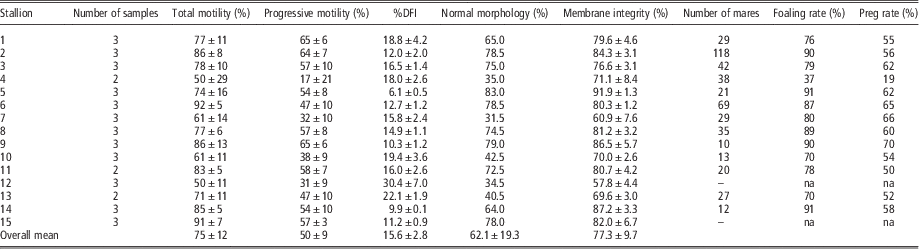
Mean (±s.d.) measurements for sperm motility, membrane integrity and %DFI for each stallion are shown in Table 1, together with the morphology results from one ejaculate per stallion. %DFI was strongly negatively correlated with SYBR living (r=−0.75; P<0.01), and positively correlated with SYBR dead sperm populations (r=0.80; P<0.01). For %DFI, the negative correlation to total motility was close to the threshold of significance (r=−0.52; P<0.1). Total and progressive motility were correlated with membrane integrity (r=0.68; P<0.05 and r=0.71; P<0.01). The foaling rate and pregnancy rate per cycle variables were highly correlated (r=0.96; P<0.0001). Total and progressive motility were correlated with pregnancy rates (r between 0.69 and 0.80; P<0.01).
Variables (means±s.d.) of sperm quality in stored semen doses for AI (15 stallions, n=42 ejaculates)

AI=artificial insemination; NA=not applicable because too few mares (<10) were inseminated with stored semen doses; DFI=DNA Fragmentation Index; foaling rate=% of foaling mares (number of foaling mares/number of mares inseminated)×100; preg rate=pregnancy rate per cycle (number of pregnancies/number of cycles in which insemination took place)×100.
Morphology was performed once for each sample.
Superoxide and hydrogen peroxide production; effects of stallion, time and menadione, and corresponding relationships
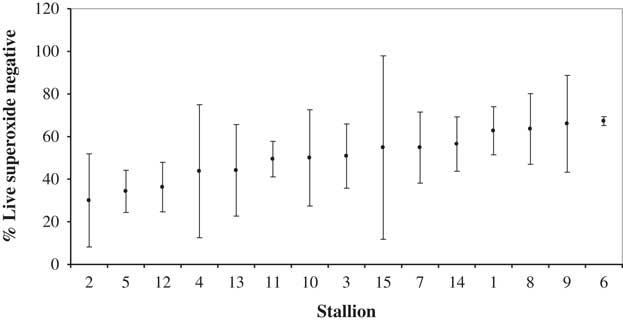
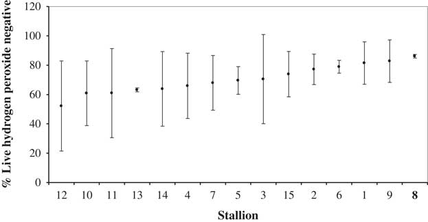
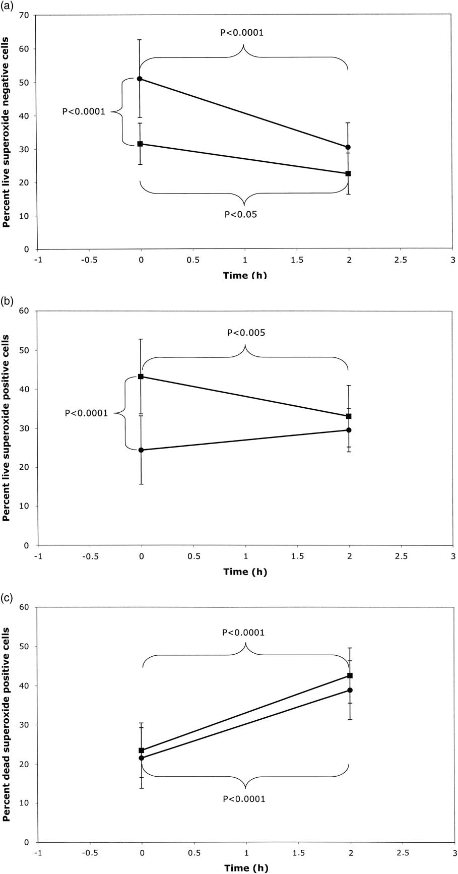
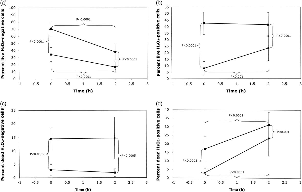
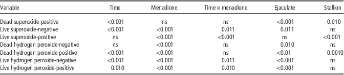
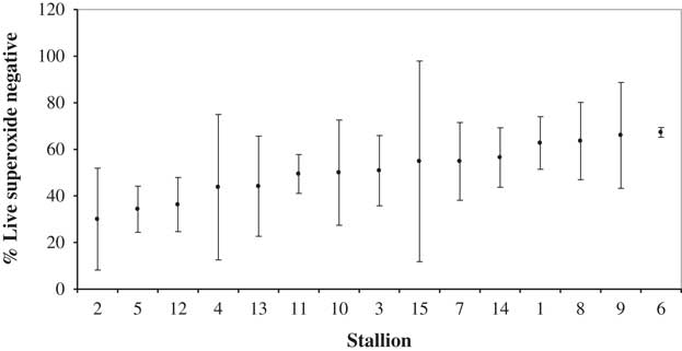
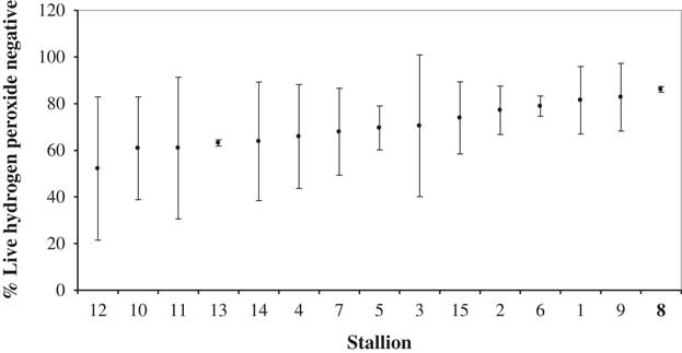
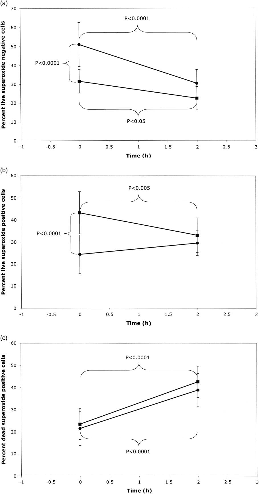
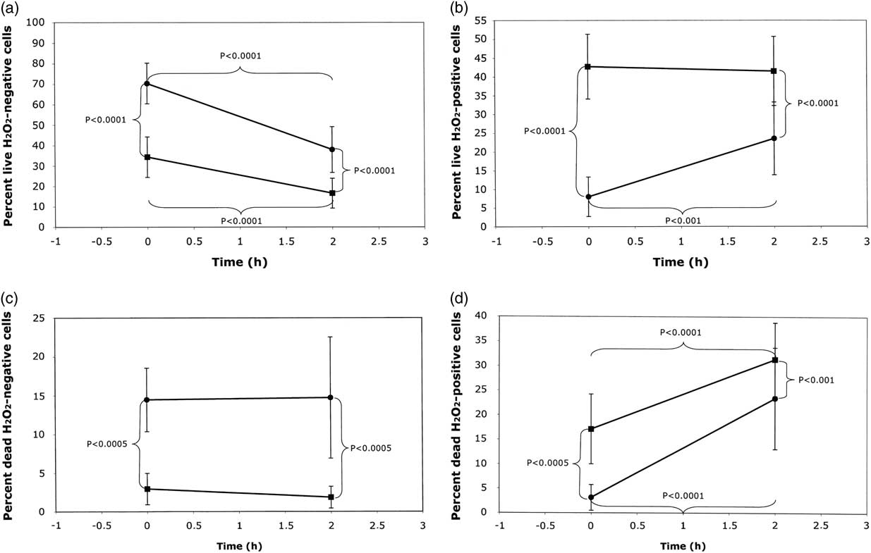
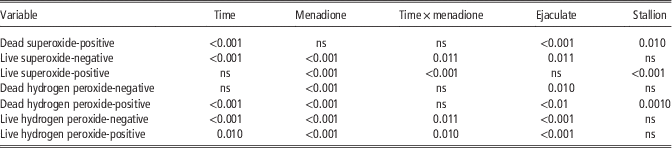
Table 2 presents a summary of the overall effects of different experimental variables on measurement parameters. No time×stallion and menadione×stallion interactions were significant. The mean levels of live superoxide-negative and live hydrogen peroxide-negative spermatozoa (Figures 1 and 2, respectively) varied widely between stallions, ranging from 14% to 50% for live superoxide-negative at 0 h, and from 52% to 86% for live hydrogen peroxide-negative spermatozoa. There was considerable variation between different ejaculates from the same stallion. The effects of menadione treatment and time on ROS production are shown in Figures 3 and 4. The 2-h incubation increased the proportions of superoxide dead from 22±8% to 39±8%, live hydrogen peroxide-positive from 8±5% to 24±10% and dead hydrogen peroxide-positive spermatozoa from 3±3% to 23±10%, while decreasing the percentages of live superoxide-negative from 51±12% to 30±7% and live hydrogen peroxide-negative spermatozoa from 70±10% to 38±11%. Menadione exposure enhanced the proportions of live hydrogen peroxide-positive from 8±5% to 43±9% and dead hydrogen peroxide-positive spermatozoa from 3±3% to 17±7%, whereas the proportions of live superoxide-negative (51±12% to 31±6%), live hydrogen peroxide-negative (71±10% to 34±10%) and dead hydrogen peroxide-negative spermatozoa (14±4% to 3±2%) were reduced. After 2-h incubation and exposure to menadione, decreased proportions of live hydrogen peroxide-negative (38±11% to 17±7%) and dead hydrogen peroxide-negative spermatozoa (15±8% to 2±1%) were seen, whereas the proportions of live hydrogen peroxide-positive (24±10% to 41±9%) and dead hydrogen peroxide-positive spermatozoa (23±10% to 41±9%) were increased. The difference in the reactions to menadione exposure between 0 and 2 h shows a somewhat conflicting picture in that after 2 h the percentage of live superoxide-positive (31±6% to 22±6%) and live hydrogen peroxide-negative cells (34±10% to 17±7%) were decreased, whereas the percentages of dead superoxide-positive (24±7% to 43±7%) and dead hydrogen peroxide-positive cells (17±7% to 31±7%) were increased. Relationships between cell populations after staining for superoxide and hydrogen peroxide at 0 h are shown in Table 3. A positive relationship was found between live superoxide-negative and live hydrogen peroxide-negative spermatozoa, whereas there was no significant relationship between live superoxide-positive and live hydrogen peroxide-positive spermatozoa.
Mean (±s.d.) values for live superoxide-negative cells for individual stallions at 0 h.
Mean (±s.d.) values for live hydrogen peroxide-negative cells for individual stallions at 0 h.
Effects of time and menadione treatment (means±s.d. for all stallions) on percentages of sperm cells found. (a) Live superoxide-negative, (b) live superoxide-positive and (c) dead superoxide-positive. ●=control samples; ■=samples treated with menadione.
Effects of time and menadione treatment (means±s.d. for all stallions) on percentages of sperm cells found. (a) Live hydrogen peroxide-negative, (b) live hydrogen peroxide-positive, (c) dead hydrogen peroxide-negative and (d) dead hydrogen peroxide-positive. ●=control samples; ■=samples treated with menadione.
Significance (P-values) of the effects of time, menadione, rank of ejaculate and stallion on % of sperm labelled for superoxide and hydrogen peroxide
The relationship between hydrogen peroxide and superoxide in cooled stored stallion spermatozoa at time 0 h

Relationships between superoxide and hydrogen peroxide production with measures of sperm quality and pregnancy rates
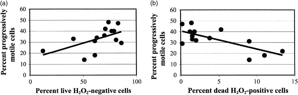
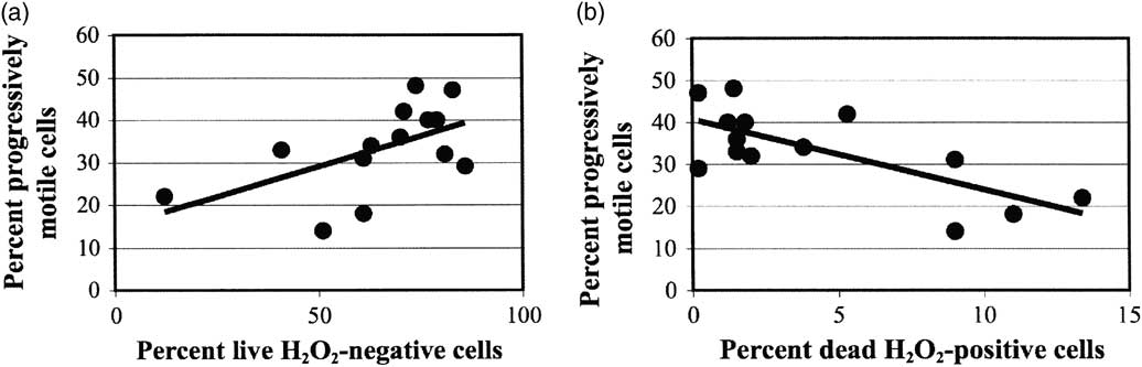
There were no significant correlations between mean values for live superoxide-positive spermatozoa and progressive motility, membrane integrity or %DFI. In contrast, the proportion of live hydrogen peroxide-negative spermatozoa was correlated with progressive motility (r=0.59; P<0.05), whereas the proportion of dead hydrogen peroxide-negative cells was negatively correlated with progressive motility ( r =−0.64; P<0.01) (Figure 5). Live hydrogen peroxide-negative spermatozoa and chromatin damage were negatively correlated (r=−0.54; P<0.05), whereas the correlation between per cent of live hydrogen peroxide-positive spermatozoa and %DFI was not significant. Finally, from non-stimulated samples at time 0, the percentages of superoxide dead and dead hydrogen peroxide-positive sperm were negatively related to the foaling rate (r=−0.52; P<0.07 and r=−0.58; P<0.05, respectively). The negative relationships were stronger when combining results from both assays for ROS (r=−0.61; P<0.05).
Correlations between percentages of (a) live hydrogen peroxide-negative cells and progressively motile cells (R 2=0.31) and (b) dead hydrogen peroxide-positive cells and progressively motile cells (R 2=0.53).
Discussion
The objectives of the present study were to investigate the naturally occurring ROS content of cells in cooled stallion semen doses for AI, including the variation in ROS production among stallions, and the relationship of ROS with other parameters of sperm quality and with pregnancy rate, for future use in a model for fertility prediction. Most of the previous studies on ROS have focused on their levels in frozen–thawed semen, or on the effect of the addition of extraneous ROS, especially hydrogen peroxide, or sources of ROS, to sperm suspensions. As such, superoxide and hydrogen peroxide content of cells in stored stallion semen doses have not been quantified previously and might explain some of the variability in fertility between individuals. It has been reported in other studies that chromatin damage and abnormal morphology can affect the likelihood of pregnancy after AI (Morrell et al., Reference Morrell, Johannisson, Dalin, Hammar, Sandebert and Rodriguez-Martinez2008). ROS are thought to cause chromatin damage in spermatozoa and therefore might be expected to affect fertility adversely; however, as yet, the relationship between ROS production in cooled semen doses and fertility after AI has not been examined.
The results presented here show that percentages of sperm cells positive for ROS and sperm quality varied considerably among individual stallions, and also among ejaculates for certain individual stallions (data not shown).
Previous studies have investigated ROS production and motility in stimulated samples, for example, Baumber et al. (Reference Baumber, Ball, Gravance, Medina and Davies-Morel2000). After 30-min incubation with a stimulator of ROS production, there was a significant decrease in total motility and progressive motility compared with the control (Baumber et al., Reference Baumber, Ball, Gravance, Medina and Davies-Morel2000). In the study reported here, ROS production did not have an adverse effect on sperm motility, although there were positive correlations between progressive motility and the proportions of non-ROS-producing spermatozoa present, possibly suggesting that either the levels produced were not high enough to damage the spermatozoa or that endogenous sources of hydrogen peroxide are not as damaging to spermatozoa as exogenous ones. Although sperm motility was not measured after a 2-h incubation in the present study, it was measured in the stored semen samples after a further 24-h cold storage (6°C). The correlations between motility and hydrogen peroxide populations obtained suggests that the presence of endogenous hydrogen peroxide in the stored samples, coupled with prolonged storage, might have been detrimental to sperm motility. There was a large variation in hydrogen peroxide production among stallions. A larger sample size, with an increased number of stallions and number of ejaculates, would be required to establish the effect of hydrogen peroxide on sperm motility in unstimulated samples.
Menadione was added to stimulate ROS production, to determine whether low ROS levels indicated a lack of ability of the spermatozoa to produce ROS. Menadione treatment was the experimental variable for which the overall effect on measurement variables was most pronounced; it is, however, interesting to note that the effect of menadione on the variable ‘superoxide dead’ was non-significant, meaning that menadione exposure does not kill the cells. Addition of menadione resulted in an increase in ROS production in all ejaculates after incubation for 0 h. After a 2-h incubation, the effects of menadione were less pronounced, although ROS content of cells was still increased in most samples. The results of menadione addition indicate that, although the stallion spermatozoa were initially more receptive to being stimulated to produce ROS, they had a lower ability to respond to menadione when stimulated after 2-h incubation, that is, when the baseline production of ROS was higher than the initial level. This was especially true with respect to production of superoxide, although the capacity for induction of hydrogen peroxide remained to a greater extent. These results suggest that spermatozoa initially producing a high level of ROS could not be stimulated to produce even higher levels, whereas those initially producing a lower level of ROS could be stimulated to produce more. More correlations were seen between different parameters for the stimulated samples than for the non-stimulated samples, for example, motility and ROS production. In the stimulated samples, it appeared that spermatozoa with higher motility had higher metabolism and produce more ROS, or alternatively, that low production of ROS in the sample does not have a negative effect on motility. An additional observation was that a low ROS production was correlated with %DFI, and that spermatozoa with a low %DFI could be stimulated to produce more ROS than those originally producing higher levels. These results suggest that incubation or stimulation of spermatozoa with menadione is associated with a change from superoxide production to hydrogen peroxide production, with an associated increase in chromatin damage.
ROS production in the stored sample can be influenced by the sperm concentration. It is standard practice in Sweden to adjust the sperm concentration to provide approximately one billion progressively motile spermatozoa per cooled semen dose: thus, semen doses with low progressive motility will have a higher sperm concentration in the resulting AI doses. It was our intention to examine ROS levels in standard semen doses as these are used for AI. The results presented here showed a positive correlation between sperm concentration and hydrogen peroxide production at 2 h, suggesting that sperm doses of lower quality may produce more hydrogen peroxide during storage, which in turn may damage the DNA of stored spermatozoa and influence pregnancy rates.
One of the mechanisms behind reduced sperm fertility after storage is lipid peroxidation (oxidative degradation of polyunsaturated lipids in the cell membrane; Alvarez et al., Reference Alvarez, Touchstone, Blasco and Storey1987; Griveau et al., Reference Griveau, Dumont, Renard, Callegari and LeLannou1995; Ortega Ferrusola et al., Reference Ortega Ferrusola, Gonzáles Fernández, Morrell, Salazar Sandoval, Macías García, Rodríguez-Martinez, Tapia and Peña2009). In the present study, membrane integrity was not affected significantly by ROS production in the sample. However, the semen extender (INRA96) used in the current study contains compounds such as HEPES (Batellier et al., Reference Batellier, Duchamp, Vidament, Arnaud, Palmer and Magistrini1998), which are thought to have an anti-oxidative effect (Ermilov et al., Reference Ermilov, Diamond, Sacco and Dozortsev1999).
A negative relationship was found between the percentage of dead cells containing ROS (either superoxide or hydrogen peroxide), and the foaling rate and the same trends were found for relationships with per cycle pregnancy rate. Stronger relationships with fertility may have been found without use of this extender containing anti-oxidative compounds. The relationships shown here could be of biological significance because of the correlation between the different ROS variables and their relationship with fertility. However, the numbers of mares inseminated per stallion were small and these results should be confirmed on a larger set of stallions and mares as inferences from fertility trials cannot be made reliably on small samples (Amann, Reference Amann2005). Interestingly, the foaling rates were mostly better than average for cooled semen, being 70% to 91% in the present study compared with 65.2% reported by Rota et al. (Reference Rota, Furzi, Panzini and Camillo2004).
In conclusion, semen quality is a complex subject involving many interacting variables, with some of the interactions being, as yet, poorly understood. The results presented here based on stored semen samples indicate that strong individual variation exists for superoxide and hydrogen peroxide measurements, that the ROS status can influence sperm quality and may be some of the factors influencing fertility. Combination of ROS variables improves the correlation with fertility, thus indicating the usefulness of including ROS variables in a future model for prediction of the fertility of semen samples. More work and a larger sample size are needed to unravel the different effects and improve systems to predict stallion fertility.
Acknowledgements
We are grateful to Flyinge AB for supplying us with semen for these experiments and to Annika Rikberg and Karin Selin-Wretling for performing the evaluation of morphology. The study was financed by the Foundation for Equine Research, Stockholm, Sweden (H1047052).